University certificate
Scientific endorser

The world's largest faculty of nursing”
Introduction to the Program
With this 100% online Postgraduate diploma, you will stay at the technological forefront of Diagnostic Imaging and optimize your procedures for preparing individuals"

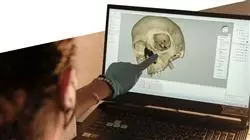
The advent of Industry 4.0 has had a significant impact on the healthcare field, especially in the field of Forensic Diagnostics. Thanks to the advancement of technologies, professionals have access to more detailed and accurate images of injuries, bone fractures and even previous diseases of individuals. Computed Tomography has become the latest trend in this field, providing a detailed view of internal injuries. In this context, it is necessary for nurses to stay at the forefront of technology in this area to optimize their clinical skills and facilitate interdisciplinary communication.
In this way, they will be highly specialized to correctly document forensic evidence and contribute to determine the nature of the injuries.
To contribute in this matter, TECH is developing a Postgraduate diploma in Forensic Diagnostic Imaging Tools for Human Skeleton Imaging Imaging. Its objective is to provide a solid understanding of the analysis of the human body through the most innovative imaging equipment. To achieve this, the academic itinerary will delve into the correct operation of machinery such as X-Ray Tubes, Ultrasound and Magnetic Resonance Imaging. This will enable graduates to provide quality care to individuals and ensure that they are in optimal positions for imaging. Likewise, the syllabus will delve into the bone structure of the human figure, emphasizing the components of the Locomotor System and its main associated pathologies. In this way, professionals will be qualified to obtain information on demographic and anthropological characteristics of human populations and take them into account for the recognition of individuals.
For this course, TECH has created a fully online educational environment, designed to meet the needs of professionals with busy schedules. In this way, they will be able to individually manage their schedules and evaluations. Furthermore, the teaching incorporates the revolutionary Relearningmethod, based on the repetition of key concepts to consolidate knowledge in an optimal way.
A unique, key, and decisive educational experience to boost your professional development”
This Postgraduate diploma in Forensic Diagnostic Imaging Tools for Human Skeleton Imaging Imaging contains the most complete and up-to-date scientific program on the market. The most important features include:
- The development of practical cases presented by experts in Forensic Radiology
- The graphic, schematic and eminently practical contents with which it is conceived gather scientific and practical information on those disciplines that are indispensable for professional practice
- Practical exercises where self-assessment can be used to improve learning
- Its special emphasis on innovative methodologies
- Theoretical lessons, questions to the expert, debate forums on controversial topics, and individual reflection assignments
- Content that is accessible from any fixed or portable device with an Internet connection
You will delve into the structure of the human skeleton to estimate important biological characteristics such as age, sex or height from radiological images"
The program’s teaching staff includes professionals from the field who contribute their work experience to this educational program, as well as renowned specialists from leading societies and prestigious universities.
The multimedia content, developed with the latest educational technology, will provide the professional with situated and contextual learning, i.e., a simulated environment that will provide immersive education programmed to learn in real situations.
This program is designed around Problem-Based Learning, whereby the professional must try to solve the different professional practice situations that arise during the course. For this purpose, students will be assisted by an innovative interactive video system created by renowned and experienced experts.
You will be able to document relevant clinical findings observed during the Diagnostic Imaging process, such as the presence of visible lesions"

With the Relearning system used by TECH, you will reduce the long hours of study and memorization. You will enjoy natural learning!"
Why study at TECH?
TECH is the world’s largest online university. With an impressive catalog of more than 14,000 university programs available in 11 languages, it is positioned as a leader in employability, with a 99% job placement rate. In addition, it relies on an enormous faculty of more than 6,000 professors of the highest international renown.

Study at the world's largest online university and guarantee your professional success. The future starts at TECH”
The world’s best online university according to FORBES
The prestigious Forbes magazine, specialized in business and finance, has highlighted TECH as “the world's best online university” This is what they have recently stated in an article in their digital edition in which they echo the success story of this institution, “thanks to the academic offer it provides, the selection of its teaching staff, and an innovative learning method aimed at educating the professionals of the future”
A revolutionary study method, a cutting-edge faculty and a practical focus: the key to TECH's success.
The most complete study plans on the university scene
TECH offers the most complete study plans on the university scene, with syllabuses that cover fundamental concepts and, at the same time, the main scientific advances in their specific scientific areas. In addition, these programs are continuously being updated to guarantee students the academic vanguard and the most in-demand professional skills. In this way, the university's qualifications provide its graduates with a significant advantage to propel their careers to success.
TECH offers the most comprehensive and intensive study plans on the current university scene.
A world-class teaching staff
TECH's teaching staff is made up of more than 6,000 professors with the highest international recognition. Professors, researchers and top executives of multinational companies, including Isaiah Covington, performance coach of the Boston Celtics; Magda Romanska, principal investigator at Harvard MetaLAB; Ignacio Wistumba, chairman of the department of translational molecular pathology at MD Anderson Cancer Center; and D.W. Pine, creative director of TIME magazine, among others.
Internationally renowned experts, specialized in different branches of Health, Technology, Communication and Business, form part of the TECH faculty.
A unique learning method
TECH is the first university to use Relearning in all its programs. It is the best online learning methodology, accredited with international teaching quality certifications, provided by prestigious educational agencies. In addition, this disruptive educational model is complemented with the “Case Method”, thereby setting up a unique online teaching strategy. Innovative teaching resources are also implemented, including detailed videos, infographics and interactive summaries.
TECH combines Relearning and the Case Method in all its university programs to guarantee excellent theoretical and practical learning, studying whenever and wherever you want.
The world's largest online university
TECH is the world’s largest online university. We are the largest educational institution, with the best and widest online educational catalog, one hundred percent online and covering the vast majority of areas of knowledge. We offer a large selection of our own degrees and accredited online undergraduate and postgraduate degrees. In total, more than 14,000 university degrees, in eleven different languages, make us the largest educational largest in the world.
TECH has the world's most extensive catalog of academic and official programs, available in more than 11 languages.
Google Premier Partner
The American technology giant has awarded TECH the Google Google Premier Partner badge. This award, which is only available to 3% of the world's companies, highlights the efficient, flexible and tailored experience that this university provides to students. The recognition as a Google Premier Partner not only accredits the maximum rigor, performance and investment in TECH's digital infrastructures, but also places this university as one of the world's leading technology companies.
Google has positioned TECH in the top 3% of the world's most important technology companies by awarding it its Google Premier Partner badge.
The official online university of the NBA
TECH is the official online university of the NBA. Thanks to our agreement with the biggest league in basketball, we offer our students exclusive university programs, as well as a wide variety of educational resources focused on the business of the league and other areas of the sports industry. Each program is made up of a uniquely designed syllabus and features exceptional guest hosts: professionals with a distinguished sports background who will offer their expertise on the most relevant topics.
TECH has been selected by the NBA, the world's top basketball league, as its official online university.
The top-rated university by its students
Students have positioned TECH as the world's top-rated university on the main review websites, with a highest rating of 4.9 out of 5, obtained from more than 1,000 reviews. These results consolidate TECH as the benchmark university institution at an international level, reflecting the excellence and positive impact of its educational model.” reflecting the excellence and positive impact of its educational model.”
TECH is the world’s top-rated university by its students.
Leaders in employability
TECH has managed to become the leading university in employability. 99% of its students obtain jobs in the academic field they have studied, within one year of completing any of the university's programs. A similar number achieve immediate career enhancement. All this thanks to a study methodology that bases its effectiveness on the acquisition of practical skills, which are absolutely necessary for professional development.
99% of TECH graduates find a job within a year of completing their studies.
Postgraduate Diploma in Forensic Diagnostic Imaging Tools for Human Skeleton Imaging
Discover at TECH Global University an exceptional opportunity to broaden your knowledge and skills in the fascinating field of forensic radiology with our Postgraduate Diploma in Postgraduate Diploma in Forensic Diagnostic Imaging Tools for Human Skeleton Imaging, specially designed for nursing professionals seeking to excel in this constantly evolving field. Our institution is proud to offer a comprehensive and up-to-date syllabus, ranging from the basic fundamentals to the most advanced diagnostic imaging techniques applied to the human skeletal system. Through our online classes, you will have the flexibility to study from anywhere and at any time, adapting your learning to your schedule and pace of life. At TECH, we understand the critical importance of having highly qualified professionals in the analysis of forensic medical images for the effective resolution of cases. Therefore, our program focuses on providing you with the theoretical and practical knowledge necessary to accurately interpret X-rays, CT scans and other images of the human skeleton.
Advance in forensic diagnostics with this online postgraduate program
From the identification of traumatic injuries to the detection of pathological abnormalities, our program will prepare you to deal with a wide range of forensic scenarios. In addition, you will explore relevant topics such as forensic documentation, chain of custody and crime scene protocols, giving you a comprehensive perspective of working in this field. By enrolling in our Postgraduate Diploma, you will not only acquire advanced knowledge, but you will also have access to a state-of-the-art educational platform. There, you will find interactive teaching resources, updated study material and personalized advice from our teachers, who will guide you through every step of your learning process. Don't miss the opportunity to differentiate yourself in your professional career and make a significant contribution to the field of forensic nursing. Enroll today in our 100% online postgraduate program and become an expert in forensic diagnostic imaging tools for the human skeleton!


