University certificate
The world's largest faculty of medicine”
Introduction to the Program
Thanks to this program, master the mechanisms to interpret the updated imaging tests aimed at detecting a traumatic injury”

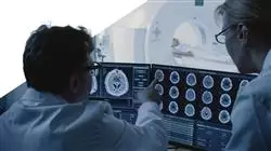
Diagnostic imaging is the best ally for doctors to quickly detect the pathology suffered by a patient who requires urgent intervention and then adapt the treatment and care to the results obtained. Given their relevance, diagnostic mechanisms have undergone an enormous revolution in recent years to enable them to be carried out in a short interval of time and to facilitate the tasks of physicians. Due to the positive impact they have on the possible recovery of the patient, the physician is obliged to master the interpretation of the most advanced tests in order to provide top quality health care.
That is why TECH has created this Hybrid professional master’s degree, in order to provide the health professional with the most updated knowledge in the field of diagnosis of diseases through imaging tests, as well as in the selection of the most appropriate tests based on the pathology in question. Throughout this academic period, students will expand their knowledge in the interpretation of images used to detect heart failure, a vascular lesion in the central nervous system or a bone fracture. Similarly, they will undertake an ultrasound evaluation in special situations such as patients suffering severe trauma, shock or stroke.
In turn, this academic itinerary is distinguished by the collaboration of an International Guest Director of maximum prestige and the best scientific and research results. This specialist is involved in the program through the development of 10 rigorous Masterclasses.
All this theoretical learning phase is developed in a 100% online mode, which enables students to complete their studies without the need to make uncomfortable trips to physical educational centers. In addition, this teaching is complemented with a 3-week internship in a reference hospital, where they will put into real work all their acquired knowledge and perfect their healthcare work.
During this Hybrid professional master’s degree, you will receive a series of rigorous Masterclasses from an internationally renowned expert in the field of Clinical Imaging”
This Hybrid professional master’s degree in Clinical Imaging for Emergency and Critical Care contains the most complete and up-to-date scientific program on the market.The most important features include:
- Development of more than 100 clinical cases presented by medical professionals with expertise in diagnostic imaging in emergency, urgent and critical care situations
- The graphic, schematic, and practical contents with which they are created, provide scientific and practical information on the disciplines that are essential for professional practice
- Updated imaging techniques for the detection of acute pathologies of the cardiovascular system
- State-of-the-art diagnostic imaging methods for detecting various head and neck injuries
- Protocols for performing urgent clinical ultrasound scans in cases of trauma, shock or respiratory failure
- All of this will be complemented by theoretical lessons, questions to the expert, debate forums on controversial topics, and individual reflection assignments
- Content that is accessible from any fixed or portable device with an Internet connection
- In addition, you will be able to carry out a clinical internship in one of the best hospital centers in the world
Complete your exquisite theoretical learning with a hospital internship of 120 hours where, surrounded by the best professionals, you will enhance your skills in diagnostic imaging”
In this proposed Master's program, of a professionalizing nature and blended learning modality, the program is aimed at updating medical experts in diagnostic imaging for patients in critical situations. The contents are based on the latest scientific evidence, and oriented in a didactic way to integrate theoretical knowledge into medical practice, and the theoretical-practical elements will facilitate the updating of knowledge and will allow decision making in patient management.
Thanks to their multimedia content developed with the latest educational technology, they will allow the medical professional to obtain situated and contextual learning, that is to say, a simulated environment that will provide immersive learning programmed to train in real situations. The design of this program is based on Problem-Based Learning, by means of which the student must try to solve the different professional practice situations that arise during the program. For this purpose, students will be assisted by an innovative interactive video system created by renowned experts.
This program will allow you to exercise in simulated environments to face with solvency all the real challenges of your profession"

Become a reference professional in the interpretation of imaging tests by enrolling in this program offered by TECH"
Why study at TECH?
TECH is the world’s largest online university. With an impressive catalog of more than 14,000 university programs available in 11 languages, it is positioned as a leader in employability, with a 99% job placement rate. In addition, it relies on an enormous faculty of more than 6,000 professors of the highest international renown.

Study at the world's largest online university and guarantee your professional success. The future starts at TECH”
The world’s best online university according to FORBES
The prestigious Forbes magazine, specialized in business and finance, has highlighted TECH as “the world's best online university” This is what they have recently stated in an article in their digital edition in which they echo the success story of this institution, “thanks to the academic offer it provides, the selection of its teaching staff, and an innovative learning method aimed at educating the professionals of the future”
A revolutionary study method, a cutting-edge faculty and a practical focus: the key to TECH's success.
The most complete study plans on the university scene
TECH offers the most complete study plans on the university scene, with syllabuses that cover fundamental concepts and, at the same time, the main scientific advances in their specific scientific areas. In addition, these programs are continuously being updated to guarantee students the academic vanguard and the most in-demand professional skills. In this way, the university's qualifications provide its graduates with a significant advantage to propel their careers to success.
TECH offers the most comprehensive and intensive study plans on the current university scene.
A world-class teaching staff
TECH's teaching staff is made up of more than 6,000 professors with the highest international recognition. Professors, researchers and top executives of multinational companies, including Isaiah Covington, performance coach of the Boston Celtics; Magda Romanska, principal investigator at Harvard MetaLAB; Ignacio Wistumba, chairman of the department of translational molecular pathology at MD Anderson Cancer Center; and D.W. Pine, creative director of TIME magazine, among others.
Internationally renowned experts, specialized in different branches of Health, Technology, Communication and Business, form part of the TECH faculty.
A unique learning method
TECH is the first university to use Relearning in all its programs. It is the best online learning methodology, accredited with international teaching quality certifications, provided by prestigious educational agencies. In addition, this disruptive educational model is complemented with the “Case Method”, thereby setting up a unique online teaching strategy. Innovative teaching resources are also implemented, including detailed videos, infographics and interactive summaries.
TECH combines Relearning and the Case Method in all its university programs to guarantee excellent theoretical and practical learning, studying whenever and wherever you want.
The world's largest online university
TECH is the world’s largest online university. We are the largest educational institution, with the best and widest online educational catalog, one hundred percent online and covering the vast majority of areas of knowledge. We offer a large selection of our own degrees and accredited online undergraduate and postgraduate degrees. In total, more than 14,000 university degrees, in eleven different languages, make us the largest educational largest in the world.
TECH has the world's most extensive catalog of academic and official programs, available in more than 11 languages.
Google Premier Partner
The American technology giant has awarded TECH the Google Google Premier Partner badge. This award, which is only available to 3% of the world's companies, highlights the efficient, flexible and tailored experience that this university provides to students. The recognition as a Google Premier Partner not only accredits the maximum rigor, performance and investment in TECH's digital infrastructures, but also places this university as one of the world's leading technology companies.
Google has positioned TECH in the top 3% of the world's most important technology companies by awarding it its Google Premier Partner badge.
The official online university of the NBA
TECH is the official online university of the NBA. Thanks to our agreement with the biggest league in basketball, we offer our students exclusive university programs, as well as a wide variety of educational resources focused on the business of the league and other areas of the sports industry. Each program is made up of a uniquely designed syllabus and features exceptional guest hosts: professionals with a distinguished sports background who will offer their expertise on the most relevant topics.
TECH has been selected by the NBA, the world's top basketball league, as its official online university.
The top-rated university by its students
Students have positioned TECH as the world's top-rated university on the main review websites, with a highest rating of 4.9 out of 5, obtained from more than 1,000 reviews. These results consolidate TECH as the benchmark university institution at an international level, reflecting the excellence and positive impact of its educational model.” reflecting the excellence and positive impact of its educational model.”
TECH is the world’s top-rated university by its students.
Leaders in employability
TECH has managed to become the leading university in employability. 99% of its students obtain jobs in the academic field they have studied, within one year of completing any of the university's programs. A similar number achieve immediate career enhancement. All this thanks to a study methodology that bases its effectiveness on the acquisition of practical skills, which are absolutely necessary for professional development.
99% of TECH graduates find a job within a year of completing their studies.
Hybrid Professional Master’s Degree in Clinical Imaging for Emergency and Critical Care
At TECH Global University, we present our Hybrid Professional Master's Degree in Clinical Imaging for Emergencies, Emergencies and Critical Care, a cutting-edge program designed for health professionals interested in acquiring advanced skills in diagnostic imaging in critical situations. If you want to improve your diagnostic skills and make a decisive contribution to patient care in emergency situations, this master's degree is ideal for you. Our program combines the advantages of online classes with direct interaction in face-to-face sessions, providing you with a complete and flexible learning experience. The online classes will allow you to access the content from anywhere and at any time, adapting to your needs and schedule. You will be able to study at your own pace and review the material as many times as you wish. In addition, you will have the support of specialized tutors who will answer your questions and provide you with personalized guidance. The in-person internship, on the other hand, offer you the opportunity to put your knowledge into practice in a simulated emergency and critical care environment. You will work alongside clinical imaging experts and healthcare professionals in realistic scenarios, where you will be able to apply advanced imaging techniques and make accurate diagnoses in highly complex situations.
Become an expert in Imaging
In our Hybrid Professional Master's Degree in Clinical Imaging for Emergency and Critical Care, you will learn about the latest technologies in diagnostic imaging, such as computed tomography, magnetic resonance imaging and advanced ultrasound. You will acquire skills in real-time image interpretation, clinical case analysis and fast and effective decision making. At the end of the program, you will be able to use imaging as a fundamental tool in the evaluation of patients in critical situations. You will contribute to the early detection of injuries, the identification of urgent pathologies and the planning of appropriate treatments. Your proficiency in clinical imaging for emergency and critical care will make you a highly valued professional prepared to face the challenges of medical care in emergency situations. Don't miss the opportunity to take your diagnostic skills to the next level. Enroll in our Hybrid Professional Master's Degree in Clinical Imaging for Emergency and Critical Care at TECH Global University and excel in your professional career.


