University certificate
The world's largest faculty of veterinary medicine”
Why study at TECH?
Advances in diagnosis and intervention in traumatology make it possible to improve the health of animals in an effective way"

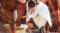
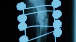
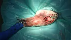
The teaching team of this Advanced master’s degree in Veterinary Traumatology has made a careful selection of the different state-of-the-art surgical techniques for experienced professionals working in the veterinary field, focusing also on medical history, physical examination of the patient, complementary medical tests and interpretation, differential diagnoses and treatment.
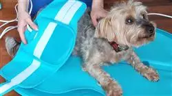
In addition to the techniques most commonly used in small animals, which are those found in traditional practices, this program also places special emphasis on larger species, so a careful selection of techniques used in the diagnosis and treatment of lameness in ruminants, camelids, suidos and equids has been programmed, including the description of musculoskeletal surgery and rehabilitation.
Throughout this specialization, the student will learn all of the current approaches to the different challenges posed by their profession. A high-level step that will become a process of improvement, not only on a professional level, but also on a personal level. Additionally, at TECH we have a social commitment: to help highly qualified professionals to specialize and to develop their personal, social and professional skills throughout the course of their studies.
We will not only take you through the theoretical knowledge we offer, but we will introduce you to another way of studying and learning, one which is simpler, more organic, and efficient. We will work to keep you motivated and to develop your passion for learning, helping you to think and develop critical thinking skills. And we will push you to think and develop critical thinking.
This Advanced master’s degree is designed to give you access to the specific knowledge of this discipline in an intensive and practical way. A great value for any professional.
In addition, as it is a 100% online specialization, it is the student himself who decides where and when to study. Without the restrictions of fixed timetables or having to move between classrooms, this course can be combined with work and family life.
Veterinarians need to update their knowledge of traumatology, as a high number of consultations pertain to this field"
This Advanced master’s degree in Veterinary Traumatology contains the most complete and up-to-date scientific program on the market. Its most notable features are:
- The latest technology in e-learning software
- Intensely visual teaching system, supported by graphic and schematic contents that are easy to assimilate and understand
- The development of practical case studies presented by practising experts
- State-of-the-art interactive video systems
- Teaching supported by telepractice
- Continuous updating and retraining systems
- Self-regulated learning that allows full compatibility with other occupations
- Practical exercises for self-assessment and learning verification
- Support groups and educational synergies: Questions to the expert, discussion and knowledge forums
- Communication with the teacher and individual reflection work
- Content that is accessible from any fixed or portable device with an Internet connection
- Supplementary documentation databases are permanently available, even after the program
A high-level scientific program, supported by advanced technological development and the teaching experience of the best professionals"
Our teaching staff is made up of working professionals. In this way we ensure that we deliver the educational update we are aiming for. A multidisciplinary team of professionals trained and experienced in different fields, who will develop the theoretical knowledge efficiently, but, above all, will put at the service of the study the practical knowledge derived from their own experience.
This mastery of the subject is complemented by the effectiveness of the methodological design of this grand master. Developed by a multidisciplinary team of e-learning experts, it integrates the latest advances in educational technology. This way, you will be able to study with a range of comfortable and versatile multimedia tools that will give you the operability you need in your training.
The design of this program is based on Problem-Based Learning, an approach that views learning as a highly practical process. To achieve this remotely, we will use telepractice. With the help of an innovative interactive video system andlearning from an expert, to acquire the knowledge as if you were facing the scenario you are learning at that moment. A concept that will allow you to integrate and fix learning in a more realistic and permanent way.
We give you the opportunity to experience a deep and complete immersion in the strategies and approaches in Advanced master’s degree"

A program created for professionals who aspire to excellence and that will allow you to acquire new skills and strategies in a fluid and effective way"
Syllabus
The contents of this Advanced master’s degree have been developed by the different experts on this course, with a clear purpose: to ensure that our students acquire each and every one of the necessary skills to become true experts in this field. The content of this Advanced master’s degree enables you to student to learn all aspects of the different disciplines involved in this field. A complete and well-structured program will take you to the highest standards of quality and success.

Through a very well compartmentalized development, you will be able to access the most advanced knowledge of the moment in Veterinary Traumatology"
Module1. Osteogenesis
1.1. History of Orthopedic Surgery
1.1.1. The 5 Steps to Learn Surgery
1.1.2. State of Orthopedic Surgery in the world
1.1.3. Why Should I Study Orthopedics?
1.2 Osteogenic Cells
1.2.1. Osteoblasts
1.2.2. Osteocytes
1.2.3. Osteoclasts
1.3. The Bone Matrix
1.4. The Growth Plate
1.4.1. Organization of the Growth Plate
1.4.2. Blood Supply of the Growth Plate
1.4.3. Structure and Function of the Growth Plate
1.4.4. Cartilaginous Components
1.4.4.1. Reserve Zone
1.4.4.2. Proliferative Zone
1.4.4.3. Hypertrophic Zone
1.4.5. Bone Components (Metaphysis)
1.4.6. Fibrous and Fibrocartilaginous Components
1.5. Diaphyseal Bone Formation
1.6. Cortical Remodelling
1.7. Bone Irrigation
1.7.1. Normal Irrigation of Young Bone
1.7.2. Normal Irrigation of Mature Bone
1.7.2.1. Afferent Vascular System
1.7.2.1.1. Physiology of the Afferent Vascular System
1.7.2.2. Efferent Vascular System
1.7.2.2.1. Physiology of the Efferent Vascular System
1.7.2.3. Intermediate Vascular System of Compact Bone
1.7.2.3.1. Physiology Intermediate Vascular System of Compact Bone
1.7.2.3.2. Bone Cell Activity
1.8. Calcium-Regulating Hormones
1.8.1. Parathyroid Hormone
1.8.1.1. Anatomy of the Parathyroid Glands
1.8.1.2. Parathyroid Hormone Biosynthesis
1.8.1.3. Control of Parathyroid Hormone Secretion
1.8.1.4. Biological Action of Parathyroid Hormone
1.8.2. Calcitonin
1.8.2.1. Thyroid C (Parafollicular) Cells
1.8.2.2. Calcitonin Secretion Regulation
1.8.2.3. Biological Action and Physiological Significance of Calcitonin
1.8.2.4. Primary and Secondary Hypercalcitoninemia
1.8.3. Cholecalciferol (vitamin D)
1.8.3.1. Metabolic Activation of Vitamin D
1.8.3.2. Subcellular Mechanisms of Action of Active Vitamin Metabolites
1.8.3.3. Effects of Hormonal Alterations on the Skeleton under Pathological Conditions
1.8.3.4. Vitamin D Deficiency
1.8.3.5. Vitamin D Excess.
1.8.3.6. Primary and Secondary Hyperparathyroidism
1.9. Biomechanics of Fractures
1.9.1. Bone as a Material
1.9.2. The Role of Bone in Bone Fracture. Basic Mechanical Concepts
1.10. Clinical-Imaging Evaluation of Fracture Repair.
1.10.1. Basic Fracture Repair
1.10.1.1. Callus formation
1.10.1.1.1. Misty Callus
1.10.1.1.2. Stratified Callus
1.10.1.1.3. Fracture Healing
1.10.2. Bone Response to Trauma
1.10.2.1. Inflammatory Phase
1.10.2.2. Repair Phase
1.10.2.3. Remodelling Phase
1.10.3. First Intention Repair
1.10.4. Second Intention Repair
1.10.5. Clinical Union
1.10.5.1. Clinical Union Ranges
1.10.5.2. Repair by Third Intention (delayed joining)
1.10.5.3. Lack of Unity
1.10.6. Bone Behaviour with Different Fixation Methods
1.10.6.1. Bone Behaviour with the Use of External Fixation (splints and bandages)
1.10.6.2. Bone Behaviour with the use of External Fixators
1.10.6.3. Bone Behaviour with the Use of Steinmann Intramedullary Nailing
1.10.6.4. Bone Behaviour with the Use of Plates and Screws
1.10.6.5. Bone Behaviour with the Use of Prosthesis
1.10.6.5.1. Cemented
1.10.6.5.2. Biological
1.10.6.5.3. Blocked
Module 2. Orthopedic Physical Examination
2.1. The Owner’s First Contact with the Hospital
2.1.1. Questions to Be Asked at Reception
2.1.2. Appointment with the Patient
2.1.3. Age, Sex, Race
2.2. Dynamic Orthopedic Physical Examination
2.2.1. Capturing Images and Video
2.2.2. Slow Motion Video
2.2.3. Front, Rear and Side Views
2.2.4. Walking, Trotting, Running
2.3. Static Orthopaedic Physical Examination
2.3.1. Methodology for its Implementation
2.3.2. Degrees of Claudication
2.3.3. Superficial Palpation
2.3.4. Superficial Palpation
2.3.5. The Anatomy that One Should Know in Each Palpated Region
2.3.6. Joint Ranges of Motion and the Goniometer
2.3.7. According to Breed and Age Which Are the 5 Most Commonly Encountered Diseases
2.4. The 20 Most Commonly Encountered Orthopedic Diseases and the Clinical Symptomatology Encountered (I)
2.4.1. Rupture of the Anterior Cruciate Ligament
2.4.2. Patellar Dislocation.
2.4.3. Elbow Dysplasia
2.4.4. Hip Dysplasia
2.4.5. Osteochondritis Dissecans of the Shoulder, Tarsus, Femur
2.4.6. Canine Panosteitis
2.5. Orthopedic Diseases (II)
2.5.1. Radius Curvature
2.5.2. Hypertrophic Osteodystrophy
2.5.3. Hypertrophic Osteoarthropathy.
2.5.4. Contracture of the Carpal Flexor Tendon
2.5.5. Scapulohumeral Instability
2.5.6. Wobbler Syndrome
2.5.7. Intervertebral Disc Disease
2.6. Orthopedic Diseases (III)
2.6.1. Hemivertebra
2.6.2. Lumbosacral Instability
2.6.3. Elbow Dislocation
2.6.4. Dislocation of the Hip
2.6.5. Avascular Necrosis of the Femoral Head (legg perthes)
2.6.5. Polyarthritis (Autoimmune, l-cell, Erlichia, Ricketsia)
2.6.6. Osteoarthritis as a Result of Disease
2.7. Performance of the Dynamic and Static Orthopedic Physical Examination for the Second Time
2.8. The Three Presumptive Diagnoses and How to Differentiate Them
2.9. Diagnostic Work
2.9.1. Radiology
2.9.2. Ultrasound
2.9.3. Laboratory Clinic
2.9.4. Tomography
2.9.5. Magnetic Resonance
2.10. Arthrocentesis
2.10.1. Preparation for Arthrocentesis
2.10.2. Arthrocentesis Approach in Different Regions
2.10.3. Shipment of Samples
2.10.4. Physical Examination of Synovial Fluid
2.10.5. Histochemistry of Synovial Fluid
2.10.6. Osteoarthritis and Prognosis to Its Treatment by Synovial Fluid Assessment
Module 3. Diagnosis of Lameness in Major Species: Ruminants, Swine and Equids
3.1. Medical History
3.1.1. Basic Information
3.1.2. Current Problem
3.1.3. Importance of Conformation
3.1.3.1. Thoracic Limbs
3.1.3.2. Pelvic Limbs
3.1.3.3. Back
3.1.3.4. Digits
3.2. Static Physical Examination
3.2.1. Observation
3.2.2. Palpitation
3.3. Dynamic Physical Evaluation
3.3.1. Basic Biomechanical Characteristics
3.3.2. Examination Protocol
3.3.3. Lameness of the Thoracic Limbs
3.3.4. Lameness of the Pelvic Limb
3.3.5. Types of Claudication
3.3.6. Compensatory Lameness
3.3.7. Classification
3.3.8. Flexion Test
3.4. Diagnostic Anesthesia
3.4.1. Types of Local Anesthetics
3.4.2. General Considerations
3.4.3. Perineural Anesthesia
3.4.4. Intrasynovial Anesthesia
3.4.5. Recommended Action Protocols
3.4.6. Interpretation of Results
3.5. Analysis and Quantification of Movement
3.5.1. Kinetic Study
3.5.2. Kinematic Study
3.6. Radiological Examination
3.6.1. General Considerations
3.6.2. Main Findings and Interpretation
3.7. Ultrasound Examination
3.7.1. General Considerations
3.7.2. Main Findings and Interpretation
3.8. Advanced Diagnostic Imaging Techniques
3.8.1. Magnetic Resonance
3.8.2. Computerized Tomography
3.8.3. Gammagraphy
3.9. Introduction to Treatment
3.9.1. Conservative Medicine Therapies
3.9.2. Surgical Management
3.10. Clinical Examination in Ruminants, Swine and Camelids
3.10.1. Ruminants (Cattle, Sheep) and Camelids (Camels, Alpacas and Llamas)
3.10.2. Swine (Pigs, Wild Boar)
Module 4. Main Musculoskeletal Pathologies in Major Species: Ruminants, Swine and Equids
4.1. Articular Pathology
4.1.1. Classification
4.1.2. Etiology
4.1.3. Main Joints Affected in Sport Horses
4.1.4. Diagnosis
4.1.5. Treatment Management
4.2. Maladaptive Bone Pathology
4.2.1. Etiology
4.2.2. Diagnosis
4.2.3. Treatment Management
4.3. Tendon Pathology
4.3.1. Etiology
4.3.2. Main Areas Affected in Sport Horses
4.3.3. Diagnosis
4.3.4. Treatment Management
4.4. Ligament Pathology
4.4.1. Etiology
4.4.2. Main Areas Affected in Sport Horses
4.4.3. Diagnosis
4.4.4. Treatment Management
4.5. Muscular Pathology
4.5.1. Etiology and Classification
4.5.2. Diagnosis
4.5.3. Treatment Management
4.6. Head, Dorsum and Pelvis Pathologies
4.6.1. Cervical Pathology
4.6.2. Thoracic-Lumbar Pathologies
4.6.3. Lumbo-Sacral Pathologies
4.6.4. Sacroiliac Pathology
4.7. Podotrochlear Pathologies. Palmar Hoof Pain
4.7.1. Etiology
4.7.2. Clinical Signs
4.7.3. Diagnosis
4.7.4. Treatment Management
4.8. Conservative Therapy and Therapeutic Farriery
4.8.1. Nonsteroidal Anti-Inflammatories
4.8.2. Corticosteroids
4.8.3. Hyaluronic Acid
4.8.4. Glycosaminoglycans and Oral Supplements
4.8.5. Bisphosphonates
4.8.6. Polyacrylamide Gel
4.8.7. Other Treatments
4.8.8. Therapeutic Farriery
4.9. Regenerative Biological Therapy
4.9.1. Use of Mesenchymal Cells
4.9.2. Autologous Conditioned Serum
4.9.3. Autologous Protein Solution
4.9.4. Growth Factors
4.9.5. Platelet-Rich Plasma
4.10. Main Musculoskeletal Pathologies in Ruminants, Camelids and Swine
4.10.1. Ruminants (Cattle, Sheep) and Camelids (Camels, Alpacas and Llamas)
4.10.2. Swine (Pigs, Wild Boar)
Module 5. Developmental Diseases: Angular and Flexural Deformities, Osteochondrosis and Subchondral Cyst in Major Species: Ruminants, Swine and Equids
5.1. Angular Deformities Etiopathogenesis
5.1.1. Anatomy
5.1.2. Hormonal Factors
5.1.3. Perinatal and Developmental Factors
5.2. Diagnosis and Preserved Treatment of Angular Deformities
5.2.1. Clinical and Radiography Diagnosis
5.2.2. Use of Splints, Resins and Fittings
5.2.3. Use of Shockwaves
5.3. Surgical Treatment of Angular Deformities
5.3.1. Bone Growth Stimulation Techniques
5.3.2. Bone Growth Delay Techniques
5.3.3. Corrective Ostectomy
5.3.4. Prognosis
5.4. Etiopathogenesis and Diagnosis of Flexural Deformities
5.4.1. Congenital
5.4.2. Acquired
5.5. Conservative Treatment of Flexural Deformities
5.5.1. Physiotherapy and Exercise Control
5.5.2. Medical Treatment
5.5.3. Use of Splints and Resins
5.6. Surgical Treatment of Flexural Deformities
5.6.1. Distal Interphalangeal Joint
5.6.2. Metacarpal/Metatarsal-Phalangeal Joint
5.6.3. Carpal Joint
5.6.4. Tarsal Joint
5.7. Osteochondrosis I
5.7.1. Etiopathogenesis
5.7.2. Diagnosis
5.7.3. Location of Lesions
5.8. Osteochondrosis II
5.8.1. Treatment
5.8.2. Prognosis
5.9. Subchondral Bone Cyst I
5.9.1. Etiopathogenesis
5.9.2. Diagnosis
5.9.3. Location of Lesions
5.10. Subchondral Bone Cyst II
5.10.1. Treatment
5.10.2. Prognosis
Module 6. Skeletal External Fixators and Circular Fixators
6.1. External Fixators
6.1.1. History of the External Skeletal Fixator
6.1.2. Description of the External Fixator
6.2. Parts Constituting the Kirschner-Ehmer Apparatus
6.2.1. Nails
6.2.1.1. Fixators
6.2.2. Connecting Bar.
6.3. Settings of the External Skeletal Fixator
6.3.1. Half Skeletal Fixation Apparatus
6.3.2. Standard Kirschner-Ehmer Apparatus
6.3.3. Modified Kirschner-Ehmer Apparatus
6.3.4. Bilateral External Fixator Model
6.4. Mixed Skeletal Fixator Apparatus
6.5. Methods of Application of the Kirschner-Ehmer Apparatus
6.5.1. Standard method
6.5.2. Modified Method
6.6. External Fixators with Dental Acrylic
6.6.1. The Use of Epoxy Resin
6.6.2. The Use of Dental Acrylics
6.6.2.1. Preparation of Acrylics
6.6.2.2. Application and Setting Time
6.6.2.3. Post-Surgery Care.
6.6.2.4. Removal of the Acrylic
6.6.3. Bone Cement for Use in Fractures of the Spine
6.7. Indications and Uses of External Fixators
6.7.1. Femur
6.7.2. Tibia
6.7.3. Tarsus
6.7.4. Humerus
6.7.5. Radio and Ulna
6.7.6. Carpus
6.7.7. Jaw
6.7.8. Pelvis
6.7.9. Spinal Column
6.8. Advantages and Disadvantages of Using External Fixators
6.8.1. Acquisition of Acrylic Material
6.8.2. Care in the Application of Acrylics
6.8.3. Toxicity of Acrylic
6.9. Postoperative Care
6.9.1. Cleaning of the Acrylic Fixator
6.9.2. Post-Operative Radiographic Studies.
6.9.3. Gradual Removal of the Acrylic
6.9.4. Care when Removing the Fixator
6.9.5. Repositioning of the Acrylic Fixator
6.10. Circular Fixators
6.10.1. History
6.10.2. Components
6.10.3. Structure
6.10.4. Application
6.10.5. Advantages and Disadvantages
Module 7. Intramedullary Nailing
7.1. History
7.1.1. Kuntcher’s Nail
7.1.2. The First Canine Patient with an Intramedullary Nail
7.1.3. The Use of the Steinmann Nail in the1970s
7.1.4. The Use of the Steinmann Nail Today
7.2. Principles of Intramedullary Nail Application
7.2.1. Type of Fractures in Which it Can Be Exclusively Placed
7.2.2. Rotational Instability
7.2.3. Length, Tip and Rope
7.2.4. Normograde and Retrograde Application. Nail Diameter to Medullary Canal Ratio.
7.2.5. Principle of the 3 Points of the Cortex
7.2.6. Behaviour of the Bone and its Irrigation after Intramedullary Nail Fixation. The Steinmann Nail and the Radius
7.3. The Use of Locks with the Steinmann Intramedullary Nail
7.3.1. Principles of Application of Fastenings and Lashings
7.3.2. Barrel Principle
7.3.3. Type of Fracture Line
7.4. Principles of Application of the Tension Band
7.4.1. Pawel’s Principle
7.4.2. Application of Engineering to Orthopedics
7.4.3. Bone Structures where the Tension Band is to Be Applied
7.5. Normograde and Retrograde Application Method of the Steinmann Nail
7.5.1. Proximal Normograde
7.5.2. Distal Normograde
7.5.3. Proximal Retrograde
7.5.4. Distal Retrograde
7.6. Femur
7.6.1. Proximal Femoral Fractures
7.6.2. Fractures of the Distal Third of the Femur
7.6.3. Supracondylar Fractures or Fracture-Separation of the Distal Epiphysis
7.6.4. Intercondylar Femoral Fracture
7.6.5. The Steinmann Intramedullary Nail and Half Kirschner Device
7.6.6. The Steinmann Intramedullary Nail with Locks or Screws
7.7. Tibia
7.7.1. Avulsion of the Tibial Tubercle
7.7.2. Fractures of the Proximal Third
7.7.3. Fractures of the Middle Third of the Tibia
7.7.4. Fractures of the Distal Third of the Tibia
7.7.5. Fractures of the Tibial Malleoli
7.7.6. The Steinmann Intramedullary Nail and Half Kirschner Device
7.7.7. The Steinmann Intramedullary Nail with Locks or Screws
7.8. Humerus
7.8.1. Steinmann Intramedullary Nail in the Humerus
7.8.2. Fractures of the Proximal Fragment
7.8.3. Fractures of the Middle Third or Body of the Humerus
7.8.4. Steinmann Intramedullary Nail Fixation
7.8.5. Steinmann Intramedullary Nail and Auxiliary Fixation
7.8.6. Supracondylar Fractures.
7.8.7. Fractures of the Medial or Lateral Epicondyle
7.8.8. Intercondylar T or Y Fractures
7.9. Ulna
7.9.1. Acromion
7.10. The Extraction of the Steinmann Intramedullary Nail
7.10.1. X-Ray Monitoring
7.10.2. Callus Formation in Steinmann Nail Fractures
7.10.3. Clinical Union
7.10.4. How to Remove the Implant
Module 8. Bone Plates and Screws
8.1. History of Metal Plates in Internal Fixing
8.1.1. The Initiation of Plates for Fracture Fixation
8.1.2. The World Association of Orthopedic Manufacturers (AO/ASIF)
8.1.2.1. Sherman and Lane Plates
8.1.2.2. Steel Plates
8.1.2.3. Titanium Plates
8.1.2.4. Plates of Other Materials
8.1.2.5. Combination of Metals for New Plate Systems
8.2. Different Fixing Systems with Plate 8 (AO/ASIF, ALPS, FIXIN)
8.2.1. AO/ASIF Plates
8.2.2. Advanced Locked Plate System. (ALPS)
8.2.2.1. FIXIN and Its Conical Block
8.3. Instrument Care
8.3.1. Disinfection
8.3.2. Cleaning
8.3.3. Rinsing
8.3.4. Drying
8.3.5. Lubrication
8.4. Instruments Used for the Fixation of Plates and Screws
8.4.1. Self-Tapping Screws and Tap Removal.
8.4.2. Depth Gages
8.4.3. Drilling Guides
8.4.4. Plate Benders and Plate Twisters
8.4.5. Screw Heads
8.4.6. Screws/Bolts
8.5. Use and Classification of Screws
8.5.1. Cancellous Bone Screws
8.5.2. Cortical Bone Screws
8.5.3. Locked Screws/Bolts
8.5.4. Fastening of Screws
8.5.4.1. Use of the Drill
8.5.4.2. Use of the Countersink
8.5.4.3. Borehole Depth Measurement
8.5.4.4. Use of the Tap
8.5.4.5. Introduction to Screws
8.6. Technical Classification of Screws
8.6.1. Big Screws
8.6.2. Small Screws
8.6.3. Mini Screws
8.7. Classification of Screws According to Their Function
8.7.1. Screw with Interfragmentary Compression Effect
8.7.2. The Cortical Bone Screw with Interfragmentary Compression Effect
8.7.3. Screw Reduction and Fixation Techniques with Interfragmentary Compression Effect
8.7.4 Locked Bolts
8.8. Bone Plates
8.8.1. Bases for Fixing with Plates
8.8.2. Classification of Plates According to Their Shape
8.8.3. Dynamic Compression Plates
8.8.3.1. Way of Action
8.8.3.2. Fixing Technique
8.8.3.3. Advantages Provided by Dynamic Compression Plates (DPC)
8.8.3.4. Disadvantages of Dynamic Compression Plates (DPC)
8.8.4. Locked Plates
8.8.4.1. Advantages and Disadvantages
8.8.4.2. Types of Locks
8.8.4.3. Way of Action
8.8.4.4. Fixing Techniques
8.8.4.3. Instruments
8.8.5. Minimum Contact Plates
8.8.6. Mini Plates
8.8.7. Special Plates
8.8.8. Classification of Plates According to Their Function
8.8.8.1. Compression Plate
8.8.8.2. Neutralization Plate
8.8.8.3. Bridge Plate.
8.9. Guide for Proper Selection of Implants
8.9.1. Biological Factors
8.9.2. Physical Factors
8.9.3. Collaboration of the Owner in the Treatment
8.9.4. Table of Implant Size According to Patients Weight
8.10. Guide to the Removal of Bone Plates
8.10.1. Fulfilled Clinical Function
8.10.2. Implant Ruptures
8.10.3. Implant Bends
8.10.4. Implant Migrates
8.10.5. Rejection
8.10.6. Infections
8.10.7. Thermal Interference
Module 9. Pelvis Fractures
9.1. Anatomy of the Pelvis
9.1.1 General Considerations
9.2. Non-Surgical Group
9.2.1. Stable Fractures
9.2.2. Weight of the Patient
9.2.3. Age of the Patient
9.3. Surgical Group
9.3.1. Intra-Articular Fracture
9.3.2. Closure of the Pelvic Canal
9.3.3. Joint Instability of a Hemipelvis
9.4. Fracture Separation of the Sacro-Iliac Joint
9.4.1. Surgical Approach for Reduction and Fixation
9.4.2. Examples of Surgically Treated Fractures
9.5. Fractures of the Acetabulum
9.5.1. Examples of Surgically Treated Fractures
9.6. Fracture of the Ilium
9.6.1. Surgical Approach to the Lateral Surface of the Ilium
9.6.2. Examples of Surgically Treated Cases
9.7. Ischial Fractures
9.7.1. Surgical Approach to the Body of the Ischium
9.7.2. Examples of Surgically Treated Cases
9.8. Pubic Symphysis Fractures
9.8.1. Surgical Approach to the Ventral Surface of the Pubic Symphysis
9.8.2. Reparation Methods
9.9. Fractures of the Ischial Tuberosity
9.9.1. Surgical Approach
9.9.2. Healed, Non-Reduced, Compressive Fractures of the Pelvis
9.10. Postoperative Management of Pelvic Fractures
9.10.1. The Use of the Harness
9.10.2. Waterbed
9.10.3. Neurological Damage
9.10.4. Rehabilitation and Physiotherapy
9.10.5. Radiographic Studies and Evaluation of the Implant and Bone Repair
Module 10. Pelvic Limb Fractures
10.1. General Overview of Pelvic Limb Fractures
10.1.1. Soft Tissue Damage
10.1.2. Neurological Assessment
10.2. Preoperative Care
10.2.1. Temporary Immobilization
10.2.2. Radiographic Studies
10.2.3. Laboratory Exams
10.3. Surgical preparation
10.3.1. Horos
10.3.2. Vpop-Pro
10.3.3. E Clean Orthoplanner
10.4. Fractures of the Proximal Femoral Proximal Third
10.4.1. Avulsion Fracture of the Femoral Head
10.4.2. Fractures of the Femoral Head. Pre-surgical Assessment.
10.4.3. Fracture Separation of the Proximal Epiphysis of the Femur
10.5. Femoral Neck Fracture
10.5.1. Fractures of the Femoral Neck, Greater Trochanter and Femoral Body
10.5.2. Of the Greater Trochanter with or without Dislocation of the Femoral Head
10.5.3. Surgical Procedure Using a Plate and Bone Screws for Fixation of Proximal Fractures
10.5.4. Complications of Femoral Head and Femoral Neck Fractures
10.5.5. Arthroplastic Excision of the Femoral Head and Neck
10.5.6. Total Hip Replacement
10.5.6.1. Cemented System
10.5.6.2. Biological System
10.5.6.3. Locked System
10.6. Fractures of the Middle Third of the Femur
10.6.1. Fractures of the Body of the Femur
10.6.2. Surgical Approach to the Femoral Body
10.6.3. Femoral Body Fracture Fixation
10.6.3.1. Steinmann Nail
10.6.3.2. Locked Nails
10.6.3.3. Plates and Screws
10.6.3.3.1. External Fixators
10.6.3.3.2. System Combinations
10.6.4 Postoperative Care
10.7. Fractures of the Distal Femoral Third
10.7.1. Fracture by Separation of the Distal Femoral Epiphysis or Supracondylar Fracture
10.7.2. Intercondylar Fractures of the Femur
10.7.3. Fracture of the Femoral Condyles. “T- or “Y-Fractures”
10.8. Fractures of the Patella
10.8.1. Surgical Technique
10.8.2. Post-Surgical Treatment
10.9. Fractures of the Tibia
10.9.1. Classification of Fractures of the Tibia and Fibula
10.9.1.1. Avulsion of the Tibial Tubercle
10.9.1.2. Fracture Separation of the Proximal Tibial Epiphysis
10.9.1.3. Fractures of the Proximal Tibia and Fibula
10.9.1.4. Fractures of the Body of the Tibia and Fibula
10.9.2. Internal Fixation
10.9.2.1. Intramedullary Nails
10.9.2.2. Intramedullary Nail and Supplementary Fixation
10.9.2.3. Skeletal External Fixator
10.9.2.4. Bone Plates
10.9.2.5. Mipo
10.9.3. Fractures of the Distal Portion of the Tibia
10.9.3.1. Separation Fracture of the Distal Epiphysis of the Tibia
10.9.3.2. Fractures of the Lateral or Medial Malleolus or Both
10.9.3.2.1. Treatment
10.10. Fractures and Dislocations of the Tarsus, Metatarsus and Phalanges
10.10.1. Calcaneal Fracture
10.10.2. Dislocation of the Intertarsal and Metatarsal Joint
10.10.3. Fracture or Dislocation of the Central Bone of the Tarsus
10.10.4. Fractures of the Metatarsal Bones and Phalanges
Module 11. Thoracic Limb Fractures
11.1. Scapula
11.1.1. Classification of Fractures
11.1.2. Conservative Treatment
11.1.3. Surgical Approach
11.1.3.1. Reduction and Fixation
11.2. Dorsal Dislocation of the Scapula
11.2.1. Diagnosis
11.2.2. Treatment
11.3. Fractures of the Humerus
11.3.1. Fractures of the Proximal Humerus
11.4. Humeral Body Fractures
11.5. Supracondylar Fractures
11.5.1. Open Reduction
11.5.1.1. Medial Approach.
11.5.1.2. Lateral Approach
11.5.2. Fixation of Supracondylar Fractures
11.5.3. Post-Surgical
11.5.4. Fractures of the Medial or Lateral aspect of the Humeral Condyle
11.5.4.1. Surgical Procedure
11.5.4.2. Post-Surgical
11.6. Intercondylar fractures, Condylar T-Fractures, and Y-Fractures
11.6.1. Surgical Procedure for the Reduction and Fixation of Intercondylar Fractures
11.6.2. Pain
11.7. Fractures of the Radius and Ulna
11.7.1. Ulna Fracture Involving the Lunate Curvature
11.7.1.1. Post-Surgical
11.7.2. Separation Fracture of the Proximal Radial Epiphysis
11.7.2.1. Surgical Procedure
11.7.3. Fracture of the Proximal Third of the Ulna and Dislocation of the Radial Head and Distal portion of the Ulna
11.7.4. Fractures of the Proximal Third of the Ulna, Dislocation of the Radial Head and Separation of the Radius and Ulna (Monteggia Fracture)
11.7.5. Fractures of the Radius and Ulna
11.7.5.1. Closed Reduction and External Fixation of the Radius and Ulna
11.7.5.1.1. Masson Splint and Other Coaptation Splints
11.7.5.1.2. Acrylic Splints or Similar Moulds
11.7.5.2. Surgical Approach to the Radius and Ulna Body
11.7.5.2.1. Craniomedial Approach to the Radius
11.7.5.2.2. Craniolateral Approach (Radius and Ulna)
11.7.5.2.3. Caudal or Post-Ulna Approach
11.7.6. Fixation
11.7.6.1. External Fixators
11.7.6.2. Circular Fixators
11.7.6.3. Intramedullary Nails
11.7.6.4. Bone Screws
11.7.6.5. Bone Plates
11.8. Fractures of the Maxilla and Mandible
11.8.1. Fixation of the Mandibular Symphysis
11.8.2. Fixation of Fractures of the Mandibular Body
11.8.2.1. Orthopedic Wire Around the Teeth
11.8.2.2. Orthopedic Wire Ties
11.8.2.3. Intramedullary Nailing
11.8.2.4. Skeletal External Fixator
11.8.2.5. Bone Plates
11.8.2.6. Fractures of the Maxilla
11.8.2.6.1. Treatment of Fractures in Young Growing Animals
11.8.2.6.2. Some Characteristic Aspects of Immature Bone
11.8.2.6.3. Primary Indications for Surgery
11.8.2.6.3.1. Intramedullary Nails
11.8.2.6.3.2. External Skeletal Fixator
11.8.2.6.3.3. Bone Plates
11.9. Distal Fractures
11.9.1. Of the Carpus
11.9.2. Of the Metacarpals
11.9.3. Of the Phalanges
11.9.4. Reconstruction of Ligaments
11.10. Fractures Resulting in Incongruence of the Articular Surface
11.10.1. Fractures Affecting the Growth Nucleus
11.10.2. Classification of the Epiphysis Based on its Type
11.10.3. Classification of Slipped or Split Fractures Involving the Growth Nucleus and Adjacent Epiphyseal Metaphysis
11.10.4. Clinical Assessment and Treatment of Damage to Nucleus Growth
11.10.5. Some of the Most Common Treatments for Premature Physis Closure
Module 12. Reparation of Fractures in Major Species: Ruminants, Swine and Equids
12.1. Bone Metabolism and Healing
12.1.1. Anatomy
12.1.2. Histological Structure
12.1.3. Bone Healing
12.1.4. Biomechanics of the Bone
12.1.5. Classification of Fractures
12.2. Stabilization of Fractures in an Emergency, Decision Making and Transport
12.2.1. Clinical Examination of a Patient With a Suspected Fracture
12.2.2. Stabilization of a Patient With Fractures
12.2.3. Transport of a Patient With a Fracture
12.2.4. Stabilization of Fractures, Decision-Making and Transport of Ruminants (Cattle, Sheep), Camelids (Camels, Alpacas and Llamas) and Swine (Pigs, Wild Boar)
12.3. External Coaptation
12.1.1. Placement of Robert Jones Bandages
12.1.2. Placement of Acrylic Casts
12.1.3. Splints, Bandages With Casts and Combinations
12.1.4. Complications of Acrylic Casts
12.1.5. Removal of Acrylic Casts
12.2. Reducing Fractures, Management of Soft Tissue in the Approach
12.2.1. Displacements of Fracture Strands
12.2.2. Objectives of the Fracture Reduction
12.2.3. Reduction Techniques
12.2.4. Evaluation of Reduction
12.2.5. Management of Soft Tissues
12.2.5.1. Histology and Blood Supply of the Skin
12.2.5.2. Physical Properties and Biomechanics of the Skin
12.2.5.3. Planning the Approach
12.2.5.4. Incisions
12.2.5.5. Wound Closure
12.3. Materials for Implants in Large Animals
12.3.1. Material Properties
12.3.2. Stainless Steel
12.3.3. Titanium
12.3.4. Material Fatigue
12.4. External Fixators
12.4.1. Transfixion Casts
12.4.2. External Fixators
12.4.3. External Fixators of Ruminants (Cattle, Sheep), Camelids (Camels, Alpacas and Llamas) and Swine (Pigs, Wild Boar)
12.5. Instruments for Inserting an Implant
12.5.1. Plate Contouring Instruments
12.5.2. Instruments for Inserting Screws
12.5.3. Instruments for Inserting Plates
12.6. Implants
12.6.1. Screws
12.6.2. Plates
12.6.3. Placement Techniques
12.6.4. Functions of Each Implant
12.6.5 Tension Band
12.7. Bone Grafts
12.7.1. Indications
12.7.2. Removal Sites
12.7.3. Complications
12.7.4. Synthetic Bone Grafts
12.8. Complications of Inserting an Implant
12.8.1. Lack of Reduction
12.8.2. Incorrect Number and Size of Implants
12.8.3. Incorrect Position of the Implant
12.8.4. Complications Related to the Compression Screw
12.8.5. Complications Related to Plates
Module 13. Musculoskeletal Injuries and Infections in Large Animals: Ruminants, Swine and Equids
13.1. Exploration and Wound Types
13.1.1. Anatomy
13.1.2. Initial Assessment, Emergency Treatment
13.1.3. Wound Classification
13.1.4. Wound Healing Process
13.1.5. Factors Influencing Wound Infection and Wound Healing
13.1.6. Primary and Secondary Intention Wound Healing
13.1.7. Particularities in Ruminants and Swine
13.2. Tissue Management, Hemostasis and Suture Techniques
13.2.1. Incision and Tissue Dissection
13.2.2. Hemostasis
13.2.2.1. Mechanical Hemostasis
13.2.2.2. Ligatures
13.2.2.3. Tourniquet
13.2.2.4. Electrocoagulation
13.2.2.5. Chemical Hemostasis
13.2.3. Tissue Management, Irrigation and Suctioning
13.3. Suturing Materials and Techniques
13.3.1. Materials Used
13.3.1.1. Instruments
13.3.1.2. Suture Material Selection
13.3.1.3. Needles
13.3.1.4. Drainages
13.3.2. Approaches to Wound Suturing
13.3.3. Suture Patterns
13.4. Acute Wound Repair
13.4.1. Wound Treatment Medication
13.4.2. Debriding
13.4.3. Hoof Wounds
13.4.4. Emphysema Secondary to Wounds
13.5. Repair and Management of Chronic and/or Infected Wounds
13.5.1. Particularities of Chronic and Infected Wounds
13.5.2. Causes of Chronic Wounds
13.5.3. Management of Severely Contaminated Wounds
13.5.4. Laser Benefits
13.5.5. Larvotherapy
13.5.6. Cutaneous Fistulas Treatment
13.6. Management and Repair of Synovial Wounds, Joint Lavage and Physitis
13.6.1. Diagnosis
13.6.2. Treatment
13.6.2.1. Systemic and Local Antibiotic Therapy
13.6.2.2. Types of Joint Lavage
13.6.2.3. Analgesia
13.6.3. Physitis
13.6.3.1. Diagnosis
13.6.3.2. Treatment
13.6.4. Particularities in Ruminants and Swine
13.7. Bandages, Dressings, Topical Treatments and Negative Pressure Therapy
13.7.1. Types and Indications of the Different Types of Bandages and Dressings
13.7.2. Topical Treatment Types
13.7.3. Ozone Therapy
13.7.4. Negative Pressure Therapy
13.8. Tendon Lacerations Management and Repair
13.8.1. Diagnosis
13.8.2. Emergency Treatment
13.8.3. Paratendinous Laceration
13.8.4. Tenorraphy
13.8.5. Avulsion and Rupture of Tendons in Ruminants
13.8.6. Ligament Lacerations in Ruminants Swine
13.9. Reconstructive Surgery and Skin Grafting
13.9.1. Principles and Techniques of Reconstructive Surgery
13.9.2. Principles and Techniques of Skin Grafts
13.10. Treatment of Exuberant Granulation Tissue Sarcoid Burns
13.10.1. Causes of the Appearance of Exuberant Granulation Tissue
13.10.2. Treatment of Exuberant Granulation Tissue
13.10.3 Sarcoid Appearance in Wounds
13.10.3.1. Wound Associated Sarcoid Type
13.10.3.2. Treatment
13.10.4. Burn Treatment
Module 14. Arthroscopy, Bursoscopy and Tenoscopy in Major Species: Ruminants, Swine and Equids
14.1. Fundamentals and of the Arthroscopy Technique. Arthroscopy Instruments and Equipment
14.1.1. Start of Veterinary Arthroscopy
14.1.2. Arthroscopy Specific Material
14.1.3. Arthroscopy Technique
14.1.3.1. Patient Preparation
14.1.3.2. Insertion and Position of Instruments
14.1.3.3. Triangulation Technique
14.1.3.4. Arthroscopic Diagnosis and Techniques
14.2. Arthroscopic Indications and Technique for the Metacarpo/Metatarsophalangeal Joint
14.2.1. Indications
14.2.2. Arthroscopic Exploration of the Dorsal Recess and Palmar/Patellar Recess
14.2.3. Arthroscopic Surgery of the Distal Dorsal Recess
14.2.3.1. Fragmentation and Osteochondral Fragments
14.2.3.2. Use of Arthroscopy in the Treatment of Condylar Fractures and First Phalangeal Fractures
14.2.3.3. Villonodular Synovitis
14.2.4. Arthroscopic Recessopalmar/Plantar Surgery
14.2.4.1. Removal of Osteochondral Fragments
14.3. Indications and Arthroscopic Technique of the Carpus
14.3.1. Indications
14.3.2. Arthroscopic Exploration of the Antebrachiocarpal: Joint (Radiocarpal)
14.3.3. Arthroscopic Examination: Intercarpal Joint
14.3.4. Arthroscopic Surgery of Antebrachiocarpal and Intercarpal Joints
14.3.4.1. Fragmentation and Osteochondral Fragments
14.3.4.2. Ligament Lacerations
14.3.4.3. Biarticular Fractures
14.3.5. Arthroscopic Examination of the Carpal Joint in Ruminants
14.4. Arthroscopic Indications and Technique for the Distal and Proximal Interphalangeal Joint
14.4.1. Indications
14.4.2. Arthroscopic Exploration of the Distal Interphalangeal Joint
14.4.3. Arthroscopic Surgery of the Distal Interphalangeal Joint
14.4.3.1. Removal of Osteochondral Fragments
14.4.3.2. Subchondral Cysts of the Third Phalange
14.4.4. Arthroscopic Examination of the Proximal Interphalangeal Joint
14.4.5. Arthroscopic Surgery of the Proximal Interphalangeal Joint
14.4.6. Arthroscopic Examination of These Joints in Ruminants
14.5. Arthroscopic Indications and Technique for the Tarsocrural Joint
14.5.1. Indications
14.5.2. Arthroscopic Examination of the Dorsal Recess and Palmar Recess
14.5.3. Arthroscopic Surgery of the Dorsal Recess and Palmar Recess
14.5.3.1. Osteochondrosisdissecans
14.5.3.2. Fractures
14.5.3.3. Collateral Ligament Injuries
14.5.4. Arthroscopic Examination of the Tarsocrural Joint in Ruminants
14.6. Arthroscopic Indications and Technique for the Patellofemoral Joint and Femorotibial Joints
14.6.1. Indications
14.6.2. Arthroscopic Examination of the Patellofemoral Joint
14.6.3. Arthroscopic Surgery of the Patellofemoral Joint
14.6.3.1. Osteochondrosisdissecans
14.6.3.2. Fragmentation of the Patella
14.6.4. Arthroscopic Examination of the Femorotibial Joints
14.6.5. Arthroscopic Surgery of the Femorotibial Joints
14.6.5.1. Cystic Lesions
14.6.5.2. Articular Cartilage Injuries
14.6.5.3. Fractures
14.6.5.4. Cruciate Ligament Injuries
14.6.5.5. Meniscal Injuries
14.6.6. Arthroscopic Exploration of the Patellofemoral Joint and Femorotibial Joints in Ruminants
14.7. Indications and Arthroscopic Technique of the Elbow, Scapulohumeral and Coxofemoral Joints
14.7.1 Indications
14.7.2 Exploration
14.7.3 Scapulohumeral Osteochondrosis
14.7.4 Fractures and Osteochondrosis Dissecans of the Elbow
14.7.5 Soft Tissue and Osteocartilaginous Lesions of the Coxofemoral Joint
14.8. Indications and Arthroscopic Technique of the Flexor Digital Sheath, Carpal and Tarsal Canal
14.8.1. Indications
14.8.2. Exploration
14.8.3. Tenoscopic Surgery
14.8.3.1. Diagnosis and Debridement of Tendon Lacerations
14.8.3.2. Demotomy of Palmar/Plantar Annular Ligament
14.8.3.3. Excision of Osteochondromas and Exostoses
14.8.3.4. Removal of the Accessory Ligament of the SDFT
14.9. Indications and Arthroscopic Technique of the Navicular, Calcaneal, and Bicipital Bursae
14.9.1. Indications
14.9.2. Examinations
14.9.3. Bursoscopic Surgery
14.9.3.1. Laceration at the Calcaneal Insertion of SDFT
14.9.3.2. Fragmentation of the Calcaneal Tuberosity
14.9.3.3. Traumatic Bicipital Bursitis
14.9.3.4. Penetrating Injuries of the Bursapodotrochlea
14.9.3.5. Lacerations of the SDFT in the Bursapodotrochlea
14.10. Postoperative Care, Complications and Rehabilitation Plans
14.10.1. Postoperative Care
14.10.2. Complications Associated with Synovial Endoscopy Techniques
14.10.3 Postoperative Management Rehabilitation Plans
Module 15. Orthopedic Diseases
15.1. Cranial Cruciate Ligament Rupture
15.1.1. Definition
15.1.2. Etiology
15.1.3. Pathogenesis
15.1.4. Clinical Signs
15.1.4.1. Diagnosis
15.1.4.2. Therapy
15.2. Patellar Dislocation and Legg Perthes Disease
15.2.1. Definition
15.2.1.1. Etiology
15.2.1.2. Pathogenesis
15.2.1.3. Clinical Signs
15.2.1.4. Diagnosis
15.2.1.5. Therapy
15.3. Hip Dysplasia and Traumatic Hip Dislocation
15.3.1. Definition
15.3.2. Etiology
15.3.3. Pathogenesis
15.3.4. Clinical Signs
15.3.5. Diagnosis
15.3.6. Therapy
15.4. Elbow Dysplasia
15.4.1. Definition
15.4.2. Etiology
15.4.3. Pathogenesis
15.4.4. Clinical Signs
15.4.5. Diagnosis
15.4.6. Therapy
15.5. Radius Curvature
15.5.1. Definition
15.5.2. Etiology
15.5.3. Pathogenesis
15.5.4. Clinical Signs
15.5.5. Diagnosis
15.5.6. Therapy
15.6. Wobbler Syndrome
15.6.1. Definition
15.6.2. Etiology
15.6.3. Pathogenesis
15.6.4. Clinical Signs
15.6.5. Diagnosis
15.6.6. Therapy
15.7. Lumbosacral Instability
15.7.1. Definition
15.7.2. Etiology
15.7.3. Pathogenesis.
15.7.4. Clinical Signs
15.7.5. Diagnosis
15.7.6. Therapy
15.8. Osteomyelitis, Osteoarthritis and Osteosarcoma
15.8.1. Definition
15.8.2. Etiology
15.8.3. Pathogenesis
15.8.4. Clinical Signs
15.8.5. Diagnosis
15.8.6. Therapy
15.9. Osteochondrosis-Osteochondritis Discordant (Ocd) and Panosteitis
15.9.1. Definition
15.9.2. Etiology
15.9.3. Pathogenesis
15.9.4. Clinical Signs
15.9.5. Diagnosis
15.9.6. Therapy
15.10. Scapulohumeral Instability
15.10.1. Definition
15.10.2. Etiology
15.10.3. Pathogenesis
15.10.4. Clinical Signs
15.10.5. Diagnosis
15.10.6. Therapy
Module 16. Preoperative Aspects in Major Species: Ruminants, Swine and Equids
16.1. Preparation for Surgery: Decision Making, Operation Risks, Patient Considerations
16.1.1. Surgical Risk
16.1.2. Preoperative Patient Evaluation
16.2. Pharmacological Management for On-Site Procedures
16.2.1. Sedation Drugs
16.2.2. Continuous Infusions
16.2.3. Local Anesthetics
16.2.4. Containment Systems, Other Considerations
16.2.5. Selection of Procedures to Be Performed On Site
16.3. General Anesthesia
16.3.1. Inhalation General Anesthesia
16.3.2. Intravenous General Anesthesia
16.4. Recovery from General Anesthesia
16.4.1. Management During Recovery
16.4.2. Factors Affecting Recovery
16.4.3. Different Techniques or Installations for Anesthetic Recovery
16.5. General Surgical Technique
16.5.1. General Aspects
16.5.2. Basic Manipulation of Surgical Instruments
16.5.3. Tissue Incision, Blunt Dissection
16.5.4. Tissue Retraction and Handling
16.5.5. Surgical Irrigation and Suction
16.6. Preparation of the Surgery, Personnel, Patient and Surgical Area
16.6.1. Presurgical Planning
16.6.2. Surgical Attire, Preparation of Surgical Equipment: Gloves, Gowns etc.
16.6.3. Preparation of the Patient and Surgical Area
16.7. Use of Diagnostic Imaging in Orthopedic Surgery
16.7.1. Diagnostic Imaging Techniques
16.7.2. Diagnostic Imaging in Preparation for Surgery
16.7.3. The Use of Intraoperative Imaging
16.8. Disinfection of Material, Sterilization
16.8.1. Cold Disinfection
16.8.2. Packaging the Material
16.8.3. Different Autoclaves and Sterilizing Products
16.9. Orthopedic Surgical Instruments in Large Animals
16.9.1. General Instruments in Orthopedics
16.9.2. Arthroscopic Instruments
16.9.3. Osteosynthesis Instruments
16.10. The Operating Room for Large Animals
16.10.1. Basic Installations
16.10.2. Importance of the Design of the Operating Room, Asepsis
16.10.3. Technical Specifications of the Advanced Surgical Equipment
Module 17. Common Orthopedic Surgeries of the Musculoskeletal System in Major Species: Ruminants, Swine and Equids Part I
17.1. Fractures of Distal Phalanx and Navicular Bone
17.1.1. Distal Phalanx
17.1.1.1. Causes
17.1.1.2. Classification
17.1.1.3. Clinical Signs
17.1.1.4. Treatment
17.1.2. Navicular Bone Fracture
17.1.2.1. Causes
17.1.2.2. Clinical Signs and Diagnosis
17.1.2.3. Treatment
17.1.3. Digital Neurectomy
17.1.4. Bovine Distal Phalanx Fracture
17.1.5. Bovine Pedal Osteitis
17.1.6. Sepsis of the Common Digital Flexor Tendon Sheath in Ruminants
17.1.6.1. Tenosynoviotomy With Resection of Affected Tissue
17.2. Middle Phalanx Fracture
17.2.1. Etiology
17.2.2. Clinical Signs
17.2.3. Diagnosis
17.2.4. Settings
17.2.4.1. Palmar/Plantar Eminence Fractures
17.2.4.1.1. Uni- and Biaxial Fractures
17.2.4.2. Axial Fractures
17.2.4.3. Comminuted Fractures
17.3. Proximal Phalangeal and Proximal Interphalangeal Joints
17.3.1. Osteoarthritis
17.3.2. Subchondral Cystic Lesions
17.3.3. Dislocations and Subluxations
17.3.4. Fracture Configurations
17.3.5. Clinical Signs
17.3.6. Diaphyseal Fractures
17.3.7. Incomplete Sagittal Fractures
17.3.8. Non-Displaced Long Incomplete Sagittal Incomplete Fractures
17.3.9. Displaced Complete Sagittal Fractures
17.3.10. Frontal Fractures
17.3.11. Comminuted Fractures
17.4. Metacarpal- Metatarsal Phalangeal Joint
17.4.1. Proximal Sesamoid Bone Fractures
17.4.1.1. Mid-Body
17.4.1.2. Basal
17.4.1.3. Abaxial
17.4.1.4. Sagittal
17.4.1.5. Biaxial
17.4.2. Osteoarthritis
17.4.3. Subchondral Cystic Lesions
17.4.4. Dislocation
17.4.5. Tenosynovitis/Desmitis/Constriction of the Annular Ligament
17.4.5.1. Mass Removal
17.4.5.2. Section of the Annular Ligament
17.4.5.3. Tendon Debridement
17.5. Metacarpal/Metatarsal Bones
17.5.1. Lateral Condylar Fractures
17.5.1.1. Signs
17.5.1.2. Diagnosis
17.5.1.3. Emergency Treatment
17.5.1.4. Surgery of Displaced Fractures
17.5.1.5. Surgery of Non-Displaced Fractures
17.5.2. Medial Condylar Fractures
17.5.2.1. Open Approach Surgery
17.5.2.2. Minimally Invasive Surgery
17.5.2.3. Post-Surgery Care
17.5.2.4. Prognosis
17.5.3. Transverse Fractures of the Distal Diaphysis of the Third Metacarpal Bone
17.5.3.1. Non-Surgical Treatment
17.5.3.2. Surgical Treatment
17.5.3.3. Prognosis
17.5.4. Diaphyseal Fractures
17.5.4.1. Non-Surgical Treatment
17.5.4.2. Surgical Treatment
17.5.4.3. Prognosis
17.5.5. Distal Physial Fractures
17.5.6. Proximal Articular Fractures
17.5.7. Dorsal Cortical Fractures
17.5.7.1. Non-Surgical Treatment
17.5.7.2. Surgical Treatment
17.5.7.3. Prognosis
17.5.8. Metacarpal/Metatarsal Bone Fractures in Ruminants (Cattle, Sheep) and Camelids Camels, Alpacas and Llamas)
17.6. Rudimentary Metacarpal/Metatarsal Bones
17.6.1. Fractures
17.6.2. Clinical Examination
17.6.3. Diagnosis
17.6.4. Proximal Fractures
17.6.4.1. Debridement
17.6.4.2. Internal Fixation
17.6.4.3. Ostectomy
17.6.4.4. Complete Removal
17.6.4.5. Prognosis
17.6.4.6. Complications
17.6.5. Mid-Body Fractures
17.6.5.1. Non-Surgical Treatment
17.6.5.2. Surgical Treatment
17.6.5.3. Prognosis
17.6.6. Distal Fractures
17.6.6.1. Non-Surgical Treatment
17.6.6.2. Surgical Treatment
17.6.6.3. Prognosis
17.6.7. Exostosis
17.6.7.1. Pathophysiology
17.6.7.2. Clinical Examination
17.6.7.3. Diagnosis
17.6.7.4. Treatment
17.6.7.4.1. Non-Surgical Treatment
17.6.7.4.2. Surgical Treatment
17.6.7.4.3. Prognosis
17.6.8. Polydactyly in Ruminants and Equidae
17.6.9. Neoplasty
17.7. Tendon and Ligament Pathologies That Can Be Resolved Surgically
17.7.1. Carporadic Extensor Carpi Radialis Tendon Rupture
17.7.1.1. Pathophysiology
17.7.1.2. Diagnosis
17.7.1.3. Treatment
17.7.1.4. Prognosis
17.7.2. Biceps Brachii Tendon and Infraspinatus Tendon Pathologies
17.7.2.1. Treatment
17.7.2.1.1. Biceps Tendon Transection
17.7.2.2. Prognosis
17.7.3. Surgery for Suspensory Ligament Desmopathy in the Forelimb
17.7.4. Surgery of Suspensory Ligament Branches
17.7.5. Suspensory Ligament Damage in Ruminants
17.7.6. Tenectomy of the Medial Head of the Deep Digital Flexor Tendon
17.7.7. Surgery for Suspensory Ligament Dismopathy of the Hind Limb
17.7.8. Intermittent Patella Fixation in Equidae
17.7.9. Patella Fixation in Ruminants
17.7.10. Tears or Avulsions of Collateral Ligaments in Ruminants
17.7.11. Cranial Cruciate Ligament Rupture in Ruminants
17.7.11.1. Peri-Surgical Planning
17.7.11.2. Imbrication of Stifle Joint
17.7.11.3. Cranial Cruciate Ligament Replacement
17.7.11.3.1. With Gluteobiceps Tendon
17.7.11.3.2. With Synthetic Material
17.7.11.3.3. Post-Surgery and Prognosis
17.7.12. Damage to Collateral Ligaments of the Stifle
17.7.12.1. Surgery
17.7.12.2. Prognosis
17.7.13. Superficial Digital Flexor Tendon Dislocation
17.8. Muscle Pathologies That Can Be Resolved Surgically
17.8.1. Fibrotic Myopathy
17.8.1.1. Pathophysiology
17.8.1.2. Diagnosis
17.8.1.3. Treatment
17.8.1.4. Prognosis
17.8.2. Arpeo (Equine Reflex Hypertonia)
17.8.2.1. Pathophysiology
17.8.2.2. Diagnosis
17.8.2.3. Treatment
17.8.2.4. Prognosis
17.8.3. Third Peroneal
17.8.3.1. Pathophysiology
17.8.3.2. Diagnosis
17.8.3.3. Treatment
17.8.3.4. Prognosis
17.8.4. Rupture and Avulsion of the Gastrocnemius Muscles
17.8.4.1. Pathophysiology
17.8.4.2. Diagnosis
17.8.4.3. Treatment
17.8.4.4. Prognosis
17.8.5. Aerophagia
17.8.5.1. Pathophysiology
17.8.5.2. Diagnosis
17.8.5.3. Treatment
17.8.5.4. Prognosis
17.8.6. Spastic Paresis
17.9. Arthrodesis
17.9.1. Equine Distal Interphalangeal Joint
17.9.2. Arthrodesis of the Distal Bovine Interphalangeal Joint
17.9.3. Proximal Interphalangeal Joint
17.9.4. Metacarpal/Metatarsophalangeal Joint
17.9.5. Of the Carpus
17.9.6. Of the Shoulder
17.9.7. Of Distal Tarsal Joints
17.9.8. Talo-Calcanea
17.10. Laminitis and Amputations in Ruminants, Swine and Equidae
17.10.1. Laminitis
17.10.1.1. Deep Digital Flexor Tendon Tenotomy
17.10.1.1.1. At Pastern Level
17.10.1.1.2. At Mid Metacarpal-Metatarsal Level
17.10.1.2. Prognosis
17.10.2. Amputations in Ruminants, Swine and Equidae
17.10.2.1. Bovine Digit Amputation
17.10.2.2. Bovine Extra Digit Amputation
17.10.2.3. Tail Amputation
17.10.2.4. Limb Amputation
17.10.2.5. Specifics in Swine
Module 18. Common Orthopedic Surgeries of the Musculoskeletal System in Major Species: Ruminants, Swine and Equids Part II
18.1. Carpus
18.1.1. Pathophysiology
18.1.2. Multifragmentary Fractures
18.1.2.1. Pathogenesis
18.1.2.2. Diagnosis
18.1.2.3. Treatment
18.1.3. Accessory Bone Fracture
18.1.3.1. Pathogenesis
18.1.3.2. Diagnosis
18.1.3.3. Treatment
18.1.3.4. Non-Surgical Treatment
18.1.3.5. Surgical Treatment
18.1.3.6. Prognosis
18.1.4. Carpal Hygroma
18.1.5. Radial Distal Exostosis
18.1.5.1. Clinical Examination
18.1.5.2. Diagnosis
18.1.5.3. Treatment
18.1.5.3.1. Non-Surgical Treatment
18.1.5.3.2. Surgical Treatment
18.1.5.4. Prognosis
18.1.6. Dislocation
18.1.6.1. Pathogenesis
18.1.6.2. Diagnosis
18.1.6.3. Treatment
18.1.6.3.1. Non-Surgical Treatment
18.1.6.3.2. Surgical Treatment
18.1.6.4. Prognosis
18.1.7. Coronation
18.1.7.1. Pathogenesis
18.1.7.2. Diagnosis
18.1.7.3. Treatment
18.1.8. Synovial Osteochondromatosis
18.1.9. Circumscribed Calcinosis
18.1.9.1. Pathophysiology
18.1.9.2. Diagnosis
18.1.9.3. Treatment
18.1.9.4. Prognosis
18.2. Radio and Ulna
18.2.1. Ulna Fracture
18.2.1.1. Anatomy
18.2.1.2. Pathogenesis.
18.2.1.3. Diagnosis
18.2.1.4. Treatment
18.2.1.4.1. Emergency Stabilization
18.2.1.4.2 Non-Surgical Treatment
18.2.1.4.3. Surgical Treatment
18.2.1.5. Prognosis
18.2.1.6. Complications
18.2.2. Radius Fractures
18.2.2.1. Anatomy
18.2.2.2. Pathogenesis.
18.2.2.3. Diagnosis
18.2.2.4. Treatment
18.2.2.4.1. Emergency Stabilization
18.2.2.4.2. Non-Surgical Treatment
18.2.2.4.3. Surgical Treatment
18.2.2.5. Prognosis
18.2.2.6. Complications
18.2.3. Radial Osteochondroma
18.2.3.1. Pathogenesis
18.2.3.2. Diagnosis
18.2.3.3. Treatment
18.2.3.4. Prognosis
18.2.4. Subchondral Cystic Lesions
18.2.5. Enostosis-Like Lesions
18.3. Humerus Fractures
18.3.1. Anatomy
18.3.2. Greater Tubercle Fracture
18.3.2.1. Diagnosis
18.3.2.2. Treatment
18.3.2.2.1. Non-Surgical Treatment
18.3.2.2.2. Surgical Treatment
18.3.2.3. Prognosis
18.3.3. Fracture of the Deltoid Tuberosity
18.3.3.1. Diagnosis
18.3.3.2. Treatment
18.3.3.3. Prognosis
18.3.4. Stress Fractures
18.3.4.1. Diagnosis
18.3.4.2. Treatment
18.3.4.3. Prognosis
18.3.5. Physiological Fractures
18.3.6. Diaphyseal Fractures
18.3.6.1. Diagnosis
18.3.6.2. Treatment
18.3.6.2.1. Non-Surgical Treatment
18.3.6.2.2. Surgical Treatment
18.3.6.3. Prognosis
18.3.7. Supraglenoid Tubercle Fractures
18.3.7.1. Treatment
18.3.7.1.1. Fragment Removal
18.3.7.1.2. Internal Fixation
18.3.7.2. Prognosis
18.4. Tarsus
18.4.1. Osteoarthritis of the Distal Intertarsal Joints
18.4.1.1. Surgical Treatment
18.4.1.2. Post-Surgery Care.
18.4.1.3. Prognosis
18.4.2. Osteoarthritis of Talocalcaneal Joint
18.4.3. Fractures of the Distal Tibia
18.4.4. Talus Bone
18.4.4.1. Trochlear Ridges
18.4.4.2. Sagittal Fractures
18.4.5. Calcaneus
18.4.5.1. Chip Fractures of the Heel Pad
18.4.6. Small Tarsal Bone Fractures
18.4.7. Tarsal Hygroma in Ruminants
18.5. Tibia and Femorotibiorotullary Joint
18.5.1. Enostosis-Like Lesions
18.5.2. Stress Fractures
18.5.2.1. Etiology
18.5.2.2. Signs
18.5.2.3. Diagnosis
18.5.2.4. Treatment
18.5.3. Tibial Fissures
18.5.3.1. Clinical Signs and Diagnosis
18.5.3.2. Treatment
18.5.4. Proximal Physial Fractures
18.5.4.1. Clinical Signs and Diagnosis
18.5.4.2. Treatment
18.5.4.3. Post-Surgery Care.
18.5.4.4. Complications
18.5.4.5. Prognosis
18.5.5. Diaphyseal Fractures
18.5.5.1. Clinical Signs and Diagnosis
18.5.5.2. Treatment
18.5.5.3. Post-Surgery Care.
18.5.5.4. Complications
18.5.5.5. Prognosis
18.5.6. Distal Physial Fractures
18.5.7. Tibial Ridge Fractures
18.5.8. Stifle
18.5.8.1. Patella Fractures
18.5.8.2. Subchondral Cystic Lesions
18.5.8.2.1. Transcondylar Screw
18.6. Femur and Pelvis
18.6.1. Head and Neck Fractures
18.6.2. Third Trochanter Fractures
18.6.3. Diaphysis Fractures
18.6.4. Distal Fractures
18.6.4.1. Prognosis
18.6.5. Pelvis Fractures
18.6.5.1. Clinical Signs
18.6.5.2. Diagnosis
18.6.5.3. Treatment
18.6.5.4. Of the Coxal Tuberosity
18.6.5.4.1. Clinical Signs
18.6.5.4.2. Diagnosis
18.6.5.4.3. Treatment
18.6.5.5. Of the Wing of the Ileum
18.6.5.6. Of the Body of the Ileum
18.6.5.7. Pubis and Ischium
18.6.5.8. Acetabulum
18.7. Dislocation and Subluxations in Ruminants and Equids
18.7.1. Distal Interphalangeal Joint
18.7.2. Proximal Interphalangeal Joint
18.7.3. Metacarpal/ Metatarsal Phalangeal Joint
18.7.4. Carpus
18.7.5. Scapulohumeral Joint
18.7.6. Coxofemoral Joint
18.7.7. Dorsal Defect of the Patella
18.7.8. Lateral Patella Dislocation in Equidae
18.7.9. Of Patella in Calves and Small Ruminants
18.7.9.1. Lateral Capsule Imbrication
18.7.9.2. Transposition of Tibial Tuberosity
18.7.9.3. Sulcoplasty
18.7.10. Of the Tarsal Joint
18.8. Head
18.8.1. Temporomandibular Joint
18.8.1.1. Condylectomy
18.8.2. Craniomaxillofacial Fractures
18.8.2.1. Incisors, Mandible and Premaxillary
18.8.2.1.1. Diagnosis
18.8.2.1.2. Surgical Management
18.8.2.1.3. Pain
18.8.3. Fractures of the Skull and Paranasal Sinuses
18.8.3.1. Clinical Signs and Diagnosis
18.8.3.2. Treatment
18.8.3.3. Post-Surgery Care.
18.8.3.4. Complications
18.8.3.5. Prognosis
18.8.4. Periorbital Fractures
18.8.4.1. Clinical Signs and Diagnosis
18.8.4.2. Treatment
18.8.4.3. Post-Surgery Care
18.8.4.4. Complications
18.8.4.5. Prognosis
18.8.5. Paranasal Sinus Fistulas
18.8.6. Dehorning
18.8.6.1. Indications
18.8.6.2. Techniques
18.8.6.3. Complications
18.8.7. Frontal Sinus Trepanation in Ruminants
18.8.7.1. Indications
18.8.7.2. Anatomy
18.8.7.3. Clinical Signs
18.8.7.4. Technique
18.8.7.5. Post-Surgery Care and Complications
18.8.8. Rostral Resection of Mandible, Premaxilla and Maxilla
18.8.8.1. Treatment
18.8.8.2. Post-Surgery Care
18.8.8.3. Complications
18.8.8.4. Prognosis
18.8.9. Campilorrinuslateralis
18.8.9.1. Treatment
18.8.9.2. Post-Surgery Care
18.8.9.3. Complications
18.8.9.4. Prognosis
18.8.10. Upper and Lower Prognathism
18.8.10.1. Treatment
18.8.10.2. Post-Surgery Care
18.8.11. Suture Periostitis
18.8.11.1. Diagnosis
18.8.11.2. Treatment
18.9. Spinal Column Surgery in Equidae
18.9.1. Considerations of the Patient and Operating Room
18.9.2. Approaches
18.9.3. Incisions Sutures
18.9.4. Anesthetic Recovery
18.9.5. Postoperative Care
18.9.6. Cervical Fractures
18.9.6.1. Atlas and Axis
18.9.6.2. Subluxation and Atlantoaxial Dislocation
18.9.6.3. From C3 to C7
18.9.7. Thoracolumbar Fractures
18.9.7.1. Dorsal Spinal Processes
18.9.7.2. Vertebral Bodies
18.9.8. Traumatic Sacral Injury
18.9.9. Traumatic Coccygeal Injury
18.9.10. Crushed Tail Head Syndrome
18.9.11. Developmental Disorders
18.9.11.1. Cervical Vertebral Stenotic Spinal Myelopathy
18.9.11.1.1. Surgical Treatment
18.9.11.1.1.1. Intervertebral Fusion
18.9.11.1.1.2. Laminectomy
18.9.11.1.2. Complications
18.9.11.2. Oxyphytoatlantoaxial Malformation
18.9.11.3. Atlantoaxial Subluxation
18.9.11.4. Atlantoaxial Instability
18.10. Neurosurgery
18.10.1. Cerebral Trauma Surgery
18.10.2. Peripheral Nerve Surgery
18.10.2.1. General Surgical Repair Techniques
18.10.2.2. Suprascapular and Axillary Nerve Damage
18.10.2.2.1. Treatment
18.10.2.2.2. Non-Surgical Treatment
18.10.2.2.3. Decompression of the Scapular Nerve
18.10.2.2.4. Prognosis
Module19. Rehabilitation of Musculoskeletal Injuries in Sport Horses
19.1. Significance of Musculoskeletal Injuries in Sport Horses
19.1.1. Introduction
19.1.2. Impact of Musculoskeletal Injuries on the Equine Industry
19.1.3. Most Common Musculoskeletal Injuries According to the Equestrian Discipline
19.1.4. Factors Associated With the Incidence of Injuries in Sport Horses
19.2. Physiotherapeutic Assessment of the Horse
19.2.1. Introduction
19.2.2. Clinical Evaluation
19.2.3. Body Alignment Assessment
19.2.4. Static Physical Assessment
19.2.4.1. Palpitation
19.2.4.2. Active Mobility Test
19.2.4.3. Passive Mobility Tests
19.3. Physiotherapeutic Assessment of the Limbs
19.3.1. Physiotherapeutic Assessment of the Thoracic Limbs
19.3.1.1. Scapula and Scapulohumeral Joint
19.3.1.2. Elbow and Forearm Joint
19.3.1.3. Carpal Joint and Shank
19.3.1.4. Distal Joints: Metacarpal/Tarso-Falangeal, Proximal Interphalangeal, Distal Interphalangeal
19.3.2. Physiotherapeutic Assessment of the Pelvic Limbs
19.3.2.1. Coxofemoral and Rump Joints
19.3.3.2. Stifle and Leg Articulation
19.3.3.3. Tarsal Joint
19.4. Physiotherapeutic Assessment of the Head of Vertebral Column
19.4.1. Physiotherapeutic Assessment of the Head
19.4.1.1. Head
19.4.1.2. Hyoid Apparatus
19.4.1.3. Temporomandibular Joint
19.4.2. Physiotherapeutic Assessment of the Vertebral Column
19.4.2.1. Cervical Region
19.4.2.2. Thoracic Region
19.4.2.3. Lumbar Region
19.4.2.4. Sacroiliac Joint
19.5. Neuromuscular Assessment of the Sport Horse
19.5.1. Introduction
19.5.2. Neurological Evaluation
19.5.2.1. Neurological Examination
19.5.2.2. Evaluation of Cranial Nerves
19.5.2.3. Evaluation of Posture and Gait
19.5.2.4. Assessment of Reflexes and Proprioception
19.5.3. Diagnostic Tests
19.5.3.1. Diagnostic Imaging Techniques
19.5.3.2. Electromyography
19.5.3.3. Cerebrospinal Fluid Analysis
19.5.4. Main Neurological Pathologies
19.5.5. Main Muscular Pathologies
19.6. Manual Therapy Techniques
19.6.1. Introduction
19.6.2. Technical Aspects of Manual Therapy
19.6.3. Considerations of Manual Therapy
19.6.4. Main Techniques of Manual Therapy
19.6.5. Manual Therapy in Limbs and Joints
19.6.6. Manual Therapy in the Spine
19.7. Electrotherapy
19.7.1. Introduction
19.7.2. Principles of Electrotherapy
19.7.3. Tissue Electrostimulation
19.7.3.1. Activation of Peripheral Nerves
19.7.3.2. Application of Electric Stimulation
19.7.4. Pain Control
19.7.4.1. Mechanism of Action
19.7.4.2. Indications of Its Use in Pain Control
19.7.4.3. Main Applications
19.7.5. Muscular Stimulation
19.7.5.1. Mechanism of Action
19.7.5.2. Indications for Use
19.7.5.3. Main Applications
19.7.6. Laser Therapy
19.7.7. Ultrasound
19.7.8. Radiofrequency
19.8. Hydrotherapy
19.8.1. Introduction
19.8.2. Physical Properties of Water
19.8.3. Physiological Response to Exercise
19.8.4. Types of Hydrotherapy
19.8.4.1. Aquatic Therapy in Flotation
19.8.4.2. Aquatic Therapy in Semi-Flotation
19.8.5. Main applications of Hydrotherapy
19.9. Controlled Exercise
19.9.1. Introduction
19.9.2. Stretching
19.9.3. Core Training
19.9.4. Cavalleti and Proprioceptive Bracelets
19.10. Rehabilitation Plans
19.10.1. Introduction
19.10.2. Tendo-Ligament Injuries
19.10.2. Muscle Injuries
19.10.3. Bone and Cartilage Lesions

A comprehensive specialized program that will take you through the necessary preparation to compete with the best in your profession”
Advanced Master's Degree in Veterinary Traumatology
.
In the field of veterinary medicine, new and less invasive techniques have emerged, which allow the execution of interventions for minor and/or serious injuries. Every day, animal health professionals receive cases of trauma that require urgent, definitive and quality solutions to preserve the life of large animal species such as camelids or swine, or domestic animals. That is why at TECH Global University we designed an Advanced Master's Degree in Veterinary Traumatology for students to understand various surgical techniques. In addition to having a comprehensive approach in which medical tests can be performed, differential diagnoses and generate a treatment according to the clinical difficulty presented.
Study an Advanced Master's Degree in Veterinary Medicine and Traumatology online
.
This program has a duration for completion of two years, in which you can see more than fifteen modules covering the topics of osteogenesis, physical examination, diagnosis of lameness in older species, major musculoskeletal pathologies in older species, developmental diseases such as deformities and flexural among others, skeletal external fixators and circular fixators, intramedullary nailing, fractures, wounds and infections and other topics developed in depth throughout the academic course. All of the above is necessary to be able to understand from the fundamental concepts, the identification and differentiation between the types of procedures that can be performed in the sector.
Get an Advanced Master's Degree online
.
The contents were created by qualified and outstanding teachers in this area of veterinary medicine, through the methodology created by TECH you will be able to fully understand the theory and practical exercises. This program is completely online and asynchronous, thus, allowing greater freedom in time and place for students to access the curriculum, either from a computer, a tablet or a smartphone. The method used in the classroom is simple and efficient to build critical thinking and obtain the best tools for the development of the profession, and thus stand out in the workplace. Finally, both individual assignments, discussion forums, questions to experts and complementary readings will help to boost assertive learning.


