University certificate
The world's largest faculty of veterinary medicine”
Why study at TECH?
Master all the techniques of minimally invasive veterinary surgery in a theoretical and practical way by taking this complete Hybrid professional master’s degree will be an easy task to accomplish"

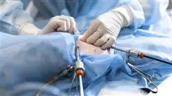
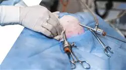
Minimally invasive techniques used in veterinary medicine for the diagnosis and treatment of various diseases found in small animals began 20 years ago and have grown exponentially in the last decade. This growth, which goes hand in hand with the growth of Human Medicine in this field, has been due to several factors: technical development, equipment and instruments, which increasingly offer higher quality images and are more affordable, the development of specific diagnostic and therapeutic techniques in this field, as well as professionals, increasingly trained, which include, preferably, the approach, through these minimally invasive techniques, most of their clinical activity, in addition to owners increasingly concerned about the health of their pets who demand more specialized clinical services, more accurate clinical diagnostics and less invasive treatments which results in less pain and shorter hospital stays for their pets.
For this reason, the Hybrid professional master’s degree in Minimally Invasive Veterinary Surgery in Small Animals develops an exhaustive, relevant and practical update on the different diseases in which these techniques can be applied. Aspects of the approach/management and newer strategies in the field of minimally invasive techniques in small animal veterinary medicine and surgery are detailed. All this through 1,500 hours of theoretical and additional content that a team of teachers specialized in the area has selected for this educational experience.
Once the assessment has been passed, the graduate will have access to a 3-week internship in a prestigious clinical center. During the stay, the professional will be able to see real cases alongside a professional team of reference in the veterinary area, applying the most innovative state-of-the-art procedures. In this way, the activities will be directed to the development and improvement of the competencies necessary for the provision of veterinary care in areas and conditions that require a high level of qualification, and are oriented to the specific program for the exercise of the activity, in a safe environment and high professional performance.
Add to your online study the surgical practices in a prestigious veterinary center with the highest quality standards and the highest level of technology"
This Hybrid professional master’s degree in Minimally Invasive Veterinary Surgery in Small Animals contains the most complete and up-to-date scientific program on the market. The most important features include:
- Development of more than 100 clinical cases presented by veterinary surgery professionals and university professors with extensive experience in minimally invasive techniques
- The graphic, schematic, and practical contents with which they are created provide scientific and practical information on the disciplines that are essential for professional practice
- Veterinary patient assessment and monitoring, the latest international recommendations in minimally invasive surgery
- Comprehensive plans for minimally invasive surgical approach for small animals
- All of this will be complemented by theoretical lessons, questions to the expert, debate forums on controversial topics, and individual reflection assignments
- Content that is accessible from any fixed or portable device with an Internet connection
- In addition, you will be able to carry out a clinical internship in one of the best veterinary centers in the world
Take an intensive 3-week internship in a first class veterinary center and acquire all the knowledge to grow personally and professionally"
In this proposal for a Master's Degree, of a professionalizing nature and hybrid learning modality, the program is aimed at updating veterinary professionals who perform their functions in minimally invasive surgery units in small animals, and who require a high level of qualification. The contents are based on the latest scientific evidence, and oriented in a teaching manner to integrate theoretical knowledge into veterinary practice, and the theoretical-practical elements will facilitate the updating of knowledge and allow decision making in patient management.
Thanks to their multimedia content developed with the latest educational technology, they will allow the veterinary professional to obtain situated and contextual learning, i.e. a simulated environment that will provide immersive learning programmed to train in real situations. This program is designed around Problem-Based Learning, whereby the professional must try to solve the different professional practice situations that arise throughout the program. For this purpose, the students will be assisted by an innovative interactive video system created by renowned and experienced experts.
Undoubtedly, having the opportunity to catch up with the most complete and cutting-edge theoretical and practical content is the best way to internalize the knowledge"

Update your knowledge through the Hybrid professional master’s degree in Minimally Invasive Veterinary Surgery in Small Animals, in a practical way and adapted to your needs"
Syllabus
TECH invests hundreds of hours in the development of each of its programs. For this reason, its qualifications are the result of the effort and perseverance of a team of experts who always strive to create the best content, adapted to the specifications of the sector, market demand and the immediate relevance of the subject matter. All of this is compiled in a convenient and accessible 100% online program that gives students the opportunity to organize their academic experience in a personalized way that is perfectly compatible with their work and personal life.

The use of the Relearning methodology in the development of the content of this program will allow you to update your praxis in a natural and progressive way: without having to invest extra hours in memorizing"
Module 1. Basic Principles in a Laparoscopy
1.1. History of Minimally Invasive Surgery
1.1.1. History of Laparoscopy and Thoracoscopy
1.1.2. Advantages and Disadvantages
1.1.3. New Perspectives
1.2. Laparoscopy Surgery Training
1.2.1. Laparoscopy Training Program
1.2.2. Skills Evalution Systems
1.3. Laparoscopy Surgery Ergonomics
1.3.1. Positioning of Surgical Equipment
1.3.2. Surgeon's Body Posture
1.4. Laparoscopy Surgical Equipment. Laparoscopy Tower
1.4.1. Insufflation Gas
1.4.2. Camera Source
1.4.3. Light Source
1.5. Laparoscopy Surgical Instruments
1.5.1. Trocars
1.5.2. Dissection, Cutting and Aspiration Instruments
1.5.3. Auxiliary Instruments
1.6. Energy Systems
1.6.1. Physical principles |
1.6.2. System Types. Monopolar, Bipolar, Sealent
1.7. Laparoscopic Suture
1.7.1. Extracorporeal Suture
1.7.2. Intracorporeal Suture
1.7.3. New Systems and Suture Materials
1.8. Access to the Abdomen and Creation of the Pneumoperitoneum
1.8.1. Access to the Abdomen
1.8.2. Creation of the Pneumoperitoneum
1.9. Laparoscopy Surgical Complications
1.9.1. Intraoperative complications
1.9.2. Immediate Postoperative Complications
1.9.3. Conversion
1.10. Single Incision Laparoscopy and NOTES
1.10.1. Basic Management and Ergonomics Principles
1.10.2. Surgical Techniques of Single Incision Laparoscopy
1.10.3. Surgical Techniques of NOTES
Module 2. Urinary, Reproductive and Digestive System Diseases
2.1. Anatomy and Physiology of the Male and Female Reproductive System
2.1.1. Anatomy of the Female Reproductive System
2.1.2. Anatomy of the Male Reproductive System
2.1.3. Reproduction Physiology
2.2. Pyometra and Stump Pyometra. Ovarian Tumors and Ovarian Remnant Syndrome
2.2.1. Pyometra
2.2.2. Stump Pyometra
2.2.3. Ovarian Remnant Syndrome
2.2.4. Ovarian Tumors
2.3. Prostate and Testicles. Prostatic Hyperplasia, Prostatic Cysts, Prostatitis and Prostatic Abscesses, Prostatic Neoplasms, Testicular Neoplasms
2.3.1. Prostatic Hyperplasia
2.3.2. Cysts, Abscesses, Prostatitis
2.3.3. Prostatic Neoplasms
2.3.4. Testicular Neoplasms
2.4. Urinaru Anatomy
2.4.1. Kidney
2.4.2. Urether
2.4.3. Bladder
2.4.4. Urethra
2.5. Urinary Stones
2.5.1. Diagnosis
2.5.2. Treatment
2.6. Urinary Incontinence, Urinary System Tumors, Ectopic Urethers
2.6.1. Urinary Incontinence
2.6.1.1. Diagnosis
2.6.1.2. Treatment
2.6.2. Urinary System Tumors
2.6.2.1. Diagnosis
2.6.2.2. Treatment
2.6.3. Ectopic Urethers
2.6.3.1. Diagnosis
2.6.3.2. Treatment
2.7. Digestive System
2.7.1. Stomach
2.7.2. Intestine
2.7.3. Liver
2.7.4. Bladder
2.8. Dilatation-Torsion Syndrome
2.8.1. Diagnosis
2.8.2. Treatment
2.9. Gastric and Intestinal Foreign Bodies
2.9.1. Diagnosis
2.9.2. Treatment
2.10. Digestive and Liver Tumors
2.10.1. Diagnosis
2.10.2. Treatment
Module 3. Splenic, Extrahepatic, Endocrine and Upper Respiratory Tract Diseases
3.1. Splenic Masses
3.1.1. Diagnosis
3.1.2. Treatment
3.2. Portosystemic Shunt
3.2.1. Diagnosis
3.2.2. Treatment
3.3. Extrahepatic Biliary Tree Diseases
3.3.1. Diagnosis
3.3.2. Treatment
3.4. Endocrine Anatomy
3.4.1. Adrenal Anatomy
3.4.2. Pancreas Anatomy
3.5. Adrenal Glands
3.5.1. Adrenal Masses
3.5.1.1. Diagnosis
3.5.1.2. Treatment
3.6. Pancreas
3.6.1. Pancreatitis
3.6.2. Adrenal Masses
3.7. Airway Anatomy
3.7.1. Nostrils
3.7.2. Nasal Cavity
3.7.3. Larynx
3.7.4. Trachea
3.7.5. Lungs
3.8. Laryngeal Paralysis
3.8.1. Diagnosis
3.8.2. Treatment
3.9. Brachycephalic Syndrome
3.9.1. Diagnosis
3.9.2. Treatment
3.10. Nasal Tumors. Nasal Aspergillosis. Nasopharyngeal Stenosis
3.10.1. Diagnosis
3.10.2. Treatment
Module 4. Thoracic Cavity Diseases. Inguinal and Perineal Hernia Laparoscopy and Thoracoscopy Anaesthesia
4.1. Tracheal Collapse
4.1.1. Diagnosis
4.1.2. Treatment
4.2. Thoracic Anatomy
4.2.1. Thoracic Cavity
4.2.2. Pleura
4.2.3. Mediastinum
4.2.4. Heart
4.2.5. Esophageal
4.3. Pericardial Effusion and Masses
4.3.1. Diagnosis
4.3.2. Treatment
4.4. Pleural Effusion and Chylothorax
4.4.1. Etiology
4.4.2. Diagnosis
4.4.3. Chylothorax
4.4.3.1. Diagnosis and Treatment
4.5. Vascular Anomalies
4.5.1. Persistent Right Aortic Arch
4.5.1.1. Diagnosis
4.5.1.2. Treatment
4.6. Pulmonary Pathologies
4.6.1. Pulmonary Tumors
4.6.2. Foreign Bodies
4.6.3. Pulmonary Lobe Torsion
4.7. Mediastinal Masses
4.7.1. Diagnosis and Treatment
4.8. Inguinal and Perineal Hernia
4.8.1. Anatomy
4.8.2. Inguinal Hernia
4.8.3. Perineal Hernia
4.9. Laparoscopy Surgery Anaesthesia
4.9.1. Considerations
4.9.2. Complications
4.10. Thoracoscopy Surgery Anaesthesia
4.10.1. Considerations
4.10.2. Complications
Module 5. Laparoscopic Techniques for the Reproductive, Endocrine, Splenic and Portosystemic Shunt Systems
5.1. Female Sterilization Technique. Ovariectomy
5.1.1. Indications
5.1.2. Trocar Positioning and Placement
5.1.3. Technique
5.2. Female Sterilization Technique. Ovariohysterectomy
5.2.1. Indications
5.2.2. Trocar Positioning and Placement
5.2.3. Technique
5.3. Laparoscopic Treatment of Ovarian Remnants
5.3.1. Indications
5.3.2. Trocar Positioning and Placement
5.3.3. Technique
5.4. Male Sterilization Technique
5.4.1. Indications
5.4.2. Trocar Positioning and Placement
5.4.3. Technique
5.5. Laparoscopic Intrauterine Insemination
5.5.1. Indications
5.5.2. Trocar Positioning and Placement
5.5.3. Technique
5.6.Excision of Ovarian Tumors
5.6.1. Indications
5.6.2. Trocar Positioning and Placement
5.6.3.Technique
5.7. Adrenalectomy
5.7.1. Indications
5.7.2. Trocar Positioning and Placement
5.7.3. Technique
5.8. Pancreatic Biopsy and Pancreatectomy
5.8.1. Indications
5.8.2. Trocar Positioning and Placement
5.8.3. Technique
5.9. Extrahepatic Shunt
5.9.1. Indications
5.9.2. Trocar Positioning and Placement
5.9.3. Technique
5.10. Splenic Biopsy and Splenectomy
5.10.1. Indications
5.10.2. Positioning
5.10.3. Technique
Module 6. Laparoscopic Techniques for the Urinary and Digestive systems
6.1. Assisted Cystoscopy by Laparoscopy
6.1.1. Indications
6.1.2. Trocar Positioning and Placement
6.1.3. Technique
6.2. Renal Biopsy
6.2.1. Indications
6.2.2. Trocar Positioning and Placement
6.2.3. Technique
6.3. Ureteronephrectomy
6.3.1. Indications
6.3.2. Trocar Positioning and Placement
6.3.3. Technique
6.4. Omentalization of Renal Cysts
6.4.1. Indications
6.4.2. Trocar Positioning and Placement
6.4.3. Technique
6.5. Ureterotomy
6.5.1. Indications
6.5.2. Trocar Positioning and Placement
6.5.3. Technique
6.6. Ureteral Reimplantation
6.6.1. Indications
6.6.2. Trocar Positioning and Placement
6.6.3. Technique
6.7. Artifical Bladder Sphincter Placement
6.7.1. Indications
6.7.2. Trocar Positioning and Placement
6.7.3. Technique
6.8. Liver Biopsy and Hepatectomy
6.8.1. Indications
6.8.2. Trocar Positioning and Placement
6.8.3. Technique
6.9. Gastropexy
6.9.1. Indications
6.9.2. Trocar Positioning and Placement
6.9.3. Technique
6.10. Extraction of Foreign Bodies from the Intestines
6.10.1. Indications
6.10.2. Trocar Positioning and Placement
6.10.3. Technique
Module 7. Laparoscopic Techniques in Extrahepatic Biliary Tree, Inguinal and Perineal Hernias. Thoracoscopic Techniques. General, Pericardium, Pleural Effusion, Vascular Rings, and Mediastinal Masses
7.1. Cholecystectomy
7.1.1. Indications
7.1.2. Trocar Positioning and Placement
7.1.3. Technique
7.2. Inguinal Hernias
7.2.1. Indications
7.2.2. Trocar Positioning and Placement
7.2.3. Technique
7.3. Perineal Hernias. Cystopexy and Colopexy
7.3.1. Indications
7.3.2. Trocar Positioning and Placement
7.3.3. Technique
7.4. Thorax Access
7.4.1. Specific Instruments
7.4.2. Animal Positioning
7.4.3. Access Technique
7.5. Thoracoscopy Surgery Complications
7.5.1. Intraoperative complications
7.5.2. Postoperative Complications
7.6. Pulmonary Biopsy and Pulmonary Lobectomy
7.6.1. Indications
7.6.2. Trocar Positioning and Placement
7.6.3. Technique
7.7. Pericardiectomy
7.7.1. Indications
7.7.2. Trocar Positioning and Placement
7.7.3. Technique
7.8. Treatment of Chylothorax
7.8.1. Indications
7.8.2. Trocar Positioning and Placement
7.8.3. Technique
7.9. Vascular Rings
7.9.1. Indications
7.9.2. Trocar Positioning and Placement
7.9.3. Technique
7.10. Mediastinal Masses
7.10.1. Indications
7.10.2. Trocar Positioning and Placement
7.10.3. Technique
Module 8. Digective Endoscopy. General Information, Techniques and Most Common Diseases
8.1. Introduction
8.1.1. History of the Digective Endoscopy
8.1.2. Patient Preparation
8.1.3. Contraindications and Complications
8.2. Equipment and Instruments
8.2.1. Equipment (flexible and rigid)
8.2.2. Accessory Instrumentation (Clamps, Baskets, Hoods, Overtubes, etc.)
8.2.3. Cleaning and Processing of Equipment
8.3. Esophagoscopy
8.3.1. Indications
8.3.2. Positioning
8.3.3. Technique
8.4. Gastroscopy
8.4.1. Indications
8.4.2. Positioning
8.4.3. Technique
8.5. Duodenal Ileostomy
8.5.1. Indications
8.5.2. Positioning
8.5.3. Technique
8.6. Colonoscopy
8.6.1. Indications
8.6.2. Positioning
8.6.3. Technique
8.7. Endoscopic Management of Foreign Bodies in the Digestive System
8.7.1. Indications
8.7.2. Technique
8.7.3. Complications and Contraindiciations
8.8. Oesophageal Stricture
8.8.1. Indications
8.8.2. Technique
8.8.3. Complications and Contraindications
8.9. Insertion of Feeding Tubes
8.9.1. Indications
8.9.2. Technique
8.9.3. Complications and Contraindiciations
8.10. Polypectomy and Mucosectomy
8.10.1. Indications
8.10.2. Technique
8.10.3. Complications and Contraindiciations
Module 9. Respiratory System Endoscopy General Information and Techniques in Most Common Diseases
9.1. Introduction
9.1.1. History of the Respiratoy Endoscopy
9.1.2. Patient Preparation
9.1.3. Contraindications and Complications
9.2. Equipment and Instruments
9.2.1. Equipment (flexible and rigid)
9.2.2. Accessory Instruments (Clamps, Baskets, etc.)
9.2.3. Cleaning and Processing of Equipment
9.3. Rhinoscopy
9.3.1. Indications
9.3.2. Positioning
9.3.3. Technique
9.4. Laryngoscopy
9.4.1. Indications
9.4.2. Positioning
9.4.3. Technique
9.5. Tracheoscopy
9.5.1. Indications
9.5.2. Positioning
9.5.3. Technique
9.6. Bronchoscopy
9.6.1. Indications
9.6.2. Positioning
9.6.3. Technique
9.7. Endoscopic Management of Foreign Bodies in the Respiratory System
9.7.1. Indications
9.7.2. Technique
9.7.3. Complications and Contraindiciations
9.8. Nasopharyngeal Stenosis
9.8.1. Indications
9.8.2. Technique
9.8.3. Complications and Contraindiciations
9.9. Tracheal and Broncheal Collapse
9.9.1. Indications
9.9.2. Technique
9.9.3. Complications and Contraindiciations
9.10. Tracheal Stenosis
9.10.1. Indications
9.10.2. Technique
9.10.3. Complications and Contraindiciations
Module 10. Urogenital System Endoscopy General Information and Techniques in Most Common Diseases
10.1. Introduction
10.1.1. History of the Urinary Endoscopy
10.1.2. Patient Preparation
10.1.3. Contraindications and Complications
10.2. Equipment and Instruments
10.2.1. Equipment (flexible and rigid)
10.2.2. Accessory Instruments (Laser, Pincers, Baskets, Fibers, Hydrophilic Guides, Stents, etc.)
10.2.3. Cleaning and Processing of Equipment
10.3. Urethrocystoscopy
10.3.1. Indications
10.3.2. Positioning
10.3.3. Technique
10.4. PCCL
10.4.1. Indications
10.4.2. Positioning
10.4.3. Technique
10.5. Percutaneous Nephroscopy
10.5.1. Indications
10.5.2. Positioning
10.5.3. Technique
10.6. Vaginoscopy
10.6.1. Indications
10.6.2. Positioning
10.6.3. Technique
10.7. UGELAB- Ultrasound-Guided Endoscopic Laser Ablation
10.7.1. Indications
10.7.2. Technique
10.7.3. Complications and Contraindiciations
10.8. Transcervical Insemination
10.8.1. Indications
10.8.2. Technique
10.8.3. Complications and Contraindiciations
10.9. Ureteral Stents
10.9.1. Indications
109.2. Technique
10.9.3. Complications and Contraindiciations
10.10. Intracorporeal Lithotripsy
10.10.1. Indications
10.10.2. Technique
10.10.3. Complications and Contraindiciations

If mastering the implantation of feeding tubes in small animals is among your priorities, this program is perfect for you. Are you going to let this great opportunity pass you by?
Hybrid Professional Master’s Degree in Minimally Invasive Veterinary Surgery in Small Animals
Minimally invasive veterinary surgery is an advanced surgical technique that uses cutting-edge technologies such as laparoscopy, arthroscopy and endoscopy, in order to minimize pain and recovery time for small animals. This technique improves the welfare and quality of life of animals, making it an increasingly popular choice in veterinary practice. Would you like to gain the knowledge necessary to specialize in this field? Then the Hybrid Professional Master's Degree in Minimally Invasive Small Animal Veterinary Surgery created by TECH Global University is ideal for you. This program focuses on advanced surgical techniques that minimize pain and recovery time for animals, which improves their well-being and quality of life. Through our curriculum, we will teach you everything you need to know about minimally invasive surgery, from basic techniques to more complex surgeries. You will have a team of experts who will guide you through a blended learning methodology that combines the convenience of online learning with hands-on classroom experience. This will allow you to interact with your classmates and professors in real time, at the same time that you attend classroom practices in our facilities.
Specialize in minimally invasive veterinary surgery in small animals
This program offered by TECH was designed under the highest quality standards, which guarantees that you will get an immersive preparation that will elevate your learning curve. Our educational model includes innovative lessons guided by experts, who will contribute their best knowledge and skills. From this, you will learn the management of laparoscopy, arthroscopy and endoscopy, in order to minimize pain and recovery time for small animals. In addition, you will develop management and leadership skills, which will allow you to lead teams and improve the quality of patient care in your workplace. Upon completion of the Hybrid Professional Master's Degree, you will be qualified to work in a variety of clinical settings, from veterinary practices to referral hospitals. Whether you wish to specialize in soft tissue, orthopedic or neurosurgical surgery, you will be prepared to provide valuable and rewarding service to patients and their owners. Enroll now and highlight your profile in the job market!


