University certificate
The world's largest faculty of medicine”
Description
With this 100% online program, you will optimize your practice with the most innovative techniques to recognize the radiological changes that occur in the bones during growth and aging"

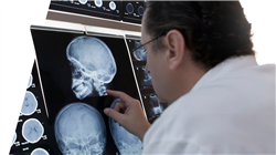
In the medical environment, Forensic Radiology applied to the child skeleton is a highly demanded field of specialization. Among the reasons for this is that radiological equipment allows professionals to detect a wide range of diseases and congenital anomalies (from bone dysplasias to malformations). In this sense, the evaluation of the anatomical characteristics and specific growth patterns of bones can provide crucial information to identify children who have died as a result of natural disasters, accidents or even homicides. In addition, in cases where skeletal remains of minors are found, imaging tools are helpful in estimating the age of the individuals at the time of the events.
For this reason, TECH is developing an avant-garde program in Forensic Radiology of the Human Skeleton at Different Stages of Biological Development. The academic itinerary will delve into the bone pathophysiology of young individuals, taking into account factors such as bone growth and the frequent acquired pathologies. Along the same lines, the syllabus will deal with the main diseases affecting the bones, among which Osteoporosis, Bone Cancer or Rickets stand out. Likewise, specialists will enhance their skills to adequately interpret radiological images derived from tools such as Computed Tomography, X-rays and Magnetic Resonance Imaging. In this way, graduates will identify evidence of trauma in children's bones and these findings will contribute to solve cases of child abuse.
As for the methodology of this program, it should be noted that it reinforces its innovative character. TECH provides students with a 100% online educational environment, therefore adapting to the needs of busy professionals who want to advance in their careers. It also employs the Relearning teaching system, based on the repetition of key concepts to fix knowledge and facilitate learning. In this way, the combination of flexibility and a robust pedagogical approach makes it highly accessible.
Assist in identifying human remains by comparing radiological features with descriptions of missing individuals"
This Postgraduate certificate in Forensic Radiology of the Human Skeleton at Different Stages of Biological Development contains the most complete and up-to-date scientific program on the market. The most important features include:
- The development of practical cases presented by experts in Forensic Radiology
- The graphic, schematic and eminently practical contents with which it is conceived gather scientific and practical information on those disciplines that are indispensable for professional practice
- Practical exercises where the self-assessment process can be carried out to improve learning
- Its special emphasis on innovative methodologies
- Theoretical lessons, questions to the expert, debate forums on controversial topics, and individual reflection assignments
- Content that is accessible from any fixed or portable device with an Internet connection
You will acquire skills to recognize radiological signs of skeletal maturation in medical images such as X-rays or CT scans"
The program’s teaching staff includes professionals from the sector who contribute their work experience to this specializing program, as well as renowned specialists from leading societies and prestigious universities.
The multimedia content, developed with the latest educational technology, will provide the professional with situated and contextual learning, i.e., a simulated environment that will provide immersive education programmed to learn in real situations.
This program is designed around Problem-Based Learning, whereby the professional must try to solve the different professional practice situations that arise during the course. For this purpose, students will be assisted by an innovative interactive video system created by renowned and experienced experts.
You will delve into Bone Growth to estimate the age of individuals and help in the identification of human remains during criminal investigations"

A syllabus, based on the revolutionary Relearning methodology, that will help you to consolidate the concepts efficiently"
Objectives
Through this university program, physicians will be highly qualified to identify radiological characteristics of skeletal maturation from radiological images. In this line, graduates will use the specific radiological characteristics of bodies to determine the chronological age of individuals at the time of death. In addition, specialists will enhance their skills to interpret images and achieve radiological findings that serve to identify human remains.

You will have a comprehensive understanding of the variability of skeletal maturation and will optimize your skills in the interpretation of radiological images”
General Objectives
- Analyze the sequence of ossification, joint development and the formation of bone structures at different stages of childhood, as well as the factors that influence bone growth, such as genetics, nutrition and chronic diseases
- Develop skills to interpret specific images of the above conditions and understand their impact on growth and musculoskeletal function
- Understand how skeletal growth and mineralization are processes that begin during fetal development and continue at different rates through childhood and adolescence until the third decade of life, when peak bone mass is reached
- Identify normal features of childhood bone anatomy, as well as signs of traumatic injuries, bone disease and pediatric orthopedic conditions, with emphasis on the importance of exposure to specific imaging techniques for children and the radiologic safety considerations for this group
Specific Objectives
- Determine the development of the bone along the growth phases, from the neonatal phase to adolescence and the respective images obtained by radiographs
- Master the morphology of healthy bone: its histology, the ossification center, the different types of bone tissues present in the bones and their dynamics during childhood
- Analyze bone factors with congenital, metabolic and infectious pathologies, distinguishing them from healthy bone and know how to apply the appropriate imaging technique to each case
- Identify the most frequent bone lesions among children and adolescents, including the establishment of the difference between accidental injuries and injuries possibly resulting from assault and abuse

The Virtual Campus will be available to you 24 hours a day, so that you can log in at the time that suits you best"
..


