Why study at TECH?

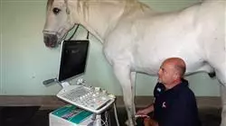
Large animals can have complex pathologies, so it is necessary to have specialized veterinarians who can treat them”
##IMAGE##

The Professional master’s degree in Internal Medicine in Large Animals incorporates innovative knowledge, based on the latest scientific evidence, that allows veterinary professionals to stay up-to-date on the newest treatments and emerging diseases that affect large animals across the world as a consequence of globalization. Specialized and advanced knowledge of these diseases is necessary since outbreaks of some diseases considered eradicated or new ones may occur in all countries of the world.

Clinical practice is a very dynamic activity, new treatments are constantly appearing in scientific publications and veterinarians must be aware of them in order to be able to offer these options to their clients. Each of the modules in this Professional master’s degree covers one of the organ systems, with emphasis on those systems that are most frequently affected in the large animals

With respect to ruminants, although their handling and the diseases they suffer from are different from those of horses, they must also be understood with sufficient scientific expertise to be able to establish adequate treatments and accurate prognoses. Camelids of the new world or South America, which include mainly llamas and alpacas as domesticated animals, are animals bred for different purposes including fiber production, pack animals or meat production in South America. Horses are animals that are used both for leisure and as companion animals, as well as in different sports disciplines, which adds an important added economic value. It is essential to have a high level of knowledge in internal medicine to be able to work with these horses, since, due to their economic value, they are not readily accessible to clinicians with little training

This Professional master’s degree is designed by professors with the highest recognized degree of specialization, thus guaranteeing quality in all aspects, both clinical and scientific, in large animals. 

Get trained with us and learn how to diagnose and treat diseases in large animals, in order to improve their quality of life" 

This Professional master’s degree in Internal Medicine in Large Animals contains the most complete and up-to-date educational program on the market. The most important features include:

  • Practical cases presented by experts in Internal Medicine in Large Animals 
  • The graphic, schematic, and practical contents with which they are created provide scientific and practical information on the disciplines that are essential for professional practice
  • Latest innovations on Internal Medicine in Large Animals
  • Practical exercises where the self-assessment process can be carried out to improve learning
  • Special emphasis on innovative methodologies in Internal Medicine in Large Animals
  • Theoretical lessons, questions to the expert, debate forums on controversial topics, and individual reflection assignments
  • Content that is accessible from any fixed or portable device with an Internet connection

With this Professional master’s degree, you will learn to establish a specific clinical approach to horses with cardiac or vascular alterations” 

Its teaching staff includes professionals belonging to the veterinary field, who contribute their expertise to this specialization, as well as renowned specialists from leading societies and prestigious universities.

The multimedia content, developed with the latest educational technology, will provide the professional with situated and contextual learning, i.e., a simulated environment that will provide immersive training programmed to train in real situations. 

This program is designed around Problem-Based Learning, whereby the specialist must try to solve the different professional practice situations that arise throughout the program. For this, the professional will have the help of an innovative interactive video system made by renowned and experienced experts in Internal Medicine in Large Animals.    

This program comes with the best educational material, providing you with a contextual approach that will facilitate your learning"

##IMAGE##

Thanks to this program you will be capable of establishing an appropriate methodology for the examination of a patient with urinary and renal problems, among others"

Syllabus

The structure of the content has been designed by the best professionals in the field of Internal Medicine in Large Animals, with extensive experience and recognized prestige in the profession, backed by the volume of cases reviewed, studied, and diagnosed, and with extensive knowledge of new technologies applied to veterinary medicine.

##IMAGE##

We have the most complete and up-to-date academic program in the market. We strive for excellence and for you to achieve it too" 
 

Module 1. Alterations of the Cardiovascular System in Large Animals

1.1. Cardiovascular Anatomy and Clinical Pathophysiology

1.1.1. Embryonic Development and Anatomy of the Heart
1.1.2. Fetal Circulation
1.1.3. Cardiac Cycle
1.1.4. Ion Channels and Action Potentials
1.1.5. Neurohormonal Control of the Heart
1.1.6. Arrhythmogenic Mechanisms

1.2. Cardiovascular Examination

1.2.1. Medical History
1.2.2. Cardiovascular Examination

1.3. Diagnostic Tests

1.3.2. Tensiometry
1.3.3. Electrocardiography
1.3.4. Echocardiography
1.3.5. Vascular Ultrasound

1.4. Congenital and Valvular Heart Disease in Horses

1.4.1. Ventricular Septal Defect
1.4.2. Tetralogy of Fallot
1.4.3. Patent Ductus Arteriosus
1.4.4. Aortic Insufficiency
1.4.5. Mitral Insufficiency
1.4.6. Tricuspid Regurgitation

1.5.  Arrhythmias in Horses

1.5.1. Supraventricular Arrhythmias
1.5.2. Ventricular Arrhythmias
1.5.3. Conduction Disturbances

1.6. Pericarditis, Myocarditis, Endocarditis and Vascular Alterations in Horses

1.6.1. Pericardial Disorders
1.6.2. Myocardial Disorders

    1.6.2.1. Endocardial Disorders
    1.6.2.2. Aorto-Cardiac and Aorto-Pulmonary Fistulas

1.7. Cardiovascular Diseases in Cattle

1.7.1. Examination of the Cardiovascular System in Cattle
1.7.2. Congenital Cardiovascular Pathologies
1.7.3. Arrhythmias
1.7.4. Heart Failure and Cor Pulmonale
1.7.5. Valvular and Endocardial Diseases
1.7.6. Myocardial Diseases and Cardiomyopathies
1.7.7. Pericardial Diseases
1.7.8. Thrombosis and Embolism
1.7.9. Neoplasty

1.8. Cardiovascular Diseases in Small Ruminants

1.8.1. Examination of the Cardiovascular System in Small Ruminants
1.8.2. Congenital Cardiovascular Pathologies
1.8.3. Acquired Cardiovascular Pathologies
1.8.4. Toxic Cardiopathies or Due to Nutritional Deficiencies
1.8.5. Vascular Diseases

1.9. Cardiovascular Diseases in Camelids

1.9.1. Examination of the Cardiovascular System in Camelids
1.9.2. Congenital Cardiovascular Pathologies
1.9.3. Acquired Cardiovascular Pathologies
1.9.4. Toxic Cardiopathies or Due to Nutritional Deficiencies
1.9.5. Anemia, Polycythemia and Erythrocytosis
1.9.6. Neoplasty.

1.10. Cardiovascular Pharmacology

1.10.1. Vasodilators
1.10.2. Positive Inotropes
1.10.3. Diuretics
1.10.4. Antiarrhythmics
1.10.5. Treatment of Heart Failure

Module 2. Alterations of the Respiratory System in Large Animals

2.1. Clinical Examination and Main Diagnostic Techniques of the Upper Respiratory Tract

2.1.1. Anamnesis and General Physical Examination
2.1.2. Examination of Upper Respiratory Tract
2.1.3. Endoscopy at Rest
2.1.4. Dynamic Endoscopy
2.1.5. Ultrasound and Radiography of Upper Respiratory Tract
2.1.6. Culture and Antibiogram

2.2. Clinical Examination and Main Diagnostic Techniques of the Lower Respiratory Tract

2.2.1. Examination of Lower Respiratory Tract
2.2.2. Thoracic Ultrasound Scan
2.2.3. Thoracic Radiography 
2.2.4. Sample Collection: Tracheal Aspirate, Bronchoalveolar Lavage and Thoracocentesis
2.2.5. Arterial Blood Gases
2.2.6. Pulmonary Function Tests
2.2.7. Pulmonary Biopsy

2.3. Diseases of the Upper Respiratory Tract in Horses

2.3.1. Ethmoidal Hematoma
2.3.2. Sinusitis
2.3.3. Sinus Cysts
2.3.4. Guttural Pouch Pathologies: Tympanism, Mycosis, Empyema
2.3.5. Lymphoid Hyperplasia
2.3.6. Epiglottic Entrapment
2.3.7. Pharyngeal Collapse
2.3.8. Dorsal Displacement of the Soft Palate
2.3.9. Recurrent Laryngeal Papillomatosis
2.3.10. Arytenoid Chondritis
2.3.11. Rostral Displacement of Palatopharyngeal Arch

2.4. Equine Respiratory Viruses

2.5.1. Influenza
2.4.2. Herpesvirus
2.4.3. Other Respiratory Viruses

2.5. Exercise-Induced Pulmonary Hemorrhage in Horses

2.5.1. Clinical Signs
2.5.2. Pathogenesis.
2.5.3. Diagnosis
2.5.4. Treatment
2.5.5. Prognosis

2.6. Pleuropneumonia and Bacterial Pneumonia in Equidae

2.6.1. Clinical Signs
2.6.2. Pathogenesis.
2.6.3. Diagnosis
2.6.4. Treatment
2.6.5. Prognosis

2.7. Severe or Acute Asthma in Equidae

2.7.1. Clinical Signs
2.7.2. Pathogenesis.
2.7.3. Diagnosis
2.7.4. Treatment
2.7.5. Prognosis

2.8. Respiratory Pathologies in Cattle

2.8.1. Examination of the Respiratory System in Cattle
2.8.2. Alterations in the Upper Respiratory Tract
2.8.3. Bovine Respiratory Syndrome
2.8.4. Interstitial Pneumonia and Other Causes of Pneumonia in Bovines
2.8.5. Alterations of the Thoracic Cavity

2.9. Respiratory Pathologies in Small Ruminants

2.9.1. Examination of the Respiratory System in Sheep and Goats
2.9.2. Alterations in the Upper Respiratory Tract
2.9.3. Pneumonia
2.9.4. Alterations of the Thoracic Cavity

2.10. Respiratory Pathologies in Camelids

2.10.1. Examination of the Respiratory System in Camelids
2.10.2. Alterations in the Upper Respiratory Tract
2.10.3. Pneumonia and Other Pulmonary and Thoracic Pathologies
2.10.4. Neoplasty.

Module 3. Neurological Alterations in Large Animals

3.1. Neurological Examination and Main Diagnostic Tests

3.1.1. Clinical Examination and Clinical Signs
3.1.2. Dynamic Evaluation and Locating the Lesion
3.1.3. Diagnostic Tests: Cerebrospinal Fluid Extraction and Analysis
3.1.4. Other Diagnostic Tests

3.2. Epilepsy, Convulsions, Congenital and Degenerative Diseases in Horses

3.2.1. Epilepsy and Convulsions
3.2.2. Sleep Disorders
3.2.3. Cerebellar Abiotrophy
3.2.4. Shivers
3.2.5. Degenerative Myeloencephalopathy
3.2.6. Polineuritis

3.3. Central Nervous System Trauma and Vestibular Syndrome in Equidae

3.3.1. Cerebral Trauma
3.3.2. Spinal Cord Trauma
3.3.3. Vestibular Syndrome

3.4. Compressive Diseases of the Spinal Cord in Horses

3.4.1. Pathogenesis and Clinical Signs
3.4.2. Diagnosis
3.4.3. Treatment and Prognosis

3.5. Viral Diseases That Affect the Central Nervous System (CNS) in Equidae

3.5.1. Equine Herpesvirus Myeloencephalopathy
3.5.2. Togavirus Encephalitis
3.5.3. West Nile Virus Encephalitis
3.5.4. Rabies
3.5.5. Bornavirus and Other Viral Encephalitides

3.6. Other Diseases that Affect the CNS

3.6.1. Equine Motor Neurone Disease (EMND)
3.6.2. Grass Sickness (Equine Dysautonomia)
3.6.3. Neoplasms
3.6.4. Metabolic Alterations That Cause Neurological Symptomology
3.6.5. Toxins
3.6.6. Headsaking
3.6.7. Lyme Disease

3.7. Tetanus and Botulism

3.7.1. Tetanus
3.7.2. Botulism

3.8. Bovine Neurological Diseases

3.8.1. Examination of the Nervous System in Cattle
3.8.2. Alterations that Mainly Affect the Brain in Cattle
3.8.3. Alterations that Mainly Affect the Brainstem in Cattle
3.8.4. Alterations that Mainly Affect the Cerebelum in Cattle
3.8.5. Alterations that Mainly Affect the Spinal Cord in Cattle
3.8.6. Alterations that Mainly Affect the Peripheral Nerves in Cattle

3.9. Neruological Diseases in Small Ruminants

3.9.1. Examination of the Nervous System in Sheep and Goats
3.8.2. Alterations that Mainly Affect the Brain in Small Ruminants
3.8.3. Alterations that Mainly Affect the Brainstem in Small Ruminants
3.8.4. Alterations that Mainly Affect the Cerebelum in Small Ruminants
3.8.5. Alterations that Mainly Affect the Spinal Cord in Small Ruminants

3.9. Neurological Diseases in Camelids

3.9.1. Examination of the Nervous System and Diagnostic Techniques in Camelids
3.9.2. Congenital Pathologies and Development of the Nervous System
3.9.3. Infectious Meningoencephalitis
3.9.4. Main, Non-infectious Neuropathologies
3.9.5. Secondary Neuropathologies
3.9.6. Myopathies and Vertebral Pathologies
3.9.7. Visual and Hearing Impairments of Neurological Origin

Module 4. Alterations of the Gastrointestinal System in Large Animals

4.1. Clinical Examination and Diagnostic Tests

4.1.1. Physical Examination
4.1.2. Imaging Techniques
4.1.3. Endoscopy
4.1.4. Absorption and Digestion Test
4.1.5. Other Tests

4.2. Alterations that Affect the Stomach of Equidae

4.2.1. Gastric Ulcer Syndrome
4.2.2. Stomach Impactions
4.2.3. Other Diseases that Affect the Stomach

4.3. Strangulation Lesions in Horses

4.3.1. Strangulation Lesions of the Small Intestine
4.3.2. Strangulation Lesions of the Large Intestine

4.4. Obstructive Lesions in Horses

4.4.1. Obstructive Lesions of the Esophagus
4.4.2. Obstructive Lesions of the Small Intestine
4.4.3. Obstructive Lesions of the Large Intestine

4.5. IBD: Inflammatory Diseases/ Malabsorption Syndrome in Equidae

4.5.1. Clinical Approach
4.5.2. Alimentary Lymphosarcoma
4.5.3. Granulomatous Enteritis
4.5.4. Eosinophilic Enterocolitis
4.5.5. Lymphocytic-Plasmocytic Enterocolitis
4.5.6. Proliferative Enteropathy
4.5.6. Others

5.6. Pathologies that Affect the Liver

4.6.1. Liver Disease and Liver Failure
4.6.2. Clinical Signs of Liver Disease
4.6.3. Acutre Liver Diseases
4.6.4. Chronic Liver Diseases
4.6.5. Vascular and Congenital Diseases

4.7. Colitis, Enteritis and Peritonitis

4.7.1. Colitis
4.7.2. Enteritis
4.7.3. Treatment Options for Horses With Acute Diarrhea
4.7.4. Peritonitis

4.8. Gastrointestinal Alterations in Cattle

4.8.1. Examination of the Gastrointestinal System in Cattle
4.8.2. Alterations of the Oral Cavity
4.8.3. Indigestion
4.8.4. Traumatic Reticuloperitonitis
4.8.5. Abomasal Displacements and Other Abomasal Alterations
4.8.6. Obstructive Intestinal Alterations
4.8.7. Diarrhea in Adult Cattle

4.9. Gastrointestinal Alterations of Small Ruminants

4.9.1. Examination of the Gastrointestinal System in Small Ruminants
4.9.2. Alterations of the Oral Cavity
4.9.3. Indigestion and Other Pre-Stomach Disturbances
4.9.5. Enterotoxemias
4.9.4. Diarrhea in Adult Sheep and Goats

4.10. Gastrointestinal Alterations in Camelids

4.10.1. Anatomy and Physiology of the Gastrointestinal Tract of Camelids
4.10.2. Diagnostic Techniques
4.10.3. Congenital Gastrointestinal Pathologies
4.10.4. Diseases of the Oral Cavity
4.10.5. Diseases of the Esophagus
4.10.6. Pathologies of Gastric and Pre-Stomach Compartments
4.10.7. Enteritis and Diarrhea
4.10.8. Acute Abdomen and Colic
4.10.9. Neoplasms of the Gastrointestinal Tract

Module 5. Alterations of the Urinary System in Large Animals

5.1. Anatomy, Physiology and Diagnostic Tests

5.1.1. Anatomy
5.1.2. Physiology

    5.1.2.1. Elimination of Nitrogenous Components
    5.1.2.2. Electrolyte Removal and Recovery (Tubular Function)
    5.1.2.3. Water Balance

5.2. Hematology and Blood Biochemistry

5.2.1. Urianalysis

    5.2.1.1. Density
    5.2.1.2. Test Strip
    5.2.1.3. Microscopy
    5.2.1.4. Enzimuria
    5.2.1.5. Excretional Fractions
    5.2.1.6. Cultures

5.2.2. Imaging Techniques

    5.2.2.1. Ultrasound
    5.2.2.2. Radiology
    5.2.2.4. Endoscopy
    5.2.2.5. Gammagraphy

5.2.3. Renal Biopsy
5.2.4. Quantification of Renal Function (Clearance)

5.3. Acute Renal Insufficiency (ARF) in Horses

5.3.1. Causes
5.3.2. Pathophysiology
5.3.3. Acute Tubular Necrosis
5.3.4. Acute Interstitial Nephritis
5.3.5. Acute Glomerulonephritis
5.3.6. Diagnosis
5.3.7. Treatment
5.3.8.  Prognosis

5.4. Chronic Renal Insufficiency in Horses

5.4.1. Predisposing Factors
5.4.2. Glomerulonephritis
5.4.3. Acute Interstitial Nephritis
5.4.4. Other Causes
5.4.5. Diagnosis
5.4.6. Treatment
5.4.7. Prognosis

5.5. Renal Tubular Acidosis in Horse

5.5.1. Pathophysiology
5.5.2. Type 1
5.5.3. Type 2
5.5.4. Type 3
5.5.5. Quantitative/ Traditional Approximation of Imbalances
5.5.6. Diagnosis
5.5.7. Treatment

5.6. Investigation and Differential Diagnosis of Polyuria/Polydipsia

5.6.1. Diagnostic Protocol
5.6.2. Causes

    5.6.2.1. Renal Insufficiency
    5.6.2.2. Cushing

5.6.2.3. Primary Polydipsia

    5.6.2.4. Excessive Consumption of Salt
    5.6.2.5. Diabetes Insipidus
    5.6.2.6. Diabetes Mellitus
    5.6.2.7. Sepsis
    5.6.2.8. Iatrogenic

5.7. Investigation and Differential Diagnosis of Pigmenturia (Renal Hemorrhage, Urolithiasis, Urethritis)

5.7.1. Urethritis/Urethral Defects
5.7.2. Cystitis
5.7.3. Pyelonephritis
5.7.4. Urolithiasis

    5.7.4.1. Urethral Calculi
    5.7.4.2. Bladder Stones

5.7.5. Idiopathic Renal Hematuria
5.7.6. Hematuria Associated with Exercise
5.7.7. Pigmenturia Caused by Systemic Pathology

5.8. Genitourinary Diseases in Cattle

5.8.1. Congenital Genitourinary Pathologies
5.8.2. Kidney Damage and Failure
5.8.3. Other Diseases of the Kidneys
5.8.4. Diseases of the Urethers, Bladder and Urethra 

5.9. Genitouriary Diseases in Small Ruminants

5.9.1. Congenital Genitourinary Pathologies
5.9.2. Kidney Damage and Failure
5.9.3. Other Diseases of the Kidneys
5.9.4. Urinary Obstruction.
5.9.5. Diseases of the Urethers, Bladder and Urethra

5.10. Genitourinary Diseases in Camelids

5.10.1. Congenital Genitourinary Pathologies
5.10.2. Kidney Damage and Failure
5.10.3. Other Diseases of the Kidneys
5.10.4. Urinary Obstruction.
5.10.5. Diseases of the Urethers, Bladder and Urethra
5.10.6. Neoplasty.

Module 6. Neonatology in Large Animals

6.1. Clinical Approach to Newborn Foals

6.1.1. Examination of Newborn Foals
6.1.2. System Evaluation
6.1.3. Examination of the Mother and the Placenta

6.2. Septicemia in Foals

6.2.1. Risk Factors
6.2.2. Clinical Signs
6.2.3. Diagnosis
6.2.4. Treatment
6.2.5. Prognosis

6.3. Neonatal Hypoxia Syndrome in Foals

6.3.1. Etiopathogenesis
6.3.2. Clinical Signs
6.3.3. Diagnosis
6.3.4. Treatment
6.3.5.  Prognosis

6.4. Respiratory Alterations in Newborn Foals

6.4.1. Choanal Atresia
6.4.2. Wry Nose
6.4.3. Pneumonia
6.4.4. Acute Respiratory Distress Syndrom
6.4.5. Rib Fracture
6.4.6. Management of Foals With Pulmonary Pathologies

6.5. Genitourinary Alterations in Newborn Foals

6.5.1. Patent Urachus
6.5.2. Uroperitoneum
6.5.3. Renal Insufficiency
6.5.4. Inguinal and Scrotal Hernias
6.5.5. Ectopic Urethers

6.6. Digestive Alterations in Newborn Foals

6.6.1. Dysphagia
6.6.2. Gastric Ulcer Syndrome
6.6.3. Approach to Newborns With Colic
6.6.4. Meconium Impaction
6.6.5. Diarrhea / Enterocolitis

6.7. Intensive Care, Treatment and Procedures

6.7.1. Monitoring of the Newborn
6.7.2. Care of the Nursing Foal
6.7.3. Fluid Therapy
6.7.4. Food for a Sick Foal
6.7.5. Antibiotherapy
6.7.6. Cardiopulmonary Resuscitation

6.8. Main Problems in Calves

6.8.1. Congenital Diseases
6.8.2. Trauma and Death During Birth
6.8.3. Prematurity, Dysmaturity and Neonatal Maladjustment
6.8.4. Diseases and Perinatal Problems
6.8.5. Diseases Associated With Reproductive Biotechnologies
6.8.6. Failure of Transfer of Colostral Immunoglobulins
6.8.7. Diarrhea in Calves

6.9. Main Problems in New Born Small Ruminants

6.9.1. Congenital Diseases
6.9.2. Prematurity, Dysmaturity and Neonatal Maladjustment
6.9.3. Perinatal Problems
6.9.4. Gastrointestinal Tract Diseases
6.9.5. Locomotor Apparatus Diseases
6.9.6.  Failure of Transfer of Colostral Immunoglobulins

6.10. Main Problems in Newborn Camelids

6.10.1. Congenital Diseases
6.10.2. Prematurity, Dysmaturity and Neonatal Maladjustment
6.10.3. Perinatal Problems
6.10.4. Gastrointestinal Tract Diseases
6.10.5. Locomotor Apparatus Diseases
6.10.6. Failure of Transfer of Colostral Immunoglobulins

Module 7. Ophthalmology in Large Animals

7.1. Anatomy and Diagnostic Tests

7.1.1. A  natomy and Physiology of the Eyeball
7.1.2. Optic Nerve Blocks
7.1.3. Ophthalmologic examination
7.1.4. Basic Diagnostic Tests
7.1.5. Tonometry
7.1.6. Direct and Indirect Ophthalmoscopy
7.1.7. Ocular Ultrasonography
7.1.8. Other Diagnostic Imaging Tests
7.1.9. Electroretinography
7.1.10. Sub-palpebral Catheter Placement

7.2. Alterations of the Eyelids, Conjunctiva and Nasolacrimal Duct in Equidae

7.2.1. Anatomy of Adnexal Tissues
7.2.2. Alterations of the Ocular Orbit
7.2.3. Eyelid Alterations
7.2.4. Alterations of the Ocular Conjunctiva
7.2.5. Alterations of the Nasolacrimal Duct

7.3. Corneal Ulcers in Horses

7.3.1. General Aspects
7.3.2. Classification of Corneal Ulcers
7.3.3. Simple, Complex and Severe Ulcers
7.3.4. Indolent Ulcer
7.3.5. Iridocele and Ocular Perforation
7.3.6. Keratomalacia (Melting)

7.4. Infectious Keratitis and Stromal Abscesses in Equidae

7.4.1. Parasitic Keratitis
7.4.2. Viral Keratitis
7.4.3. Fungal Keratitis
7.4.4. Bacterial Keratitis
7.4.5. Stromal Abscess
7.4.6. Corneal Surgery

7.5. Immune-Mediated Diseases and Idiopathic Nonulcerative Keratitis of the Cornea in Equidae

7.5.1. General Aspects. Classification
7.5.2. Superficial Immune-Mediated Keratitis
7.5.3. Deep-Mid-Stromal Immune-Mediated Keratitis
7.5.4. Endolethial Immune-Mediated Keratitis
7.5.5. Other Immune-Mediated Diseases of the Cornea

7.6. Equine Recurrent Uveitis and Other Uveal Disorders in Equidae

7.6.1. Anatomy and Physiology of the Uveal Tract
7.6.2. Congenital Diseases of the Uvea
7.6.3. Acute Uveitis
7.6.4. Equine Recurrent Uveitis

7.7. Other Ocular Alterations of Equidae

7.7.1. Crystalline Lens Alterations
7.7.2. Alterations of the Retina and Glaucoma
7.7.3. Ocular Neoplasms and the Adjacent Structures

7.8. Ocular Alterations in Cattle

7.8.1. Infectious Keratoconjunctivitis
7.8.2. Ocular Carcinoma
7.8.3. Other Alterations of the Eyelids, Conjunctiva and Adjacent Tissue
7.8.4. Other Ocular Alterations

7.9. Ocular Alterations in Small Ruminants

7.9.1. Diseases of the Ocular Orbit
7.9.2. Infectious Keratoconjunctivitis
7.9.3. Parasitic Keratitis
7.9.4. Retinal Degeneration
7.9.5. Blindness

7.10. Ocular Alterations in Camelids

7.10.1. Congenital Diseases
7.10.2. Ulcerative Keratitis
7.10.3. Parasitic Keratitis

Module 8. Endocrinology and Dermatology in Large Animals

8.1. Clinical Approach and Diagnostic Tests in Equine Dermatology

8.1.1. Medical History
8.1.2. Sampling and Main Diagnostic Methods
8.1.3. Other Specific Diagnostic Techniques

8.2. Infectious, Immune-Mediated and Allergic Diseases in Horses

8.2.1. Viral Diseases
8.2.2. Bacterial diseases
8.2.3. Fungal Diseases
8.2.4. Parasitic diseases
8.2.5. Hypersensitivity Reactions: Types
8.2.6. Insect Sting Allergy
8.2.7. Other Types of Allergies and Skin Reactions

8.3. Cutaneous Neoplasms in Equidae

8.3.1.Sarcoids
8.3.2. Melanoma
8.3.3. Squamous Cell Carcinoma
8.3.4. Other Skin Tumors

8.4. Alterations to the Thyroid and Adrenal Gland in Equidae

8.4.1. Functions of the Thyroids
8.4.2. Factors Affecting the Measurement of Thyroid Hormones
8.4.3. Diagnostic Test for Thyroid Function
8.4.4. Hypothyroidism
8.4.5. Hyperthyroidism
8.4.6. Neoplasms of the Thyroids
8.4.7. Adrenal Insufficiency in Adults
8.4.8. Adrenal Insufficiency in Foals
8.4.9. Hyperadrenocorticism

    8.4.10. Adrenocortical Neoplasms

8.5. Dysfunction of the Pars Intermedia of the Pituitary in Equidae

8.5.1. Etiopathogenesis
8.5.2. Clinical Signs
8.5.3. Diagnosis
8.5.4. Treatment

8.6. Equine Metabolic Syndrome

8.6.1. Etiopathogenesis
8.6.2. Clinical Signs
8.6.3. Diagnosis
8.6.4. Treatment

8.7. Alterations in the Metabolism of Calcium, Phosphorus and Magnesium in Horses. Anhidrosis

8.7.1. Alterations That Occur With Hypocalcemia
8.7.2. Alterations That Occur With Hypercalcemia
8.7.3. Diseases That Occur With Hypophosphatemia
8.7.4. Pathologies Associated with Hyperphosphatemia
8.7.5. Hypomagnesemia
8.7.6. Hypermagnesemia
8.7.7. Anhidrosis

8.8. Dermatological, Metabolic and Endocrine Alterations in Cattle

8.8.1. Congenital Dermatological Pathologies
8.8.2. Skin and Fur Diseases
8.8.3. Diseases of the Subcutis
8.8.4. Diseases of Hooves and Horns
8.8.5. Dutaneous Neoplasms
8.8.6. Ketosis
8.8.7. Calcium, Magnesium and Phosphorous Disorders
8.8.8. Other Endocrinopathies

8.9. Dermatological and Endocrine Alterations in Small Ruminants

8.9.1. Congenital Dermatological Pathologies
8.9.2. Infectious Dermatitis
8.9.3. Vesicular and Mucocutaneous Junction Diseases
8.9.4. Parasitic Diseases of Hair and Wool
8.9.5. Caseous Lymphadenitis
8.9.6. Skin and Adnexal Diseases Associated with Toxicity and Nutritional Problems
8.9.7. Neoplasty.
8.9.8. Pregnancy Toxemia
8.9.9. Stump, Rickets

8.10. Dermatological and Endocrine Alterations in Camelids

8.10.1.Infectious Dermatitis
8.10.2. Mucocutaneous Junction Diseases
8.10.3. Diseases Affecting Fiber Quality

Module 9. Laboratorial Diagnosis in Equidae. Alterations of the Hematopoietic System and Immunology in Large Animals

9.1. Hematology in Adult Horses: Alterations in the Red Series

9.1.1. Physiology of Red Blood Cells and Platelets
9.1.2. Interpretation of Alterations in the Red Series
9.1.3. Iron Metabolism
9.1.4. Thrombocytopenia/Thrombocytosis
9.1.5. Polycythemia
9.1.6. Anemia

    9.1.6.1. Losses: Bleeding
    9.1.6.2. Destruction

        9.1.6.2.1. Infectious and Parasitic Diseases That Cause Anemia:
                        Piroplasmosis, EIA and Other Diseases
        9.1.6.2.2. Immune-mediated Hemolysis

            9.1.6.2.3. Neonatal Isoerythrolisis

        9.1.6.2.4. Oxidative Damage

    9.1.6.3. Lack of Production

        9.1.6.3.1. Anemia Chronic Inflammation
        9.1.6.3.2. Myeloptisis/Aplasia Medular

9.1.7. Physiology of the White Series

    9.1.7.1 Neutrophils
    9.1.7.2 Eosinophils
    9.1.7.3 Basophils
    9.1.7.4 Lymphocytes
    9.1.7.5 Mast cells
    9.1.7.6 Leukemias

9.2. Biochemistry in Adult Horses

9.2.1. Renal Profile
9.2.2. Liver Profile
9.2.3. Acute Phase Proteins
9.2.4. Muscular Profile
9.2.5. Other Determinants

9.3. Hematology and Biochemistry in Foals/ Geriatric Horses

9.3.1. Differences in Hematology
9.3.2. Differences in Biochemistry

    9.3.2.1.Differences in Renal Function
    9.3.2.2 Differences in Liver Function
    9.3.2.3 Differences in Muscular Profile

9.4. Immune Response of Foals and Geriatric Horses

9.4.1. Peculiarities of the Immune System of Neonatal Foals
9.4.2. Evolution of the Immune Response During the First Year of Age
9.4.3. Senecundity: Peculiarities of the Geriatric Immune System

9.5. Hypersensitivity Reactions. Immune-Mediated Diseases

9.5.1. Hypersensitivity Type 1
9.5.2. Hypersensitivity Type 2
9.5.3. Hypersensitivity Type 3
9.5.4. Hypersensitivity Type 4
9.5.5. Immunocomplexes Manifestations of Immune-Mediated Diseases

9.6. Hemostasis Disorders

9.6.1. Primary Hemostasis
9.6.2. Secondary Hemostasis
9.6.3. Coagulation Based on Intrinsic and Extrinsic Pathways vs. Cell-Based Coagulation Model (Initiation, Propagation and Amplification)
9.6.4. Anticoagulation
9.6.5. Fibrinolysis/Antifibrinolysis
9.6.6. Disseminated Intravascular Coagulation
9.6.7. Hemorrhagic Purpura
9.6.8. Hereditary Problems
9.6.9. Procoagulant and Anticoagulant Treatments

9.7. Basic Principles of Acid-Base Equilibrium. Fluid Therapy

9.7.1. Introduction. Why is Acid-Base Equilibrium Important?
9.7.2. Basic Concepts
9.7.3. Protection Mechanisms: Short and Long-Term Slope Compensations
9.7.4. Interpreting Methods
9.7.5. Step by Step. How to Interpret the Acid-Base to Obtain Maximum Information

    9.7.5.1. Lactate
    9.7.5.2. Electrolytes

        9.7.5.2.1. Hypernatremia ( >145mmol/l)
        9.7.5.2.2. Hyponatremia (Horse<134 mmol/l)
        9.7.5.2.3. Hyperpotassemia or Hyperkalemia (>4,5mmol/l)
        9.7.5.2.4. Hypotassemia or Hypokalemia (<3.5 mmol/l)
        9.7.5.2.5. Hyperchloremia (>110 mmol/l)
        9.7.5.2.6. Hypochloremia (<90 mmol/l)

    9.7.5.3. SIDm
    9.7.5.4. ATO
    9.7.5.5. SIG

9.7.6. Classification of the Alterations
9.7.7. Basic Principles of Fluid Therapy
9.7.8. Body Composition of Fluids and Electrolytes
9.7.9. Estimation of Dehydration
9.7.10. Types of Fluid

    9.7.10.1. Crystaloid Solutions

        9.7.10.1.1. Ringer's Lactate
        9.7.10.1.2. Isofundin®
        9.7.10.1.3. Saline Solution (0.9% NaCl)
        9.7.10.1.4. Sterovet®
        9.7.10.1.5. Bicarbonate
        9.7.10.1.6. Glucosaline 0,3/3,6%

    9.7.10.1.7. Hypertonic Saline Solution (7.5% NaCl)
    9.7.10.2. Colloidal Solutions

        9.7.10.2.1. IsoHes®
        9.7.10.2.2. Plasma

9.8. Interpretation of Laboratory Analysis and Immunological and Hematopoietic Alterations in Cattle

9.8.1. Blood Count
9.8.2. Blood Biochemistry
9.8.3. Allergies
9.8.4. Immune-Mediated Anemia
9.8.5. Thrombocytopenia

9.9. Interpretation of Laboratory Analysis and Immunological and Hematopoietic Alterations in Small Ruminants

9.9.1. Blood Count
9.9.2. Anemia and the FAMACHA System
9.9.3. Blood Biochemistry

9.10. Interpretation of Laboratory Analysis and Immunological and Hematopoietic Alterations in Camelids

9.10.1. Blood Count
9.10.2. Anemia
9.10.3. Blood Biochemistry

Module 10. Infectious and Parasitic Diseases in Large Animals

10.1. Prevention and Control of Infectious Diseases

10.1.1. Laboratory Diagnostic Tests
10.1.2. Antimicrobial Tests and Resistances
10.1.3. Use of Vaccines
10.1.4. Biosecurity and Control Measures

10.2. Main Infectious and Contagious Diseases in Horses

10.2.1. Notifiable Diseases
10.2.2. Diseases Caused by Bacteria
10.2.3. Viral diseases
10.2.4. Diseases Caused by Fungi

10.3. Main Infectious and Contagious Diseases in Cattle

10.3.1. Notifiable Diseases
10.3.2. Diseases Caused by Bacteria
10.3.3. Viral diseases
10.3.4. Diseases Caused by Fungi
10.3.5. Diseases Caused by Prions

10.4. Main Infectious and Contagious Diseases in Small Ruminants

10.4.1. Notifiable Diseases
10.4.2. Diseases Caused by Bacteria
10.4.3. Viral diseases
10.4.4. Diseases Caused by Fungi
10.4.5. Diseases Caused by Prions

10.5. Main Infectious and Contagious Diseases in Camelids

10.5.1. Notifiable Diseases
10.5.2. Diseases Caused by Bacteria
10.5.3. Viral diseases
10.5.4. Diseases Caused by Fungi

10.6. Main Parasites Affecting Horses

10.6.1. Hemoparasites
10.6.2. Small Strongyls or Cyathostomes
10.6.3. Big Strongyls
10.6.4. Ascarids
10.6.5. Other Nematodes
10.6.6. Cestodes

10.7. Main Parasites Affecting Cattle

10.7.1. Hemoparasites
10.7.2. Gastrointestinal Nematodes
10.7.3. Nematodes That Affect the Respiratory Tract
10.7.4. Cestodes
10.7.5. Trematodes
10.7.6. Coccidia

10.8. Main Parasites Affecting Small Ruminants

10.8.1. Hemoparasites
10.8.2. Gastrointestinal Nematodes
10.8.3. Nematodes That Affect the Respiratory Tract
10.8.4. Cestodes
10.8.5. Trematodes
10.8.6. Resistance to Anthelmintics in Small Ruminants
10.8.7. Management, Treatment and Control Programs (FAMACHA)

10.9. Main Parasites Affecting Camelids

10.9.1. Hemoparasites
10.9.2. Coccidia
10.9.3. Nematodes
10.9.4. Cestodes

10.10. Prevention and Treatment of Parasitic Diseases

10.10.1. Diagnostic Techniques
10.10.2. Therapeutic Principles
10.10.2. Resistance Development
10.10.3. Management and Control Programs

##IMAGE##

This training will allow you to advance in your career comfortably"

Professional Master's Degree in Internal Medicine in Major Species

.

In order to provide adequate care and maintain full health in major species such as equids (horses, donkeys, mules), bovids (bulls, cows, antelopes, goats, sheep) and camelids (llamas, alpacas), it is essential that veterinarians perform medical checkups periodically in order to detect any abnormality in time. An in-depth study in this area is necessary, since it makes it easier for personnel to act at the time of treating any outbreak or specific pathology. This helps to monitor the health status of the animals and provide immediate attention if necessary. With the motivation to offer professionals the latest clinical developments for the proper care of animals, TECH designed a Professional Master's Degree in Internal Medicine in Major Species. A high quality program that provides the most updated knowledge on the therapeutic currents that help treat the diseases present in these species. Throughout 12 months of preparation in virtual mode, students will develop specific skills to promptly recognize certain abnormal symptoms; thus, they will be able to establish protocols and procedural guidelines to provide timely and effective treatments.

Take a Professional Master's Degree in Internal Care of the Elderly

.

At TECH you will receive intensive training focused on acquiring the necessary skills to provide medical assistance to these animals. You will have at your fingertips an organic learning system, which will allow you to specialize in identifying all clinical signs associated with cardiovascular, neurological, gastrointestinal and respiratory diseases; as well as ophthalmological, dermatological, endocrine, urinary and renal diseases. Therefore, you will be an expert in designing from diagnostic protocols with complementary tests; to the specific and appropriate approach in the exploration of each patient. As a result, you will not only specialize in treating congenital, immune-mediated, infectious and parasitic pathologies, but you will also be able to establish health guidelines for the correct care of animals in neonatal and geriatric stages. Thanks to our online methodology you will be able to access the course at the time, place and schedule that best suits you, to do so you only need to have some device connected to the internet.