University certificate
The world's largest faculty of veterinary medicine”
Why study at TECH?
Veterinarians must continue their specialization to adapt to new developments in this field"

This training is the best option you can find to specialize in Fracture Fixation Methods "
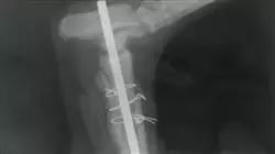
The teaching team of this Fracture Fixation Methods Expert has made a careful selection of the different state-of-the-art techniques for experienced professionals working in the veterinary field. Specifically, this specialization focuses on the study of skeletal external fixators and circular fixators, intramedullary nailing, and bone plates and screws.
External fixation of fractures is the use of a rigid support placed outside the body and connected to the bone by means of needles through the skin (transcutaneous). The placement technique with respect to other methods of internal osteosynthesis shows that external fixation improves the biological environment preserves soft tissue and irrigation, accelerates healing, decreases the risk of infection and reduces surgical time.
The external fixator provides stable fixation of the bony ends without the need for implants in the fracture line or immobilization of neighboring joints, and is therefore particularly suitable for open, exposed or infected fractures. It allows to compress, neutralize or distract the bony ends depending on the need of the pathology.
Fracture fixation with intramedullary (IM) pins in dogs and cats began in the 1940s. Its popularity increased due to advances in anesthesia, aseptic techniques, antibiotics and the awareness on the part of veterinarians and animal owners that, in most cases treated, there was a satisfactory repair.
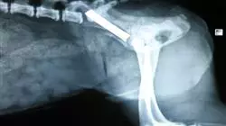
Thus, the intramedullary nail, for a long time, has been the most widely used implant in veterinary medicine because it is placed in the medullary cavity and becomes resistant to bending in all directions. Its strength is related to its diameter and its ability to restrict the movement of the fractured bone fragments. It is the most commonly used fastening system for dogs and cats.
In the last 20 years, the fixation of fractures with the use of rigid internal fixation implants, such as plates, has evolved enormously. One could speak of eight or nine different systems of fixation, more recognized, of fractures by means of plates. In this case, the specailziation will focus on the most widely used worldwide.
The teachers in this training are university professors with between 10 and 50 years of classroom and hospital experience. They are professors from schools on different continents, with different ways of doing surgery and with world-renowned surgical techniques. This makes this Postgraduate diploma a unique specialization program, different from any other that may be offered at this moment in the rest of the universities.
Do not miss the opportunity to take this Postgraduate diploma in Fracture Fixation Methods with us. It's the perfect opportunity to advance your career"
This Postgraduate diploma in Fracture Fixation Methods contains the most complete and up to date educational program on the market. The most important features of the program include:
- The development of case studies presented by experts in Fracture Fixation Methods
- The graphic, schematic, and eminently practical contents with which they are created provide scientific and practical information on the disciplines that are essential for professional practice
- Developments on Fracture Fixation Methods
- Practical exercises where the self-assessment process can be carried out to improve learning
- Special emphasis on innovative methodologies in the control of Fracture Fixation Methods
- Theoretical lessons, questions to the expert, debate forums on controversial topics, and individual reflection assignments
- Content that is accessible from any fixed or portable device with an Internet connection
This Postgraduate diploma is the best investment you can make in selecting a refresher program to update your knowledge in Fracture Fixation Methods "
Its teaching staff includes professionals from the veterinary field, who bring the experience of their work to this training, as well as recognised specialists from leading societies and prestigious universities.
Its Multimedia Content, elaborated with the latest Educational Technology, will allow the Professional a situated and contextual learning, that is to say, a Simulated Environment that will provide an immersive specialization programmed to train in real situations.
This program is designed around Problem Based Learning, whereby the specialist must try to solve the different professional practice situations that arise during the academic year. For this purpose, the professional will be assisted by an innovative interactive video system created by renowned and experienced Postgraduate diploma in Fracture Fixation Methods .
This specialisation comes with the best didactic material, providing you with a contextual approach that will facilitate your learning"

Incorporate the latest developments in Traumatology and Orthopedic Surgery in your daily practice, with this specialization of high scientific rigor"
Syllabus
The structure of the contents has been designed by the best professionals in the field of Veterinary Traumatology and Orthopedic Surgery, with extensive experience and recognized prestige in the profession, backed by the volume of cases reviewed, studied and diagnosed, and with extensive knowledge of new technologies applied to veterinary medicine.

This Postgraduate diploma in Fracture Fixation Methods contains the most complete and up to date scientific program on the market"
Module 1. Skeletal External Fixators and Circular Fixators
1.1. External Fixators
1.1.1. History of the External Skeletal Fixator
1.1.2. Description of the External Fixator
1.2. Parts Constituting the Kirschner-Ehmer Apparatus
1.2.1. Key
1.2.1.1. Fasteners
1.2.2. Connector bar
1.3. Settings of the External Skeletal Fixator
1.3.1. Half Skeletal Fixation Device
1.3.2. Standard Kirschner-Ehmer Apparatus
1.3.3. Modified Kirschner-Ehmer Apparatus
1.3.4. Bilateral External Fixator Model
1.4. Mixed Skeletal Fixator Apparatus
1.5. Methods of Application of the Kirschner-Ehmer Apparatus
1.5.1. Standard Method
1.5.2. Modified Method
1.6. External Fixators with Dental Acrylic
1.6.1. The Use of Epoxy Resin
1.6.2. The Use of Dental Acrylics
1.6.2.1. Preparation of Acrylics
1.6.2.2. Application and Setting Time
1.6.2.3. Post-Surgery Care
1.6.2.4. Removal of the Acrylic
1.6.3. Bone Cement for Use in Fractures of the Spine
1.7. Indications and Uses of External Fixatives
1.7.1. Femur
1.7.2. Tibia
1.7.3. Tarsus
1.7.4. Humerus
1.7.5. Radio and Ulna
1.7.6. Carpus
1.7.7. Jaw
1.7.8. Pelvis
1.7.9. Spinal Column
1.8 Advantages and Disadvantages of Using External Fixators
1.8.1. Acquisition of Acrylic Material
1.8.2. Care in the Application of Acrylics
1.8.3. Toxicity of Acrylic
1.9. Post-surgical care
1.9.1. Cleaning of the Fixative with Acrylic
1.9.2. Post-Operative Radiographic Studies
1.9.3. Gradual Removal of the Acrylic
1.9.4. Care when Removing the Fixative
1.9.5. Repositioning of the Acrylic Fixative
1.10. Circular fasteners
1.10.1. History
1.10.2. Components
1.10.3. Structure
1.10.4. Applications
1.10.5. Advantages and Disadvantages
Module 2. Intramedullary Enclave
2.1. History
2.1.2. Kuntcher's Nail
2.1.3. The First Canine Patient with an Intramedullary Nail
2.1.4. The Use of the Steinmann Nail in the 1970s
2.1.5. The Use of the Steinmann Nail Today
2.2. Principles of Intramedullary Nail Application
2.2.1. Type of Fractures in Which it Can Be Exclusively Placed
2.2.2. Rotational Instability
2.2.3. Length, Tip and Rope
2.2.4. Normograde and Retrograde Application. Nail Diameter to Medullary Canal Ratio
2.2.5. Principle of the 3 Points of the Cortex
2.2.6. Behaviour of the Bone and its Irrigation after Intramedullary Nail Fixation
2.2.6.1. The Steinmann Nail and Radium
2.3. The Use of Locks with the Steinmann Intramedullary Nail
2.3.1. Principles of Application of Fastenings and Lashings
2.3.1.1. Barrel Principle
2.3.1.2. Type of Fracture Line
2.4. Principles of Application of the Tension Band
2.4.1. Pawel's Principle
2.4.2. Application of Engineering to Orthopedics
2.4.3. Bone Structures where the Tension Band is to Be Applied
2.5. Normograde and Retrograde Application Method of the Steinmann Nail
2.5.1. Proximal Normograde
2.5.2. Normograde distal
2.5.3. Proximal Retrograde
2.5.4. Retrograde Distal
2.6. Femur
2.6.1. Proximal Femoral Fractures
2.6.2. Fractures of the Distal Third of the Femur
2.6.3. Supracondylar Fractures or Fracture-Separation of the Distal Epiphysis
2.6.4. Intercondylar Femoral Fracture
2.6.5. The Steinmann Intramedullary Nail and Half Kirschner Device
2.6.6. The Steinmann Intramedullary Nail with Locks or Screws
2.7. Tibia
2.7.1. Avulsion of the Tibial Tubercle
2.7.2. Fractures of the Proximal Third
2.7.3. Fractures of the Middle Third of the Tibia
2.7.4. Fractures of the Distal Third of the Tibia
2.7.5. Fractures of the Tibial Malleoli
2.7.6. The Steinmann Intramedullary Nail and Half Kirschner Device
2.7.7. The Steinmann Intramedullary Nail with Locks or Screws
2.8. Humerus
2.8.1. Steinmann Intramedullary Nail in the Humerus
2.8.2. Fractures of the Proximal Fragment
2.8.3. Fractures of the Middle Third or Body of the Humerus
2.8.4. Steinmann Intramedullary Nail Fixation
2.8.5. Clavo intramedular de Steinmann y fijación auxiliar
2.8.6. Supracondylar Fractures
2.8.7. Fractures of the Medial or Lateral Epicondyle
2.8.8. Intercondylar T- or Y-Fractures
2.9. Ulna
2.9.1. Acromion
2.10. The Extraction of the Steinmann Intramedullary Nail
2.10.1. Radiographic Follow-up
2.10.2. Callus Formation in Steinmann Nail Fractures
2.10.3. Clinical Union
2.10.4. How to Remove the Implant
Module 3. Bone Plates and Screws
3.1. History of Metal Plates in Internal Fixing
3.1.1. The Initiation of Plates for Fracture Fixation
3.1.2. The World Association of Orthopedic Manufacturers (AO/ASIF)
3.1.2.1. Sherman and Lane plates
3.1.2.2. Steel Plates
3.1.2.3. Titanium Plates
3.1.2.4. Plates of Other Materials
3.1.2.5. Combination of Metals for New Plate Systems
3.2. Different Fixing Systems with Plate 8 (AO/ASIF, ALPS, FIXIN)
3.2.1. AO/ASIF Plates
3.2.2. Advanced Locked Plate System. (ALPS)
3.2.2.1. FIXIN and its Conical Block
3.3. Instrument Care
3.3.1. Disinfection
3.3.2. Cleaning
3.3.3. Rinse
3.3.4. Drying
3.3.5. Lubrication
3.4. Instruments Used for the Fixation of Plates and Screws
3.4.1. Self-Tapping Screws and Tap Removal
3.4.2. Depth Gauges
3.4.3. Drilling Guides
3.4.4. Plate Benders and Plate Twisters
3.4.5. Screw Heads
3.4.6. Screws / Bolts
3.5. Use and Classification of Screws
3.5.1. Cancellous Bone Screws
3.5.2. Cortical Bone Screws
3.5.3. Locked Screws/Bolts
3.5.4. Fastening of Screws
3.5.4.1. Use of the Drill
3.5.4.2. Use of the Countersink
3.5.4.3. Borehole Depth Measurement
3.5.4.4. Use of the Tap
3.5.4.5. Introduction to Screws
3.6. Technical Classification of Screws
3.6.1. Big Screws
3.6.2. Small Screws
3.6.3. Mini Screws
3.7. Classification of Screws According to their Function
3.7.1. Screw with Interfragmentary Compression Effect
3.7.2. The Cortical Bone Screw with Interfragmentary Compression Effect
3.7.3. Reduction Techniques and Screw Fixation with Compression Effect. Interfragmentary
3.7.4. Locked Bolts
3.8. Bone Plates
3.8.1. Bases for Fixing with Plates
3.8.1.1. Classification of Plates According to Their Shape
3.8.1.2. Dynamic Compression Plates
3.8.1.2.1. Way of Action
3.8.1.2.2. Fixing Technique
3.8.1.2.3. Advantages Provided by Dynamic Compression Plates (DPC)
3.8.1.2.4. Disadvantages of Dynamic Compression Plates (DPC)
3.8.3. Locked Plates
3.8.3.1. Advantages and Disadvantages
3.8.3.2. Types of Locks
3.8.3.3. Way of Action
3.8.3.4. Fixing Techniques
3.8.3.5. Instruments
3.8.4. Minimum Contact Plates
3.8.5. Mini Plates
3.8.6. Special Plates
3.8.7. Classification of Plates According to Their Function
3.8.7.1. Compression Plate
3.8.7.2. Neutralization Plate
3.8.7.3. Bridge Plate
3.9. Guidance for Proper Selection of Implants
3.9.1. Biological Factors
3.9.2. Physical Factors
3.9.3. Collaboration of the Owner in the Treatment
3.9.4. Table of Implant Size According to Patient's Weight
3.10. Guide to the Removal of Bone Plates
3.10.1. It Fulfilled its Clinical Function
3.10.2. The Implant Breaks
3.10.3. The Implant Folds
3.10.4. The Implant Migrates
3.10.5. Rejection
3.10.6. Infection
3.10.7. Thermal Interference

A unique specialization program that will allow you to acquire advanced training in this field"
Postgraduate Diploma in Fracture Fixation Methods
Fracture fixation is one of the areas of veterinary medicine that, due to the nature of its processes and the extensive field of application of its practices, has presented major methodological advances in recent years. This situation has generated a growing interest, on the part of the professionals specialized in the area, for the approach of academic updating programs that allow an optimal approach to the new implementations in the area. Understanding this situation, in TECH Global University we have designed our program of Postgraduate Diploma in Fracture Fixation Methods focused on the development of new models of skeletal external fixators, contemplating the possibilities present in their use. Likewise, this postgraduate course will pay special attention to updating the professional on the following topics: the identification of the main advantages and disadvantages of the use of circular fixators; and the particularities to be considered in the use of the Steinmann intramedullary nail in the management of cases of intercondylar femur fracture.
Study an online Postgraduate Diploma course
The great importance of the various processes of the area in the modern veterinary context lead this sector to stand out as one of the most demanding with respect to the degree of knowledge and preparation of its professionals. In our Specialization program, Fracture Fixation will be approached from the identification of the new scopes of the sector, deepening, in turn, in the modernization of the following aspects: the different possibilities present in the use of external fixators with dental acrylic; and the identification of the new instrumentation used for the fixation of plates and screws.


