University certificate
The world's largest faculty of veterinary medicine”
Introduction to the Program
You will have the experience of expert professionals who will contribute their experience in this area to the program, making this training a unique opportunity for professional growth”

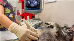
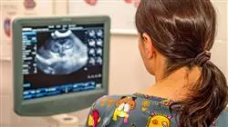
Ultrasound scanning is a universal, non-invasive, real-time technique providing very accurate diagnostic information. Ultrasound examinations are gaining great importance in everyday practice and it is increasingly common among veterinary medicine professionals to include them in their diagnostic protocols.
Ultrasound scans provide the veterinary professional with moving images of the structures being studied, as well as information on the condition of the different tissues. It also allows samples to be taken and uses contrast to refine diagnoses.
It is an operator-dependent technique, so in order to perform an adequate ultrasound examination and obtain the best results, it is necessary to be meticulous and protocolized. Therefore, it is necessary to master basic criteria prior to performing the ultrasound examination, such as: the general anatomy of the region to be explored, the specific anatomy of each viscera, to locate each structure properly and recognize its physiological ultrasound image which will allow us to identify the pathological image. It is also necessary to understand the specific physiology, to correlate the ultrasound findings with clinical signs, and to establish differential diagnoses (and sometimes definitive) with clinical sense and criteria.
Given the online format of this program, you will develop confidence, assurance and greater knowledge of pathologies and differential diagnoses when it comes to providing relevant and necessary information in daily ultrasound practice.
As it is an online program, the student is not conditioned by fixed schedules, nor do they need to move to physically move to another location. All of the content can be accessed at any time of the day, so you can balance your working or personal life with your academic life.
This specialization offers the bases and tools for you to become an expert in veterinary ultrasound with the help of renowned professionals with extensive experience in the field”
This Postgraduate diploma in Abdominal Ultrasound for Small Animals offers you the advantages of a high-level scientific, teaching, and technological course. These are some of its most notable features:
- The latest technology in online teaching software
- Intensely visual teaching system, supported by graphic and schematic content that is easy to assimilate and understand
- Practical cases presented by practising experts
- State-of-the-art interactive video systems
- Teaching supported by telepractice
- Continuous updating and recycling systems
- Self-regulating learning: full compatibility with other occupations
- Practical exercises for self-evaluation and learning verification
- Support groups and educational synergies: questions to the expert, discussion forums and debates
- Communication with the teacher and individual reflection work
- Content available from any fixed or portable device with internet connection
- Supplementary documentation databases are permanently available, even after the course
Immerse yourself in this training of the highest educational quality, which will allow you to face future challenges that may arise during daily practice in abdominal ultrasound”
Our teaching staff is made up of professionals from different fields related to this specialty. In this way, we ensure that we provide you with the training update we are aiming for. A multidisciplinary team of professionals trained and experienced in different environments, who will cover the theoretical knowledge in an efficient way, but, above all, will put the practical knowledge derived from their own experience at the service of the course: one of the differential qualities of this course.
This mastery of the subject is complemented by the effectiveness of the methodological design of this Postgraduate diploma in Abdominal Ultrasound for Small Animals. Developed by a multidisciplinary team of e-learning experts, it integrates the latest advances in educational technology. In this way, you will be able to study with a range of easy-to-use and versatile multimedia tools that will give you the necessary skills you need for your specialization.
The design of this program is based on Problem-Based Learning: an approach that conceives learning as a highly practical process. To achieve this remotely, we will use telepractice: with the help of an innovative interactive video system, and learning from an expert, you will be able to acquire the knowledge as if you were actually dealing with the scenario you are learning about. A concept that will allow you to integrate and fix learning in a more realistic and permanent way.
Learn from real cases with this highly effective educational Postgraduate diploma and open up new paths to your professional progress"

As the course is online, you will be able to train wherever and whenever you want, balancing your personal and professional life"
Syllabus
The contents of this Postgraduate diploma course have been developed by the different experts on this course, with a clear purpose: to ensure that our students acquire each and every one of the necessary skills to become true experts in this field.
A complete and well-structured program that will take you to the highest standards of quality and success.

We have the best content of the moment, developed according to the current teaching quality criteria"
Module 1. Ultrasound Diagnosis
1.1. Ultrasound Scanners
1.1.1. Frequency (F)
1.1.2. Depth
1.1.3. Acoustic Impedance
1.1.4. Physical Phenomena
1.1.4.1. Reflection
1.1.4.2. Refraction
1.1.4.3. Absorption
1.1.4.4. Dispersion
1.1.4.5. Attenuation
1.1.5. Transduction and Transducer
1.2. Operation of an Ultrasound Scanner
1.2.1. Patient Selection and Data Entry
1.2.2. Types of Exam (Preset)
1.2.3. Transducer Position
1.2.4. Freeze, Save, or Pause Image
1.2.5. Cineloop
1.2.6. Image Mode Selection
1.2.7. Depth
1.2.8. Zoom
1.2.9. Focus
1.2.10. Gain
1.2.11. Frequency (F)
1.2.12. Sector Size
1.3. Types of Probe
1.3.1. Sectorial
1.3.2. Lineal
1.3.3. Microconvex
1.4. Ultrasound Modes
1.4.1. M-Mode
1.4.2. Two-dimensional Mode
1.4.3. Transesophageal Echocardiogram
1.5. Doppler Ultrasound
1.5.1. Physical Principles
1.5.2. Indications
1.5.3. Types
1.5.3.1. Spectral Doppler
1.5.3.2. Pulsed Doppler
1.5.3.3. Continuous Doppler
1.6. Harmonic and Contrast Ultrasound
1.6.1. Harmonic Ultrasound
1.6.2. Contrast Ultrasound
1.6.3. Utilities
1.7. Patient Preparation
1.7.1. Prior Preparation
1.7.2. Positioning
1.7.3. Sedation?
1.8. Ultrasounds on the Patient
1.8.1. How Do Ultrasound Waves Behave When Passing Through Tissue?
1.8.2. What Can We See in the Image?
1.8.3. Echogenicity
1.9. Image Orientation and Expression
1.9.1. Orientation
1.9.2. Terminology
1.9.3. Examples:
1.10. Artifacts
1.10.1. Reverberation
1.10.2. Acoustic Shadow
1.10.3. Lateral Shadow
1.10.4. Posterior Acoustic Enhancement
1.10.5. Margin Effect
1.10.6. Mirror or Specular Image
1.10.7. Scintillation Artefact
1.10.8. Aliasing
Module 2. Abdominal Ultrasound Scan I
2.1. Scanning Technique
2.1.1. Introduction
2.1.2. Methodology
2.1.3. Systematization
2.2. Retroperitoneal Cavity
2.2.1. Introduction
2.2.2. Limits
2.2.3. Ultrasound Approach
2.2.4. Pathologies of the Retroperitoneal Cavity
2.3. Urinary Bladder
2.3.1. Introduction
2.3.2. Anatomy
2.3.3. Ultrasound Approach
2.3.4. Pathologies of the Urinary Bladder
2.4. Kidneys
2.4.1. Introduction
2.4.2. Anatomy
2.4.3. Ultrasound Approach
2.4.4. Kidney Pathology
2.5. Ureters
2.5.1. Introduction
2.5.2. Ultrasound Approach
2.5.3. Ureter Pathology
2.6. Urethra
2.6.1. Introduction
2.6.2. Anatomy
2.6.3. Ultrasound Approach
2.6.4. Urethral Pathologies
2.7. Female Genital System
2.7.1. Introduction
2.7.2. Anatomy
2.7.3. Ultrasound Approach
2.7.4. Pathologies of the Female Reproductive System
2.8. Pregnancy and Post-partum
2.8.1. Introduction
2.8.2. Pregnancy Diagnosis and Estimation of Gestation Time
2.8.3. Pathologies
2.9. Male Genital System
2.9.1. Introduction
2.9.2. Anatomy
2.9.3. Ultrasound Approach
2.9.4. Pathologies of the Female Reproductive System
2.10. Adrenal Glands
2.10.1. Introduction
2.10.2. Anatomy
2.10.3. Ultrasound Approach
2.10.4. Pathologies of the Adrenal Gland
Module 3. Abdominal Ultrasound Scan II
3.1. Peritoneal Cavity
3.1.1. Introduction
3.1.2. Methodology
3.1.3. Pathologies of the Peritoneal Cavity
3.2. Stomach
3.2.1. Introduction
3.2.2. Anatomy
3.2.3. Ultrasound Approach
3.2.3. Stomach Pathologies
3.3. Small Intestine
3.3.1. Introduction
3.3.2. Anatomy
3.3.3. Ultrasound Approach
3.3.4. Pathologies of the Small Intestine
3.4. Large Intestine
3.4.1. Introduction
3.4.2. Anatomy
3.4.3. Ultrasound Approach
3.4.4. Pathologies of the Large Intestine
3.5. Bladder
3.5.1. Introduction
3.5.2. Anatomy
3.5.3. Ultrasound Approach
3.5.4. Pathologies of the Spleen
3.6. Liver
3.6.1. Introduction
3.6.2. Anatomy
3.6.3. Ultrasound Approach
3.6.4. Pathologies of the Liver
3.7. Gallbladder
3.7.1. Introduction
3.7.2. Anatomy
3.7.3. Ultrasound Approach
3.7.4. Gallbladder Pathologies
3.8. Pancreas
3.8.1. Introduction
3.8.2. Anatomy
3.8.3. Ultrasound Approach
3.8.4. Pathologies of the Pancreas
3.9. Abdominal Lymph Nodes
3.9.1. Introduction
3.9.2. Anatomy
3.9.3. Ultrasound Approach
3.9.4. Pathologies of the Abdominal Lymph Nodes
3.10. Abdominal Masses
3.10.1. Ultrasound Approach
3.10.2. Localisation
3.10.3. Possible Causes/Origins of Abdominal Masses

This Postgraduate diploma in Abdominal Ultrasound for Small Animals will take you through different teaching approaches which will allow you to learn in a dynamic and efficient way”
Postgraduate Diploma in Abdominal Ultrasound for Small Animals
.
If you are a veterinary professional passionate about diagnostic medicine and want to expand your knowledge in the field of abdominal ultrasound in small animals, the Postgraduate Diploma in Abdominal Ultrasound for Small Animals from TECH Global University is the perfect program for you. In this online course, you will learn the fundamental techniques and principles of abdominal ultrasound applied to dogs, cats and other small animals. You will acquire the necessary knowledge to perform detailed and accurate ultrasound scans of the abdomen, identify internal structures and organs, and diagnose specific diseases and pathologies. The program is designed by experts in the field of veterinary ultrasound and is based on the latest advances in technology and methodology. You will learn how to use state-of-the-art ultrasound equipment and how to interpret the images obtained to obtain accurate diagnoses. In addition, you will explore different clinical applications of abdominal ultrasound, including the study of abdominal organs, evaluation of the urinary system, detection of masses and tumors, and much more.
Acquire the skills to provide accurate diagnosis in small animals
.
By studying online, you'll enjoy the flexibility to tailor your study schedule to your personal and professional needs. You will have access to interactive learning materials, multimedia resources and hands-on activities that will help you develop your skills in ultrasound image interpretation. In addition, you will have the support of specialized professors who will guide you throughout the program. The Postgraduate Diploma in Abdominal Ultrasound for Small Animals is aimed at veterinarians and animal health professionals who wish to specialize in small animal abdominal ultrasound. It is also suitable for veterinary students who wish to acquire advanced knowledge in this field. No previous knowledge in ultrasound is required, as the program covers from the fundamentals to the most advanced techniques. Become an expert in small animal abdominal ultrasound and improve your diagnostic skills in the field of veterinary medicine.


