Description

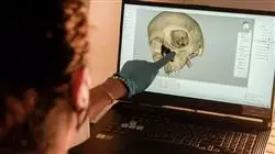
You will specifically detail visual findings on the physical condition of the victims and will contribute to the specialists' ability to determine the causes of their deterioration after completing this 100% online Professional master’s degree”
##IMAGE##

Technological advances are completely transforming the field of Forensic Radiology, making it possible to obtain accurate images that serve to identify and record injuries or diseases in the bodies of deceased persons. As a result, nursing professionals must be prepared to actively collaborate with medical personnel and other authorities to determine the cause of death or injury. In particular, they need to be up to date on advances in imaging techniques and tools that make it possible to recognize victims and even reconstruct crime scenes. However, to perform their work with maximum efficiency, these professionals need to constantly acquire new skills for the analysis of the results obtained. 

In this context, TECH implements a program in Forensic Radiology for Nursing. Developed by references in this field, the syllabus will provide graduates with the latest techniques in Diagnostic Imaging. Therefore, graduates will acquire practical skills to handle state-of-the-art machinery such as X-rays, MRI and CT scans. In this way, they will be able to capture detailed images of the deceased to analyze fractures, foreign objects or other relevant evidence for forensic investigations. The syllabus will also delve into Bone Physiopathology in order to correctly interpret the characteristics of traumatic injuries, determining both their nature and possible mechanism of injury. Additionally, the materials will investigate casuistry such as wounds caused by sharp elements and firearms.

On the other hand, the university program is based on the effective Relearninglearning method. Thanks to this, students will reduce the hours of study and will solidly consolidate the concepts addressed during this academic itinerary. Moreover, since it is taught 100% online, the only thing students will need is a device with Internet access to view the academic content hosted on the virtual platform. Undoubtedly, an ideal opportunity to reconcile daily activities with a high quality educational experience.

A highly skilled university program created to boost your career as a nurse and place you at the forefront of competitiveness in the Forensic Radiology industry"

This Professional master’s degree in Forensic Radiology for Nursing contains the most complete and up-to-date scientific program on the market. The most important features include: 

  • The development of practical cases presented by experts in Forensic Radiology
  • The graphic, schematic and eminently practical contents with which it is conceived gather scientific and practical information on those disciplines that are indispensable for professional practice
  • Practical exercises where self-assessment can be used to improve learning
  • Its special emphasis on innovative methodologies 
  • Theoretical lessons, questions to the expert, debate forums on controversial topics, and individual reflection assignments
  • Content that is accessible from any fixed or portable device with an Internet connection

You will provide comprehensive support during forensic procedures such as virtual autopsies or image-guided tissue sampling, ensuring procedures are safe"

The program’s teaching staff includes professionals from the field who contribute their work experience to this educational program, as well as renowned specialists from leading societies and prestigious universities. 

The multimedia content, developed with the latest educational technology, will provide the professional with situated and contextual learning, i.e., a simulated environment that will provide immersive education programmed to learn in real situations. 

This program is designed around Problem-Based Learning, whereby the professional must try to solve the different professional practice situations that arise during the course. For this purpose, students will be assisted by an innovative interactive video system created by renowned and experienced experts.  

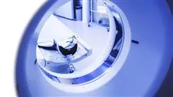
You will handle tools such as Computed Tomography to visualize in detail the internal structures of the human body and detect bone fractures"
##IMAGE##

TECH's distinctive Relearning system will allow you to update your knowledge without depending on external teaching conditions"  

Syllabus

This university program will provide nurses with specialized knowledge on Forensic Radiology, providing them with the technical skills required to provide and assist patients during diagnostic imaging procedures. Therefore, the academic itinerary will deepen in the handling of the most innovative technological tools in this context, among which Computed Tomography, X-Ray Tube or Magnetic Resonance Imaging stand out. In this sense, the syllabus will analyze the different anatomical positions and the conditions for obtaining photographs, so that the graduates will be able to appreciate in detail the trauma or pathologies.  

##IMAGE##

A high level syllabus that covers the latest scientific postulates in Bone Physiopathology in the forensic field”

Module 1. Diagnostic Imaging Techniques and Tools in the Forensic Context

1.1. Radiological Physics and its Application in the Forensic Context

1.1.1. Physics Applied to Forensic Radiology
1.1.2. Radiological Characterization in the Forensic Context
1.1.3. Structure of Matter

1.2. Operation of Equipment in the Forensic Context

1.2.1. X-ray Imaging System
1.2.2. X-ray Tube
1.2.3. Diagnostic Ultrasound

1.3. Forensic Use of Radiology

1.3.1. Computed Tomography (CT)
1.3.2. Conventional X-rays (RX)
1.3.3. Ultrasound (UI)
1.3.4. Magnetic Resonance

1.4. Forensic Radiobiology

1.4.1. Human Biology
1.4.2. Radiobiology
1.4.3. Molecular and Cellular Radiobiology

1.5. Dosimetric Quantities in Forensic Contexts

1.5.1. Radiation Protection
1.5.2. Ionization
1.5.3. Arousal
1.5.4. Fluorescence

1.6. Digital Imaging in Forensics

1.6.1. The Digital Image
1.6.2. Visualization and Understanding of Images in the Forensic Field
1.6.3. Artifacts

1.7. Forensic Computed Tomography

1.7.1. Operation
1.7.2. Scope
1.7.3. Terminology

1.8. Conventional Forensic Radiobiology Equipment

1.8.1. Operation
1.8.2. Scope
1.8.3. Terminology

1.9. Ultrasound in Forensic Medicine

1.9.1. Operation
1.9.2. Scope
1.9.3. Terminology

1.10. Magnetic Resonance in Expert Investigation

1.10.1. Operation
1.10.2. Scope
1.10.3. Terminology

Module 2. Forensic Radiology of the Non-Pathological and Non-Traumatic Human Skeleton

2.1. Forensic Radiology of the Locomotor System

2.1.1. Muscular System
2.1.2. Articular System
2.1.3. Skeletal System

2.2. Forensic Radiology of the Human Skeleton

2.2.1. Axial Skeleton
2.2.2. Appendicular Skeleton
2.2.3. Upper and Lower Extremities

2.3. Anatomical Plans and Axes of Movement in Forensic Investigation

2.3.1. Coronal Plan
2.3.2. Sagittal Plan
2.3.3. Transverse Plan
2.3.4. Bone Classification

2.4. Forensic Radiology of the Human Skull

2.4.1. Facial Bones
2.4.2. Neurocranium
2.4.3. Associated Pathologies

2.5. Forensic Radiology of the Spine

2.5.1. Cervical Vertebrae
2.5.2. Thoracic Vertebrae
2.5.3. Lumbar Vertebrae
2.5.4. Sacral Vertebrae
2.5.5. Associated Pathologies and Traumas

2.6. Forensic Radiology of the Coxal Bones

2.6.1. Ilium/Ischium/Sacral Complex
2.6.2. Public Symphysis
2.6.3. Associated Pathologies and Traumas

2.7. Forensic Upper Extremity Radiology

2.7.1. Long Bones
2.7.2. Bone Complexes of the Hands
2.7.3. Pathologies and Traumas

2.8. Forensic Radiology of the Lower Extremities

2.8.1. Long Bones
2.8.2. Bone Complexes of the Feet
2.8.3. Pathologies and Traumas

2.9. Forensic Pathologies and Traumas through Diagnostic Imaging

2.9.1. Congenital Pathologies
2.9.2. Acquired Pathologies
2.9.3. Trauma and its Variants

2.10. Interpretation of Radiographic Images in the Forensic Field

2.10.1. Radiolucent Bodies
2.10.2. Radiopaque Bodies
2.10.3. Gray Scales

Module 3. Forensic Radiology of the Human Skeleton in Phases of Biological Maturation

3.1. Bone Physiopathology in the Forensic Context

3.1.1. Functions
3.1.2. Composition - Bone Tissue
3.1.3. Cellular Component

3.1.3.1. Bone-Forming Cells (Osteoblasts)
3.1.3.2. Bone Destroyers (Osteoclasts)
3.1.3.3. Mature Bone Cells (Osteocytes)

3.2. Osteogenesis in Individuals in the Forensic Context

3.2.1. Membranous Ossification Pathway
3.2.2. Chondral Ossification Pathway
3.2.3. Periosteum

3.3. Bone Vascularization in the Forensic Context

3.3.1. Main Pathway
3.3.2. Epiphyseal Pathway
3.3.3. Metaphyseal Pathway
3.3.4. Periosteal Arterial Pathway

3.4. Bone Growth in the Forensic Context

3.4.1. Width
3.4.2. Length
3.4.3. Associated Pathologies

3.5. Forensic Radiology of Pathologies in Developing Individuals

3.5.1. Congenital Diseases
3.5.2. Acquired Pathologies
3.5.3. Trauma and its Variants

3.6. Bone Diseases Through Diagnostic Imaging in the Forensic Context

3.6.1. Osteoporosis
3.6.2. Bone Cancer
3.6.3. Osteomyelitis
3.6.4. Osteogenesis Imperfecta
3.6.5. Rickets

3.7. Forensic Radiology of the Child Skull

3.7.1. Embryonic, Fetal and Neonatal Formation.
3.7.2. Fontanelles and Fusion Phases
3.7.3. Facial and Dental Development

3.8. Forensic Radiobiological Osteology in the Adolescent

3.8.1. Sexual Dimorphism and Bone Growth
3.8.2. Bone Changes Resulting from Hormonal Action
3.8.3. Juvenile Growth Retardation and Metabolic Problems

3.9. Trauma and Categories of Childhood Fractures in Forensic Diagnostic Imaging

3.9.1. Frequent Traumas in Infantile Long Bones
3.9.2. Frequent Traumas in Infantile Flat Bones
3.9.3. Trauma Resulting from Aggression and Mistreatment

3.10. Radiology and Diagnostic Imaging Techniques in Forensic Pediatrics

3.10.1. Radiology for Neonates and Infants
3.10.2. Radiology for Children in Early Childhood
3.10.3. Radiology for Adolescents and Juveniles

Module 4. Maxillofacial Forensic Radiology

4.1. Forensic Radiological Interpretation of Head and Neck: Skull Bones

4.1.1. Forensic Radiological Interpretation of the External Paired Bones: Temporal and Parietal
4.1.2. Forensic Radiological Interpretation of the External Odd Bones: Frontal, Occipital
4.1.3. Forensic Radiological Interpretation of the Internal Odd Bones: Ethmoid and Sphenoid.

4.2. Forensic Radiological Interpretation of Head and Neck: Bones of the Face

4.2.1. Forensic Radiological Interpretation of the Vomer
4.2.2. Forensic Radiologic Interpretation of the Inferior Turbinate
4.2.3. Forensic radiological Interpretation of the Zygomatic or Malar Bone.
4.2.4. Forensic Radiological Interpretation of the Nasal Lachrymal Bone

4.3. Forensic Radiological Interpretation of Head and Neck: Oral Cavity Bones

4.3.1. Forensic Radiological Interpretation of the Upper Jaw.
4.3.2. Forensic Radiological Interpretation of the Lower Maxilla or Mandible
4.3.3. Forensic Radiological Interpretation of the Dental Parts

4.4. Radiological Interpretation of Head and Neck: Sutures

4.4.1. Forensic Radiological Interpretation of the Upper Jaw.
4.4.2. Forensic Radiological Interpretation of the Lower Maxilla or Mandible
4.4.3. Forensic Radiological Interpretation of the Dental Parts

4.5. Forensic Radiological Interpretation of Head and Neck: Facial Buttresses Sutures.

4.5.1. Forensic Radiological Interpretation of the Horizontal Buttresses
4.5.2. Forensic Radiological Interpretation of Vertical Buttresses
4.5.3. Abnormalities

4.6. Forensic Radiography of the Head and Neck: Extraoral Radiographs

4.6.1. Lateral Radiographs
4.6.2. Fronto-Occipital Radiographs
4.6.3. Occipito-Frontal Radiographs
4.6.4. Orthopantomography

4.7. Forensic Radiography of Head and Neck Anatomical Accidents: Intraoral Radiographs

4.7.1. Occlusal Radiographs
4.7.2. Periapical Radiographs
4.7.3. Bitewing Radiographs
4.7.4. Relevant Elements Observed in Intraoral Radiographs

4.8. Forensic Radiographic Interpretation of Head and Neck Anatomical Features: Extraoral Radiography

4.8.1. Lateral Radiography
4.8.2. Fronto-Occipital Radiography
4.8.3. Occipito-Frontal Radiography
4.8.4. Orthopantomography

4.9. Forensic Radiographic Interpretation of Head and Neck Anatomical Features: Intraoral Radiography

4.9.1. Occlusal Radiography
4.9.2. Periapical Radiography
4.9.3. Bitewing Radiograph

4.10. Forensic Radiographic Interpretation of Head and Neck Anatomical Features: Other Radiographic Techniques

4.10.1. Computerized Axial Tomography
4.10.2. CBCT
4.10.3. MRI

Module 5. Forensic Radiology in Human Identification

5.1. Human Identification in the Forensic Context

5.1.1. In Police Cases
5.1.2. In Judicial Cases
5.1.3. In Crimes Against Humanity and War Crimes
5.1.4. In Major Disasters

5.2. The Human Skeleton and Biological Identification (I): Osteological Sexual Characterization in Adults

5.2.1. Sexual Characterization Through the Skull
5.2.2. Sexual Characterization Through the Hip
5.2.3. Osteological Sex Characterization from Other Bones

5.3. The Human Skeleton and Biological Identification (II): Osteological Sexual Characterization in Individuals in Maturing Stages.

5.3.1. Sexual Characterization Through the Skull
5.3.2. Sexual Characterization Through the Hip
5.3.3. Osteological Sex Characterization from Other Bones

5.4. The Human Skeleton and Biological Identification (III): Determination of Age at Death in Adult Individuals

5.4.1. Age Determination from the Closure of Bone Epiphyses and Cranial Sutures 
5.4.2. Age Determination from Cartilage Ossification
5.4.3. Age Determination from the Modification of Bone Regions

5.5. The Human Skeleton and Biological Identification (IV): Age Determination at Death in Maturing Individuals

5.5.1. Determination of age from Morphometrics
5.5.2. Age Determination by Bone Birth
5.5.3. Age Determination by Epiphyseal and Fontanel Closure 

5.6. The Human Skeleton and Biological Identification (V): Determination of Stature and Muscular Build

5.6.1. Estimation of Stature of Anatomical Nature
5.6.2. Estimation of Stature of Physiological Nature
5.6.3. Bone Biomechanics and Adaptation to Physical Activity
5.6.4. Development of Muscular Complexion

5.7. Human Dentition for the Calculation of Age at Death

5.7.1. The Dentition in Maturing Individuals
5.7.2. Dentition in Adult Individuals
5.7.3. Dental Alterations and Pathologies

5.8. Biomechanics and Mechanical Forces Applied to Bone Trauma

5.8.1. Osteological Growth and Development
5.8.2. Mechanical Forces Applied to the Human Skeleton
5.8.3. Bone Adaptation to Exercise

5.9. Bone Trauma due to Temporality

5.9.1. Characterization of AntemortemTraumas
5.9.2. Characterization of PerimortemTraumas
5.9.3. Characterization of PostmortemTrauma

5.10. Trauma by Type of Injury

5.10.1. Classification by Type of Injury
5.10.2. Classification by Type of Weapon
5.10.3. Classification by Type of Object and Structure

Module 6. Radiodiagnosis of Pathologies Related to Forensic Investigation

6.1. Classification of Traumatic Fractures in the Forensic Context

6.1.1. Classification According to Skin Condition
6.1.2. Classification According to Location
6.1.3. Classification According to Fracture Trace

6.2. Stages of Bone Repair in the Forensic Context

6.2.1. Inflammatory Phase
6.2.2. Repair Phase
6.2.3. Remodeling Phase

6.3. Child Maltreatment and its Radiodiagnosis in a Forensic Context

6.3.1. Simple Radiography
6.3.2. Axial Tomography
6.3.3. Magnetic Resonance

6.4. Illegal Transport of Narcotics and Radiodiagnostics in a Forensic Context

6.4.1. Simple Radiography
6.4.2. Axial Tomography
6.4.3. Magnetic Resonance

6.5. Simple Radiographic Technique for Identification of Alterations within a Forensic Context

6.5.1. Cranial Pathologies
6.5.2. Thoracic Pathologies
6.5.3. Extremity Pathologies

6.6. Ultrasound Technique for Identification of Pathologies within a Forensic Context

6.6.1. Abdominal
6.6.2. Obstetric
6.6.3. Thoracic

6.7. Computed Tomography and Identification of Pathologies in a Forensic Context

6.7.1. Cranial
6.7.2. Thoracic
6.7.3. Ultrasound

6.8. Magnetic Resonance Imaging and Pathology Identification in a Forensic Context

6.8.1. Cranial
6.8.2. Thoracic
6.8.3. Ultrasound

6.9. Diagnostic Angiography in a Forensic Context

6.9.1. Cranial
6.9.2. Abdominal
6.9.3. Extremities

6.10. Virtopsia, Radiology in Forensic Medicine

6.10.1. MRI
6.10.2. Tomography
6.10.3. Radiography

Module 7. Forensic Radiological Techniques of Bone and Dental Trauma with Blunt 

7.1. Classification of Blunt Profile Injury Elements

7.1.1. Blunt Weapons
7.1.2. Blunt Objects
7.1.3. Blunt Mechanical Force Injuries
7.1.4. Structural Injuries
7.1.5. Short Blunt Injuries

7.2. Injury Mechanics of Blunt Elements

7.2.1. Blunt Weapons
7.2.2. Blunt Objects
7.2.3. Blunt Mechanical Force Injuries
7.2.4. Injuries Through Structures
7.2.5. Short Blunt Injuries

7.3. Injury Typologies of Blunt weapons

7.3.1. Superficial Injuries
7.3.2. Deep Injuries
7.3.3. Total or Partial Amputation Injuries

7.4. Types of Injuries Caused by Blunt Objects

7.4.1. Superficial Injuries
7.4.2. Deep Injuries
7.4.3. Total or Partial Amputation Injuries

7.5. Injury Typologies Due to Blunt Injury Mechanics

7.5.1. Superficial Injuries
7.5.2. Deep Injuries
7.5.3. Total or Partial Amputation Injuries

7.6. Injury Typologies of Blunt Structures and Short-Contusive Elements

7.6.1. Superficial Injuries
7.6.2. Deep Injuries
7.6.3. Total or Partial Amputation Injuries

7.7. Marks on the Skeleton of Injuries Due to Blunt Mechanics

7.7.1. Blunt Weapons
7.7.2. Blunt Objects
7.7.3. Blunt Mechanical Force Injuries
7.7.4. Injuries Through Structures
7.7.5. Short Blunt Injuries

7.8. Radiological Techniques for the Study of Blunt Force Injuries

7.8.1. X-Ray
7.8.2. Computerized Axial Tomography
7.8.3. Other Radiographic Techniques

7.9. Radiobiological Techniques for the Study of Injuries of Blunt Objects and Structures

7.9.1. X-Ray
7.9.2. Computerized Axial Tomography
7.9.3. Other Radiographic Techniques

7.10. Radiobiological Techniques for the Study of Blunt Mechanical Injuries and Short Blunt Elements

7.10.1. X-Ray
7.10.2. Computerized Axial Tomography
7.10.3. Other Radiographic Techniques

Module 8. Forensic Radiology of Trauma with Sharps and Blunt Elements

8.1. Classification of Sharp Weapons

8.1.1. Cutting Weapons
8.1.2. Sharp Weapons
8.1.3. Sharps

8.2. Injurious Mechanics of Edged Weapons

8.2.1. Cutting Weapons
8.2.3. Sharp Weapons
8.2.4. Sharps

8.3. Types of Injuries Caused by Cutting Weapons

8.3.1. Superficial Injuries
8.3.2. Deep Injuries
8.3.3. Total or Partial Amputation Injuries

8.4. Injury Typologies of Sharp-Edged Weapons by Sharp Weapons

8.4.1. Superficial Injuries
8.4.2. Deep Injuries
8.4.3. Total or Partial Amputation Injuries

8.5. Injury Typologies of Sharp-Edged Weapons by Sharps

8.5.1. Superficial Injuries
8.5.2. Deep Injuries
8.5.3. Total or Partial Amputation Injuries

8.6. Skeletal Marks from Sharp-Edged Weapon Injuries

8.6.1. Cutting Weapons
8.6.2. Sharp Weapons
8.6.3. Sharps

8.7. Radiological Techniques for the Study of Cutting Weapon Injuries

8.7.1. X-Ray
8.7.2. Computerized Axial Tomography
8.7.3. Other Radiographic Techniques

8.8. Radiological Techniques for the Study of Sharps Injuries

8.8.1. X-Ray
8.8.2. Computerized Axial Tomography
8.8.3. Other Radiographic Techniques

8.9. Radiological Techniques for the Study of Sharps Injuries

8.9.1. X-Ray
8.9.2. Computerized Axial Tomography
8.9.3. Other Radiographic Techniques

8.10. Analysis of Lesions at the Maturation Stage and in Animals

8.10.1. Cutting Lesions in Individuals in Early Stages of Maturation.
8.10.2. Cut Marks on Individuals in Late Stages of Biological Maturation
8.10.3. Cutting Injuries in Animals

Module 9. Radiology of Firearms and Explosives Trauma in Forensic Investigation

9.1. Firearms and Projectiles

9.1.1. Classification of Firearms
9.1.2. Elements that Compose a Firearm
9.1.3. Structure of the Firearm
9.1.4. Firearm Projectiles

9.2. Characterization of Wounds and Firearm Projectile Trajectory

9.2.1. Entrance Orifice
9.2.2. Trajectory
9.2.3. Outlet Orifice

9.3. X-ray Technique and Firearm Projectiles

9.3.1. Number of Projectiles
9.3.2. Probable Trajectory
9.3.3. Probable Caliber
9.3.4. Type of Firearm

9.4. Axial Tomography and Firearm Projectiles

9.4.1. Number of Projectiles
9.4.2. Trajectory
9.4.3. Type of Weapons Used

9.5. Ultrasound and Firearm Projectile

9.5.1. Number of Projectiles
9.5.2. Trajectory
9.5.3. Type of Weapons Used

9.6. Virtual Autopsy in Deaths Caused by Firearm Projectile Wounds 

9.6.1. Simple Radiography
9.6.2. Computerized Axial Tomography
9.6.3. Magnetic Resonance

9.7. Explosives

9.7.1. Typologies of Explosive Elements
9.7.2. Categorization
9.7.3. Mechanics of Explosions

9.8. Classification of Blast Injuries

9.8.1. Primary
9.8.2. Secondary
9.8.3. Tertiary
9.8.4. Quaternary

9.9. Radiodiagnostic Imaging in the Search for and Retrieval of Evidence

9.9.1. Simple Radiography
9.9.2. Computerized Axial Tomography
9.9.3. Magnetic Resonance

9.10. Radiological Assessment of Blast Injuries

9.10.1. Cranial
9.10.2. Cervical
9.10.3. Chest
9.10.4. Abdomen
9.10.5. Extremities

Module 10. Forensic Radiodiagnosis of Maxillofacial Trauma

10.1. Forensic Maxillofacial Trauma: Fractures of the Upper Third of the Face

10.1.1. Fractures of the Frontal Bone
10.1.2. Fractures of the Walls of the Frontal Sinuses
10.1.3. Fractures of the Temporal/Parietal Bone

10.2. Forensic Maxillofacial Trauma: Fractures of the Middle Third of the Face

10.2.1. Nasal Fractures
10.2.2. Orbital Fractures
10.2.3. Fractures of the Naso-Orbito-Ethmoidal Complex
10.2.4. Fractures of the Zygomatic Bone

10.3. Forensic Maxillofacial Trauma: Fractures of the Lower Third of the Face.

10.3.1. Fracture of the Mandibular Symphysis / Parasymphysis
10.3.2. Fracture of the Mandibular Body
10.3.3. Mandibular Angle Fracture
10.3.4. Mandibular Ramus Fracture
10.3.5. Fracture of the Mandibular Condyle

10.4. Forensic Maxillofacial Trauma: Le Fort Fractures

10.4.1. Le Fort I Fractures
10.4.2. Le Fort II Fractures
10.4.3. Le Fort III Fractures
10.4.4. Le Fort IV Fractures

10.5. Forensic Maxillofacial Trauma: Alveolodental Fractures

10.5.1. Coronary Fracture
10.5.2. Corono-Radicular Fracture
10.5.3. Root Fracture
10.5.4. Alveolar Fracture
10.5.5. Avulsion

10.6. Radiographic Techniques for the Study of Maxillofacial Trauma in the Forensic Context.

10.6.1. X-Ray
10.6.2. Computerized Axial Tomography
10.6.3. Other Radiographic Techniques

10.7. Radiographic Techniques for the Study of Alveolar Tooth Trauma in the Forensic Context

10.7.1. X-Ray
10.7.2. Computerized Axial Tomography
10.7.3. Other Radiological Techniques

10.8. Radiographic Interpretation of Maxillofacial Trauma in the Forensic Context: Isolated Fractures.

10.8.1. Radiographic Interpretation of Trauma to the Upper Third of the Face.
10.8.2. Radiographic Interpretation of Trauma of the Middle Third of the Face
10.8.3. Radiographic Interpretation of Trauma of the Lower Third of the Face 

10.9. Radiographic Interpretation of Maxillofacial Trauma Within the Forensic Context  Le Fort Fractures

10.9.1. Radiographic Interpretation in Le Fort I Fractures.
10.9.2. Radiographic Interpretation in Le Fort II Fractures.
10.9.3. Radiographic Interpretation in Le Fort III Fractures.
10.9.4. Radiographic Interpretation in Le Fort IV Fractures.

10.10. Radiographic Techniques for the Study of Alveolar Tooth Trauma in the Forensic Context

10.10.1. Coronary Fracture
10.10.2. Corono-Radicular Fracture
10.10.3. Alveolar Fracture
10.10.4. Root Fracture 
10.10.5. Avulsion  

##IMAGE##

A flexible university course, with no prearranged schedules and content available 24 hours a day. Enroll now!”

Professional Master's Degree in Forensic Radiology for Nursing

In a world where precision and accuracy are paramount, the role of forensic nursing is increasingly relevant. The ability to interpret radiological images with detail and accuracy is a crucial skill in the investigation and resolution of forensic cases. That is why at TECH we have developed this comprehensive postgraduate program to meet the needs of nursing professionals who wish to specialize in this field. Our Professional Master's Degree in Forensic Radiology offers a comprehensive approach that combines theory and practice to provide students with an in-depth understanding of the radiological principles and techniques needed in the forensic setting. Through our online classes, participants will have access to up-to-date educational content and expert guidance from leading professionals in the field.

Study at the world's largest School of Nursing

The program covers a wide range of topics, from radiological anatomy and physiology to advanced interpretation of forensic medical images. Students will learn to identify traumatic injuries, evaluate radiographic evidence, and collaborate effectively with other professionals in the investigation and resolution of complex cases. In addition, our hands-on approach ensures that students acquire real-world skills that they can apply in their professional practice. Through real-world case studies, simulations and supervised practice, participants will develop the confidence and skills necessary to successfully meet the challenges of the forensic field. Upon completion of this Professional Master's Degree in Nursing, graduates will be prepared to perform with excellence in a variety of forensic settings, including hospitals, forensic laboratories, law enforcement agencies and more. Take the opportunity to advance your career and make a difference in the field of forensic nursing. Enroll in our master's program today and take the next step toward a future filled with opportunity and professional fulfillment!