Why study at TECH?

The emergence of COVID-19 has forced specialists to keep up to date on the main respiratory therapies. Enroll on this Professional Master's Degree now and receive the training that will enable you to use the most up-to-date and effective techniques” 
##IMAGE##

Population aging, air pollution and smoking are all factors that lead to an increase in chronic respiratory pathologies, such as Chronic Obstructive Pulmonary Disease (COPD), which have a considerable impact on the population. However, the discovery and widespread use of new treatments has changed the prognosis and evolution of other respiratory diseases, such as Interstitial Lung Disease (ILD), lung cancer and cystic fibrosis, which has opened up a field of research and clinical management that, until recently, was rather limited.

Likewise, the COVID-19 pandemic has forced pulmonologists and other medical specialists to update their knowledge of infectious diseases and has highlighted the usefulness of advanced respiratory therapies such as high-flow oxygen therapy and non-invasive mechanical ventilation for managing respiratory failure.

This Professional master’s degree in Pulmonology at TECH Global University aims to provide physicians with an update on the latest scientific evidence available in published guidelines, scientific articles and systematic reviews. As such, the syllabus presented is particularly relevant today, as it includes improvements in diagnostic and therapeutic methods that can change previous paradigms in managing these patients. The syllabus also covers pathophysiological fundamentals and incorporates images that illustrate the latest diagnostic tests. Additionally, the scientific evidence on recently incorporated therapies will be reviewed in depth.

One of the main advantages of this program is that it is taught in a 100% online format, so students will have access to all the contents available in the virtual classroom from the moment they enroll. They will be able to manage their study time independently and, in addition, a self-learning approach will be favored, which will enable them to handle respiratory pathology in an ever-changing era with complete confidence.

Thanks to your specialized help, patients with pulmonary diseases will be able to improve their quality of life” 

This Professional master’s degree in Pulmonology contains the most complete and up-to-date scientific program on the market. Its most notable features are:

  • Practical cases presented by experts in Pulmonology
  • The graphic, schematic, and practical contents with which they are created, provide scientific and practical information on the disciplines that are essential for professional practice
  • Practical exercises where self-assessment can be used to improve learning
  • Special emphasis is placed on innovative methodologies in the approach to  pulmonological affections
  • Theoretical lessons, questions to the expert, debate forums on controversial topics, and individual reflection assignments
  • Content that is accessible from any fixed or portable device with an Internet connection

With the latest educational methodology and a first-rate syllabus, you will have the opportunity to update your knowledge to grow professionally and offer more personalized care”

The program’s teaching staff includes professionals from the sector who contribute their work experience to this training program, as well as renowned specialists from leading societies and prestigious universities.

The multimedia content, developed with the latest educational technology, will provide the professional with situated and contextual learning, i.e., a simulated environment that will provide immersive training programmed to train in real situations.  

This program is designed around Problem Based Learning, whereby the professional must try to solve the different professional practice situations that arise during the academic year. For this purpose, the student will be assisted by an innovative interactive video system created by renowned and experienced experts.   

A 100% online program, essential to apply the latest techniques in the field of Pulmonology"
##IMAGE##

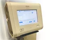
Learn to use the latest diagnostic tools for early detection of the main respiratory pathologies"

Syllabus

Pulmonology deals with the study of the physiology and pathology of the respiratory system, as well as the diagnostic tests and preventive and therapeutic measures necessary to manage these diseases. Thanks to this Professional master’s degree, students will have access to the latest information in the field, which will provide them with the key tools and skills to apply the treatments that provide the best results for patients. 

##IMAGE##

Access fully updated content on respiratory diseases and discover the latest advances in the field”  

Module 1. Interstitial Pulmonary Diseases

1.1. ILDs

1.1.1. ILD Classification and Epidemiology
1.1.2. Diagnostic Approximation

    1.1.2.1. Medical History: Physical Exploration
    1.1.2.2. Clinical Laboratory and Pulmonary Function Laboratory
    1.1.2.3. Radiodiagnosis: Chest X-Ray TACAR Radiological Patterns
    1.1.2.4. Invasive Techniques: Bronchoalveolar Lavage (BAL), Transbronchial Biopsy (TBB) and Cryobiopsy Surgical Biopsy Indications and Pathologic Patterns
    1.1.2.5. Multidisciplinary Diagnosis

1.1.3. Cellular Aging, Genetics and Biomarkers in ILDs

    1.1.3.1. Pathogenesis of Cellular Aging
    1.1.3.2. Characteristics, Value, Prognosis and Treatment of Telomeric Disorders
    1.1.3.3. Family Pulmonary Fibrosis: Biomarkers Diagnostic, Prognostic and Therapeutic Utility

1.2. Idiopathic Pulmonary Fibrosis (IPF)

1.2.1. Epidemiology
1.2.2. Risk factors
1.2.3. Natural History and Prognosis
1.2.4. Diagnostic Approximation

    1.2.4.1. Clinical Manifestations Physical Exploration
    1.2.4.2. Radiological Criteria
    1.2.4.3. Histopathological Criteria
    1.2.4.4. Biomarkers Useful in IPF

1.2.5. Treatment
1.2.6. IPF Exacerbation

1.3. Idiopathic Non-Specific Interstitial Pneumonia (NSIP) ILD Associated with Systemic Autoimmune Diseases (I): ILD Associated with Rheumatoid Arthritis (ILD- RA) and ILD Associated with Systemic Sclerosis (ILD-SS)

1.3.1. Idiopathic NSIP

    1.3.1.1. Histopathological Forms
    1.3.1.2. Diagnostic tests
    1.3.1.3. Treatment
    1.3.1.4. Prognosis

1.3.2. ILD Associated with Systemic Autoimmune Diseases

    1.3.2.1. ILD-RA
    1.3.2.2. ILD-SS

1.4. ILD Associated with Systemic Autoimmune Diseases (II)

1.4.1. Dermato / Polymyositis
1.4.2. Sjögren's Syndrome
1.4.3. Mixed Connective Tissue Disease: Overlap Syndrome
1.4.4. Interstitial Pneumonia with Autoimmune Features (IPAF)

1.5. Sarcoidosis

1.5.1. Pathophysiology
1.5.2. Histology
1.5.3. Diagnostic Approximation
1.5.4. Evolution and Prognosis
1.5.5. Treatment

1.6. Hypersensitivity Pneumonitis

1.6.1. Etiology
1.6.2. Pathophysiology
1.6.3. Classification: Clinical Forms
1.6.4. Diagnostic Criteria: Differential Diagnosis
1.6.5. Natural History and Prognosis
1.6.6. Treatment

1.7. Lung Diseases

1.7.1. Linfangioleiomyomatosis (LAM)

    1.7.1.1. Clinical Manifestations
    1.7.1.2. Diagnostic Approximation
    1.7.1.3. Treatment

1.7.2. Pulmonary Langerhans Cell Histiocytosis (PLCH)

    1.7.2.1. Clinical Manifestations
    1.7.2.2. Diagnostic Approximation
    1.7.2.3. Treatment

1.7.3. Lymphocytic Interstitial Pneumonia (LIP)

    1.7.3.1. Clinical Manifestations
    1.7.3.2. Diagnostic Approximation
    1.7.3.3. Treatment

1.8. Cryptogenic Organized Pneumonia (COP)

1.8.1. Pathogenesis
1.8.2. Clinical Manifestations
1.8.3. Radiological Patterns
1.8.4.    Diagnostic Approximation
1.8.5. Natural History
1.8.6. Treatment

1.9. Occupational Diseases

1.9.1. Asbestos-Related Diseases

    1.9.1.1. Varieties of Asbestos: Sources of Exposure
    1.9.1.2. Pleural Fibrosis: Clinical Forms and Radiological Diagnosis
    1.9.1.3. Asbestosis: Clinical and Radiological Findings, Diagnostic Criteria and Treatment

1.9.2. Silicosis
1.9.3. Coal Pneumoconiosis

1.10. Pulmonary Eosinophilias: Drug-Associated ILDs Other Rare ILDs: Pleuropulmonary Fibroelastosis Alveolar Microlithiasis Alveolar Proteinosis

1.10.1. Acute Eosinophilic Pneumonia

    1.10.1.1. Epidemiology and Risk Factors
    1.10.1.2. Pathogenesis
    1.10.1.3. Clinical, Radiological, Functional and Pathological Diagnosis
    1.10.1.4. Treatment

1.10.2. Drug-Associated ILDs

    1.10.2.1. Epidemiology
    1.10.2.2. Pathogenesis and Risk Factors
    1.10.2.3. Diagnostic Approximation
    1.10.2.4. Main Causal Agents

1.10.3. Differential Diagnosis of Pulmonary Eosinophilias
1.10.4. Other Rare ILD: Pleuropulmonary Fibroelastosis, Alveolar Microlithiasis and Alveolar Proteinosis: Diagnostic Approach, Evolution and Management

Module 2. Chronic Obstructive Pulmonary Disease

2.1. Aetiopathogenesis

2.1.1. Epidemiology
2.1.2. Risk factors
2.1.3. Pathogenesis

2.2. EPOC Pathophysiology and Clinical Presentation

2.2.1. Pathophysiology
2.2.2. Clinical Manifestations

2.3. Diagnosis and Characterization

2.3.1. Diagnosis: Anamnesis, Physical Examination, Imaging Tests, Clinical Analyses and Respiratory Functional Examination
2.3.2. Characterization

    2.3.2.1. Degree of Pulmonary Obstruction
    2.3.2.2. Clinical Types: Emphysema and Chronic Bronchitis
    2.3.2.3. Risk of Exacerbation
    2.3.2.4. Symptoms

2.4. COPD Classification according to COPD Guidelines: (The Spanish COPD Guidelines) and GOLD (Global Iniciative for Chronic Obstructive Lung Disease)

2.4.1. GOLD

    2.4.1.1. GOLD A
    2.4.1.2. GOLD B
    2.4.1.3. GOLD C
    2.4.1.4. GOLD D
    2.4.1.5. Monitoring

2.5. Maintenance Pharmacological Treatment

2.5.1. Treatment Objectives
2.5.2. Drugs

    2.5.2.1. Inhaled Treatment

    2.5.2.1.1. Bronchodilators
    2.5.2.1.2. Inhaled Corticosteroids

    2.5.2.2. Oral Treatment

    2.5.2.2.1. Theophylline
    2.5.2.2.2. Roflumilast
    2.5.2.2.3. Azithromycin

2.6. Approach to Smoking in COPD

2.6.1. Epidemiology
2.6.2. The Diagnosis of Tobacco Use in COPD
2.6.3. Non-Pharmaceutical Therapeutic Interventions
2.6.4. Pharmacological Therapeutic Interventions

2.7. Non-Pharmacological Treatment

2.7.1. Oxygen Therapy and NIMV
2.7.2. Vaccines
2.7.3. Nutrition
2.7.4. Palliative Treatment of Dyspnea
2.7.5. Lung Volume Reduction by Bronchoscopy
2.7.6. Surgery: Volume Reduction and Lung Transplantation

2.8. COPD Exacerbation

2.8.1. Etiology and Pathogenesis
2.8.2. Severity Classification
2.8.3. Treatment

2.9. Comorbidities

2.9.1. Prevalence
2.9.2. Impact on Mortality
2.9.3. Screening and Managment

2.10. Rehabilitation and Physical Exercise in COPD

2.10.1. Rehabilitation in COPD

    2.10.1.1. Benefits
    2.10.1.2. Indications
    2.10.1.3. Rehabilitation Program Structure
    2.10.1.4. Rehabilitation after COPD Exacerbation
    2.10.1.5. Special Situations

2.10.2. Physical Activity

    2.10.2.1 Measuring
    2.10.2.2 Interventions

Module 3. Asthma

3.1. Aetiopathogenesis

3.1.1. Epidemiology
3.1.2. Risk factors
3.1.3. Pathogenesis

3.2. Diagnosis

3.2.1. Clinical symptoms
3.2.2. Spirometry and Bronchodilator Test
3.2.3. Bronchial Provocation Tests
3.2.4. Fractional Exhaled Nitric Oxide (FeNO) Determination
3.2.5. Induced Sputum
3.2.6. Electronic Nose 
3.2.7. Volatile Organic Compounds in Exhaled Air
3.2.8. Diagnostic Algorithm

3.3. Control and Severity Classification

3.3.1. Control
3.3.2. Severity

3.4. Maintenance Treatment

3.4.1. Treatment Objectives
3.4.2. Drugs
3.4.3. Step Treatment
3.4.4. Avoiding Allergens and Environment
3.4.5. Education and Written Action Plans

3.5. Asthma Exacerbation Treatment

3.5.1. Risk factors
3.5.2. Severity Assessment
3.5.3. Treatment according to Severity
3.5.4. Emergency Discharge Criteria
3.5.5. Hospitalization Criteria
3.5.6. Hospital Discharge Criteria
3.5.7. Outpatient Monitoring after Exacerbation

3.6. Severe Uncontrolled Asthma

3.6.1. Epidemiology
3.6.2. Diagnostic Procedure
3.6.3. Severe Asthma Phenotypes
3.6.4. Treatment Algorithm

3.7. Occupational Asthma

3.7.1. Causative Agents
3.7.2. Classification
3.7.3. Diagnosis
3.7.4. Treatment
3.7.5. Asthma Aggravated by Work

3.8. Nasal Pathology Associated with Asthma

3.8.1. Rhinitis

    3.8.1.1. Diagnosis
    3.8.1.2. Classification
    3.8.1.3. Treatment

3.8.2. Rhinosinusitis and Nasal Polyposis

    3.8.2.1. Diagnosis
    3.8.2.2. Treatment

3.9. Pulmonary Eosinophilias Associated with Asthma

3.9.1. Chronic Eosinophilic Pneumonia
3.9.2. Allergic Bronchopulmonary Aspergillosis
3.9.3. Eosinophilic Granulomatosis with Polyangiitis

3.10. Special Situations

3.10.1. Asthma and COPD Overlap (ACO)
3.10.2. Respiratory Disease Exacerbated by Acetylsalicylic Acid
3.10.3. Asthma and Pregnancy
3.10.4. Exercise-Induced Asthma
3.10.5. Pseudoasthmas

Module 4. Respiratory Infections and Related Diseases

4.1. Community-Acquired Pneumonia (CAP)

4.1.1. Epidemiology
4.1.2. Risk factors
4.1.3. Comorbidities and Risks in CAP
4.1.4. Etiology
4.1.5. Clinical Manifestations
4.1.6. Diagnosis
4.1.7. Assess the Severity of CAP
4.1.8. Treatment
4.1.9. Clinical Response
4.1.10. Complications
4.1.11. Prevention: Vaccination

4.2. Nosocomial Pneumonia (Hospital-Acquired Pneumonia and Ventilator-Associated Pneumonia)

4.2.1. Pathogenesis
4.2.2. Risk factors
4.2.3. Intrahospital Pneumonia
4.2.4. Ventilator-Associated Pneumonia
4.2.5. Etiology
4.2.6. Diagnosis
4.2.7. Treatment
4.2.8. Preventive Measures

4.3. Pulmonary Abscess

4.3.1. Pathogenesis
4.3.2. Differences with Necrotizing Pneumonia
4.3.3. Microbiology
4.3.4. Clinical Manifestations
4.3.5. Diagnosis
4.3.6. Differential Diagnosis
4.3.7. Treatment

4.4. Coronavirus: COVID-19

4.4.1. The 2019 Pandemic
4.4.2. Epidemiology
4.4.3. Pathogenesis
4.4.4. Clinical Symptoms
4.4.5. Diagnosis
4.4.6. Treatment
4.4.7. Complications
4.4.8. Prevention

    4.4.8.1. Hygienic and Social Distancing Measures
    4.4.8.2. Vaccines

4.5. Non‐Cystic Fibrosis Bronchiectasis

4.5.1. Epidemiology and Costs
4.5.2. Pathophysiology
4.5.3. Etiology
4.5.4. Diagnosis
4.5.5. Differential Diagnosis
4.5.6. Microbiology
4.5.7. Severity and Prognostic Factors
4.5.8. Treatment
4.5.9. Monitoring
4.5.10. Consensus Treatment of Inflammatory Breast Cancer (IBC), Chronic Obstructive Pulmonary Disease (COPD) and Bronchiectasis

4.6. Cystic fibrosis

4.6.1. Aetiopathogenesis
4.6.2. Epidemiology
4.6.3. Clinical Manifestations
4.6.4. Diagnosis
4.6.5. Quality of Life Associated with Health
4.6.6. Treatment

    4.6.6.1. Aggravation
    4.6.6.2. Chronic Bronchial Infection
    4.6.6.3. Bronchial Inflammation
    4.6.6.4. Mucociliary Clearance
    4.6.6.5. New Drugs (Conventionally Fractionated Radiation Therapy (CFRT))

4.6.7. Rehabilitation
4.6.8. Nutritional Treatment
4.6.9. Treating Complications

4.7. Pulmonary Tuberculosis: Epidemiology, Clinical Practice, Diagnosis, Complications and Prognosis

4.7.1. Epidemiology
4.7.2. Etiology
4.7.3. Pathogenesis and Physiopathology
4.7.4.    Clinical Manifestations
4.7.5. Diagnosis: Concept of Infection and Tuberculous Disease

    4.7.5.1. Tuberculous Infection
    4.7.5.2. Tuberculous Disease

    4.7.5.2.1. Clinical-Radiological Diagnosis
    4.7.5.2.2. Anatomo-Pathological Diagnosis
    4.7.5.2.3. Microbiological Diagnosis

4.7.6. Complications and Prognosis

4.8. Pulmonary Tuberculosis: Treatment Chemoprophylaxis

4.8.1. Types of Bacillary Populations
4.8.2. Standard Treatment: Proper Drug Combination Selection
4.8.3. Treatment in Special Situations

    4.8.3.1. Immunodeficiencies
    4.8.3.2. Pregnancy and Breastfeeding
    4.8.3.3. Advanced Chronic Liver Failure
    4.8.3.4. Chronic Advanced Kidney Disease

4.8.4. Adverse Effects
4.8.5. Interrupting the Treatment
4.8.6. Resistance
4.8.7. Chemoprophylaxis: Latent Tuberculous Infection Treatment
4.8.8. Therapeutic Regimens for Treating Multidrug-Resistant or Extensively Drug-Resistant Pulmonary TB

4.9. Atypical Mycobacteria

4.9.1. Taxonomy and Epidemiology
4.9.2. Pathogenesis and Host Susceptibility
4.9.3. Clinical Forms
4.9.4. Diagnostic Criteria for Atypical Mycobacterial Disease
4.9.5. Treatment

4.10. Pulmonary Aspergillosis and Other Mycoses

4.10.1. Pulmonary Aspergillosis
4.10.2. Candidiasis Broncopulmonar
4.10.3. Cryptococcosis
4.10.4. Mucormycosis
4.10.5. Pneumocystis

Module 5. Bronchopulmonary Neoplasms

5.1. Epidemiology

5.1.1. Lung Cancer Incidence and Prognosis
5.1.2. Risk Factors: Tobacco, Occupations, Other Carcinogens
5.1.3. Screening

5.2. Solitary Pulmonary Nodule

5.2.1. Etiology
5.2.2. Factors Associated with Malignancy

    5.2.2.1. Malignancy Estimate
    5.2.2.2. Sequential Evaluation: Management Algorithm

5.3. Classification

5.3.1. Histological Subtypes

    5.3.1.1. Non-Small Cell: Adenocarcinoma, Epidermoid, Large Cell
    5.3.1.2. Small Cell

5.3.2. Biomarkers of Diagnostic and Therapeutic Value

5.4. Diagnosis

5.4.1. Symptoms and Signs

    5.4.1.1. Paraneoplastic Syndromes

5.4.2. Radiodiagnostics
5.4.3.    Invasive Diagnostic Methods

5.5. Staging

5.5.1. General Aspects
5.5.2. TNM Classification, 8th Edition

5.6. Multidisciplinary Evaluation of Therapeutic Approaches

5.6.1. Operability Criteria
5.6.2. Resectability Criteria

    5.6.2.1. Resectable
    5.6.2.2. Unresectable
    5.6.2.3. Potentially Resectable

5.7. Treatment in Initial Stages

5.7.1. Surgical Management

    5.7.1.1. Lobectomy Plus Lymphadenectomy
    5.7.1.2. Pneumonectomy
    5.7.1.3. Atypical Resections

5.7.2. Adjuvant

5.8. Local Advanced Disease Treatment

5.8.1. Neoadjuvant
5.8.2. Radical Chemoradiotherapy Treatment

5.9. Advanced Disease

5.9.1. Oligometastatic Disease
5.9.2. Chemotherapy
5.9.3. Immunotherapy
5.9.4. Targeted Treatments

5.10. Support Treatments

5.10.1. Radiotherapy
5.10.2. Airway-Related Complication Management: Dyspnea, Superior Vena Cava Syndrome, Hemoptysis, Endobronchial Resection
5.10.3. Other complications

Module 6. Pleural and Mediastinal Disease

6.1. Pleura

6.1.1. Anatomy
6.1.2. Histology

6.2. Pleura Physiopathology

6.2.1. Pleural Position
6.2.2. Pleural Fluid Formation
6.2.3. Pleural Fluid Absorption

6.3.    Definition and Epidemiology of Pleural Diseases

6.3.1. Pleural Effusion
6.3.2. Hemothorax
6.3.3. Chylothorax.
6.3.4. Pneumothorax
6.3.5. Solid Pleural Pathology

6.4. Clinical Diagnosis of Pleural Pathology

6.4.1. Symptoms
6.4.2. Physical Exploration

6.5. Diagnostic Imaging of Pleural Pathology

6.5.1. Chest X-ray
6.5.2. Chest CT Scan
6.5.3. Thoracic Ultrasound Scan

6.6. Invasive Diagnostic Techniques for Pleural Effusion

6.6.1. Diagnostic Thoracentesis
6.6.2. Closed Pleural Biopsy
6.6.3. Medical Thoracoscopy

6.7. Solid Pleural Pathology

6.7.1. Pleural Fibrous Tumor
6.7.2. Pleural Pathology Caused by Asbestos
6.7.3.    Mesothelioma
6.7.4. Metastatic Cancer

6.8. Pleural Effusion Patient Management

6.8.1. Diagnostic Approximation
6.8.2. Etiological Diagnosis
6.8.3. Treatment

6.9. Pneumothorax Patient Management

6.9.1. Classification
6.9.2. Diagnosis
6.9.3. Treatment

6.10. Mediastinal Diseases

6.10.1. Anatomy
6.10.2. Epidemiology
6.10.3. Mediastinitis
6.10.4. Mediastinal Tumors
6.10.5. Diagnostic Approach to Mediastinal Masses

Module 7. Pulmonary Circulation

7.1. Pulmonary Circulation Pathophysiology

7.1.1. Anatomical-Functional Recall
7.1.2. Physiological Changes with Age and Exercise
7.1.3. Pathophysiology

7.2. Acute Pulmonary Thromboembolism

7.2.1. Epidemiology and Etiopathogenesis of Acute Pulmonary Thromboembolism
7.2.2. Clinical Presentation and Probability
7.2.3. Diagnosis of Pulmonary Thromboembolism
7.2.4. Prognostic Stratification

7.3. Therapeutic Management of Acute Pulmonary Thromboembolism

7.3.1. Treatment of Acute Pulmonary Thromboembolism
7.3.2. Venous Thromboembolic Disease Prophylaxis
7.3.3. Pulmonary Embolism in Special Situations

    7.3.3.1. Pulmonary Embolism in Oncology Patients
    7.3.3.2. Pulmonary Embolism in Pregnant Women

7.4.    Pulmonary Arterial Hypertension

7.4.1. Epidemiology
7.4.2. Diagnosis and Clinical Evaluation of Pulmonary Hypertension

7.5. Classification and Types of Pulmonary Hypertension

7.5.1. ERS/ESC Rating of Pulmonary Hypertension
7.5.2. Group 1 - Pulmonary Arterial Hypertension

    7.5.2.1. Pulmonary Veno-Occlusive Disease / Pulmonary Capillary Hemangiomatosis
    7.5.2.2. Persistent Pulmonary Hypertension in Newborns

7.5.3. Group 2 - Pulmonary Hypertension Secondary to Left-Sided Heart Disease
7.5.4. Group 3 - Pulmonary Hypertension Secondary to Pulmonary Diseases
7.5.5. Group 4 - Chronic Thromboembolic Pulmonary Hypertension and Other Pulmonary Artery Obstructions
7.5.6. Group 5 - Unestablished and / or Multifactorial Mechanism Pulmonary Hypertension

7.6. Therapeutic Management of Pulmonary Arterial Hypertension

7.6.1. Pulmonary Hypertension (PH) Group 1
7.6.2. Pulmonary Hypertension (PH) Group 2
7.6.3. Pulmonary Hypertension (PH) Group 3
7.6.4. Pulmonary Hypertension (PH) Group 4
7.6.5. Pulmonary Hypertension (PH) Group 5

7.7. Hemoptysis

7.7.1. Epidemiology, Etiology
7.7.2. Differential Diagnosis
7.7.3.    Diagnostic Management
7.7.4. Treatment
7.7.5. Prognosis

7.8. Pulmonary Vasculitis

7.8.1. Epidemiology and Etiopathogenesis
7.8.2. Classification: Specific Vasculitis According to CHCC 2012 Classification
7.8.3. Diagnosis
7.8.4. Treatment
7.8.5. Prophylaxis
7.8.6. Prognosis

7.9. Alveolar Hemorrhage

7.9.1. Alveolar Hemorrhage Diagnosis

    7.9.1.1. Pathologic Anatomy/Pathogenesis
    7.9.1.2. Differential Diagnosis

7.9.2. Treatment

7.10. Intrapulmonary Shunts

7.10.1. Hepatopulmonary Syndrome
7.10.2. Arteriovenous Fistulae

Module 8. Sleep-Related Breathing Disorders

8.1. Physiology and Epidemiology

8.1.1. Sleep Disorders Classification
8.1.2. Obstructive Sleep Apnea (OSA)
8.1.3. Pathophysiology
8.1.4. Epidemiology
8.1.5. OSA as a Public Health Problem

8.2. OSA Risk Factors

8.2.1. Age and Sex
8.2.2. Obesity
8.2.3. Menopause
8.2.4. Craniofacial Anatomy and Heredity
8.2.5. Tobacco, Alcohol and Drugs
8.2.6. Supine Position

8.3. OSA and Comorbidities

8.3.1. OSA and Respiratory Diseases
8.3.2. AHT and cardiovascular risk
8.3.3. Endocrine Alterations
8.3.4. Neurological Alterations
8.3.5. Cancer

8.4. OSA Clinical Manifestations

8.4.1. Symptoms and Signs
8.4.2. Physical Exploration
8.4.3. Complementary Evaluations
8.4.4. Referral Criteria to the Sleep Unit

8.5. Diagnosis

8.5.1. Medical History
8.5.2. Polysomnography
8.5.3. Respiratory Polygraphy
8.5.4. Simplified Methods
8.5.5. Other Complementary Tests

8.6. Treatment

8.6.1. General Measures
8.6.2. Continuous Positive Airway Pressure (CPAP) Treatment
8.6.3. Other Positive Pressure Modalities: BiPAP and Servoventilator
8.6.4. Different Options to Positive Pressure

8.7. OSA in Special Population Groups

8.7.1. Children and Adolescents
8.7.2. The Elderly
8.7.3. Women
8.7.4. OSA and Pregnancy

8.8. Central Apnea Syndrome

8.8.1. Clinical Manifestations
8.8.2. Diagnosis
8.8.3. Treatment

8.9. Hypoventilation Syndromes

8.9.1. Alveolar Hypoventilation Syndromes Classification
8.9.2. Hypoventilation Obesity Syndrome
8.9.3. Idiopathic Central Alveolar Hypoventilation
8.9.4. Congenital Central Alveolar Hypoventilation Syndrome
8.9.5. Drug or Substance Induced Hypoventilation during Sleep
8.9.6. Medical Disorder Induced Hypoventilation during Sleep

8.10. Other Sleep Disorders

8.10.1. Hypersomnias
8.10.2. Parasomnias and Restless Legs Syndrome
8.10.3. Insomnia and Somnolence

Module 9. Respiratory Failure: Non-Invasive Mechanical Ventilation High-Flow Oxygen Therapy

9.1. Respiratory Failure

9.1.1. Pathophysiology-Specific (Partial, Global, Postoperative or Hypoperfusion / Shock)

    9.1.1.1. Onset-Time-Specific (Acute, Chronic and Accutely Chronic)
    9.1.1.2. Alveolar-Arterial Gradient-Specific (Normal or Elevated)
    9.1.1.3. Pathophysiological Mechanisms

9.1.2.    Oxygen Partial Pressure Decrease

    9.1.2.1. Shunt Presence
    9.1.2.2. Ventilation/Perfusion Imbalance (V/Q)
    9.1.2.3. Alveolar Hypoventilation
    9.1.2.4. Difussion Alteration

9.2. Diagnosis

9.2.1. Clinical symptoms
9.2.2. Arterial Blood Gas Analysis Interpretation
9.2.3. Pulse Oximetry
9.2.4. Imaging Tests
9.2.5. Others: Respiratory Function Tests, ECG, Blood Analysis, etc
9.2.6. Respiratory Failure Etiology
9.2.7. Respiratory Failure Treatment

    9.2.7.1. General Measures
    9.2.7.2. Oxygen Therapy, NIV and HFO (See Subsequent Sections)

9.3. Conventional Oxygen Therapy

9.3.1. Acute Oxygen Therapy Indications
9.3.2. Chronic Home Oxygen Therapy Indications
9.3.3. Administrative Systems and Sources
9.3.4. Oxygen Sources
9.3.5. Special Situations: Flying

9.4. Non-Invasive Mechanical Ventilation (NIMV)

9.4.1. Physiopathological Effects

    9.4.1.1. On the Respiratory System
    9.4.1.2. On the Cardiovascular System

9.4.2. Components

    9.4.2.1. Interfaces
    9.4.2.2. Interphase Complications: Skin Lesions, Leaks, etc
    9.4.2.3. Accessories

9.4.3. Monitoring

9.5. Indications and Contraindications for NIMV

9.5.1. Acute Phase

    9.5.1.1. Urgent Situations prior to Diagnostic Certainty
    9.5.1.2. Acute Hypercapnic Respiratory Failure (Acute COPD, OHS Patient Decompensation, Respiratory Center Depression, etc.)
    9.5.1.3. De Novo Hypoxemic ARF / ARDS / Immuno-Compromised
    9.5.1.4. Neuromuscular Diseases
    9.5.1.5. Postoperative Care
    9.5.1.6. Weaning and Extubation
    9.5.1.7. Patients Ordered Not to Intubate

9.5.2. Chronic Phase

    9.5.2.1. COPD
    9.5.2.2. Restrictive Diseases (Chest Wall, Diaphragm, Neuromuscular, etc.)
    9.5.2.3. Palliative Care

9.5.3. Contraindications
9.5.4. NIMV Failure

9.6. Basic Concepts of NIMV

9.6.1. Ventilator Respiratory Parameters

    9.6.1.1. Trigger
    9.6.1.2. Cycles
    9.6.1.3. Slope
    9.6.1.4. Inspiratory Positive Airway Pressure (IPAP)
    9.6.1.5. Expiratory Positive Airway Pressure (EPAP)
    9.6.1.6. Pressure Support
    9.6.1.7. Positive End-Expiratory Pressure (PEEP)
    9.6.1.8. Inspiration / Expiration (I/E) Ratio

9.6.2. Respiratory Curves Interpretation

9.7. Predominant Ventilation Modalities

9.7.1. Pressure Limits

    9.7.1.1. Continuous Positive Airway Pressure (CPAP)
    9.7.1.2. Bilevel Positive Airway Pressure (BiPAP)

9.7.2. Volume Limits
9.7.3. New Modalities: AVAPS, IVAPS, NAVA, Autotrack

9.8. Main Asynchronies

9.8.1. Leakage-Induced

    9.8.1.1. Self-Cycling
    9.8.1.2. Prolonged Inspiration

9.8.2. Ventilator-Induced

    9.8.2.1. Short Cycle
    9.8.2.2. Double Trigger
    9.8.2.3. Ineffective Effort

9.8.3. Patient-Induced

    9.8.3.1. AutoPEEP
    9.8.3.2. Reverse Trigger

9.9. High-Flow Nasal Cannula Therapy (HFNCT)

9.9.1. Components
9.9.2. Clinical Effects and Action Mechanisms

    9.9.2.1. Oxygenation Improvement
    9.9.2.2. Dead Space Flushing
    9.9.2.3. PEEP Effect
    9.9.2.4. Decreased Respiratory Work
    9.9.2.5. Hemodynamic Effects
    9.9.2.6. Comfort

9.10. Clinical Applications and Contraindications for Tenofovir Alafenamide (TAF)

9.10.1. Clinical Applications

    9.10.1.1. Acute Hypoxemic Respiratory Failure / ARDS / Immunocompromised
    9.10.1.2. Hypercapnic Respiratory Failure in COPD
    9.10.1.3. Acute Heart Failure and Acute Pulmonary Edema
    9.10.1.4. Surgical Setting: Invasive (Fibrobronchoscopy) and Postoperative Procedures
    9.10.1.5. Pre-Oxygenation before Intubation and Post-Extubation Respiratory Failure Prevention
    9.10.1.6. Palliative Patients

9.10.2. Contraindications
9.10.3. Complications

Module 10. Lung Transplantation

10.1. Lung Transplantation

10.1.1. Historical Recollection
10.1.2. Evolution in Recent Years: Demographic Review, Analysis by Pathologies and Survival

10.2. Receptor Selection

10.2.1. Absolute Contraindications
10.2.2. Relative Contraindications
10.2.3. Pathology-Related Referral Indications to Lung Transplant Units

    10.2.3.1. Common Interstitial Pneumonia / Non-Specific Interstitial Pneumonia
    10.2.3.2. Chronic Obstructive Pulmonary Disease
    10.2.3.3. Cystic fibrosis
    10.2.3.4. Pulmonary Hypertension

10.2.4. Lung Transplant Waiting List Indications by Pathology

    10.2.4.1. Common Interstitial Pneumonia / Non-Specific Interstitial Pneumonia
    10.2.4.2. Chronic Obstructive Pulmonary Disease
    10.2.4.3. Cystic fibrosis
    10.2.4.4. Pulmonary Hypertension

10.3. Donor Selection

10.3.1. Brain-Dead Donor
10.3.2. Asystole Donor
10.3.3. Exvivo Evaluation System

10.4. Surgical Technique

10.4.1. Affected Lung Explant
10.4.2. Bench Surgery
10.4.3. Graft Implant

10.5. Cardiorespiratory Assistance

10.5.1. ECMO as a Bridge to Transplantation
10.5.2. Intraoperatory ECMO
10.5.3. Postoperative ECMO

10.6. Lung Transplantation Complications

10.6.1. Hyperacute Rejection
10.6.2. Primary Graft Dysfunction
10.6.3. Surgical Complications
10.6.4. Perioperative Infections

10.7. Postoperative Care

10.7.1. Immunosuppressive Treatment
10.7.2. Infectious Prophylaxis
10.7.3. Monitoring

10.8. Late Complications in Lung Transplantation

10.8.1. Acute Cell Rejection (Early and Late)
10.8.2. Chronic Graft Dysfunction: Chronic Lung Allograf Disfunction (CLAD)

    10.8.2.1. Types
    10.8.2.2. Treatment

10.8.3. Tumours

    10.8.3.1. Cutaneous Tumors
    10.8.3.2. Post-Transplant Lymphoproliferative Syndrome
    10.8.3.3. Solid Tumors
    10.8.3.4. Kaposi's Sarcoma

10.8.4. Infections
10.8.5. Other Common Complications

    10.8.5.1. Diabetes Mellitus
    10.8.5.2. Hyperlipidemia
    10.8.5.3. High Blood Pressure
    10.8.5.4. Acute and Chronic Kidney Failure

10.9. Quality of Life and Survival

10.9.1. Quality of Life Analysis
10.9.2. Survival Data; Evaluation by Subgroups

10.10. Retransplantation

10.10.1. Indications and Limitations
10.10.2. Survival and Quality of Life

##IMAGE##

A unique, key and decisive training experience to boost your professional development"

Professional Master's Degree in Pneumology.

Pneumology is a medical specialty dedicated to the diagnosis, treatment and prevention of respiratory diseases. It is a discipline that requires advanced knowledge and constant updating in order to offer patients the best possible treatment. If you are interested in becoming a Postgraduate Diploma in this specialty, our Professional Master's Degree in Pneumology at TECH Global University is the perfect option for you. In our Professional Master's Degree in Pneumology, we offer comprehensive and up-to-date training in the most recent advances in the field of pulmonology. Through a rigorous academic program, you will learn about the most common pathologies in the respiratory system, as well as the most innovative diagnostic and therapeutic techniques to address them.

Become an expert in pneumology with our Professional Master's Degree in Pneumology.

Our teaching team is made up of professionals of recognized prestige in the specialty, with extensive experience in the diagnosis and treatment of respiratory diseases. This Professional Master's Degree in Pneumology is a unique opportunity to improve your clinical skills and become a Postgraduate Diploma in the specialty. You will be prepared to face the most demanding challenges in pulmonology and provide your patients with effective and quality treatment. At TECH Global University, we are committed to academic excellence and professional development. If you want to become a leader in the specialty of pulmonology, do not hesitate to enroll in our Professional Master's Degree in Pneumology!