Why study at TECH?

TECH introduces a program specialized in Locoregional Anesthesia so that you can work on updating your clinical practice from wherever you want, thanks to its convenient 100% online format” 
##IMAGE##

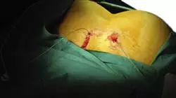
The palliative treatment of herniated discs, lumbar, inguinal, femoral, etc.; the reduction of pain in people suffering from diseases associated with the muscle and bone region; or the inhibition of the nerve root of the different areas in which a surgical intervention is to be performed, are the main areas of action of Locoregional Anesthesia. This is a medical specialty whose progress has helped thousands of people to improve their quality of life, through a considerable reduction of the discomforts they suffered in previous centuries. A quite significant example of this technique is the cervical or neuroaxial blocks, in which the vertebral facets are targeted through minimally invasive anesthetic therapies that contribute to a significant reduction of pain.

However, as in General Anesthesia, this type of procedures must be subject to an exhaustive control of the techniques, as well as of the considerations to be taken into account to avoid side effects harmful to health depending on the type of patients (children, elderly, people with various pathologies, pregnant women, etc.). For this reason, TECH Global University has developed a complete program with which, in just 12 months, you will be able to get up to date on all clinical and therapeutic developments in Locoregional Anesthesia. This Professional master’s degree will cover from the most innovative therapies to non-invasive clinical and surgical strategies for the different body regions. Furthermore, it will focus on pain-inhibiting palliative care in various types of patients, taking into account their physiological characteristics. All of this is based on the use of the latest drugs that have been tested with guarantees and are applicable at an international clinical level.

In order to achieve this, the professional will have 1,500 hours of theoretical and practical material, designed exclusively for this degree by a teaching team specialized in Anesthesiology, Resuscitation and Pain Therapy. Moreover, its convenient 100% online format will allow you to update your practice from wherever and whenever you want, in a way that is compatible with your professional activity. Therefore, it is a unique opportunity to work on perfecting your medical skills with the endorsement of the largest medical faculty in the world.

You will work on the latest developments in the application of anesthesia in the upper extremities, lower extremities, head and neck, delving into the most innovative clinical strategies for each case”

This Professional master’s degree in Locoregional Anesthesia contains the most complete and up-to-date scientific program on the market. The most important features include:

  • Practical cases presented by experts in Locoregional Anesthesiology
  • The graphic, schematic, and practical contents with which they are created, provide scientific and practical information on the disciplines that are essential for professional practice
  • Practical exercises where self-assessment can be used to improve learning
  • Its special emphasis on innovative methodologies
  • Theoretical lessons, questions to the expert, debate forums on controversial topics, and individual reflection assignments
  • Content that is accessible from any fixed or portable device with an Internet connection

Would you like to get up-to-date on what's new in Major Outpatient Surgery for anesthesiologists? If the answer is yes, this program is perfect for you"

The program’s teaching staff includes professionals from the sector who contribute their work experience to this educational program, as well as renowned specialists from leading societies and prestigious universities.

Its multimedia content, developed with the latest educational technology, will allow the professional a situated and contextual learning, that is, a simulated environment that will provide an immersive education programmed to prepare in real situations.

This program is designed around Problem-Based Learning, whereby the professional must try to solve the different professional practice situations that arise during the academic year For this purpose, the student will be assisted by an innovative interactive video system created by renowned and experienced experts.   

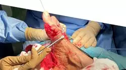
Due to its convenient format and the hundreds of hours of additional material included in the program, you will be able to delve into the latest advances in critical treatments through Regional Anesthesia"
##IMAGE##

A program that will give you the keys to provide pain relief to your patients, through the most effective and innovative clinical guidelines of current Anesthesiology"

Syllabus

The syllabus of this Professional master’s degree includes 1,500 hours of the best theoretical and practical material presented in different formats: detailed videos, research articles, complementary readings and much more. All these resources have been designed by the teaching team exclusively for this program and presented in a 100% online format to guarantee an up-to-date knowledge accessible from anywhere with an internet connection. In this way, professionals in the area of Clinical Anesthesiology will not have to worry about schedules or face-to-face classes, attending an academic experience that meets not only their needs, but also the demands of Modern Medicine. 

##IMAGE##

The theoretical content of this program has been developed based on the Relearning methodology, so that you don't have to spend extra time memorizing while you improve your knowledge”  

Module 1. Regional Anesthesia

1.1. Temporal Development

1.1.1. History of Regional Anesthesia
1.1.2. Historical Evolution of regional technique
1.1.3. Importance of Regional Anesthesia in the 21st Century

1.2. Fundamentals of Anthrozoology

1.2.1. Introduction
1.2.2. Principles of Neurostimulation

1.2.2.1. Physiology of Nerve Conduction
1.2.2.2. Physical Principles
1.2.2.3. Electrical Current Features
1.2.2.4. Device Characteristics

1.2.3. Neurostimulation Techniques

1.2.3.1. Preparation Phase
1.2.3.2. Location and Approach Phase
1.2.3.3. Infusion Phase
1.2.3.4. Implementation Phase

1.2.4. Error Sources

1.2.4.1. Neurostimulation
1.2.4.2. Electrodes
1.2.4.3. Needles
1.2.4.4. Patients

1.2.5. Equipment Maintenance
1.2.6. The Neurostimulation Industry Today

1.3. Fundamentals of Anthrozoology

1.3.1. Physical Principles of Ultrasound

1.3.1.1. Sounds and Ultrasound
1.3.1.2. Image Formation
1.3.1.3. Fabric Classification

1.3.2. Ultrasound Components

1.3.2.1. Processing System
1.3.2.2. Ultrasound/Transducers
1.3.2.3. Devices
1.3.2.4. Ultrasound Parameters

1.4. Ultrasound applied to Regional Anesthesia

1.4.1. Ultrasound of fundamental structures

1.4.1.1. Vessels
1.4.1.2. Bone
1.4.1.3. Muscle
1.4.1.4. Tendons
1.4.1.5. Pleura
1.4.1.6. Thyroid and Trachea

1.4.2. Artefacts

1.4.2.1. Acoustic Shadow
1.4.2.2. Posterior Acoustic Enhancement
1.4.2.3. Comet Tail
1.4.2.4. Refraction:
1.4.2.5. Mirror Image
1.4.2.6. Anisotropic Reflectors

1.4.3. Systematic handling of the ultrasound scanner

1.4.3.1. Probe orientation
1.4.3.2. Approaches
1.4.3.3. Factors Influencing Nutrition
1.4.3.4. Ultrasound imaging of local anesthetics and catheters

1.4.4. Preparation of an ultrasound training model

1.5. Local anesthetics

1.5.1. Structure and Classification
1.5.2. Pharmacology

1.5.2.1. Pharmacokinetics
1.5.2.2. Pharmacodynamics
1.5.2.3. Coadjuvants

1.5.3. Mechanism of Action
1.5.4. Choice of local anesthetic
1.5.5. Toxicity

1.6. Local Anesthetic Poisoning

1.6.1. Introduction
1.6.2. Pathophysiology
1.6.3. Risk Factors
1.6.4. Symptoms
1.6.5. Management

1.7. Coadjuvant drugs in Regional Anesthesia

1.7.1. Introduction
1.7.2. Steroids

1.7.2.1. Neuroaxial steroids
1.7.2.2. Perineural steroids

1.7.3. Alpha Agonists

1.7.3.1. Clonidine:
1.7.3.2. Dexmedetomidine

1.7.4. Opioids

1.7.4.1. Neuroaxial opioids
1.7.4.2. Perineural opioids

1.7.5. Other adjuvants
1.7.6. Future Strategies

1.8. Sedation in Regional Anesthesia

1.8.1. Pharmacology of Hypnotics
1.8.2. Total Intravenous Anesthesia Technique (TIVA-TCI)
1.8.3. Sedation as an adjunct to Regional Anesthesia

1.9. Materials

1.9.1. Introduction
1.9.2. Basic Monitoring
1.9.3. Asepsis and Sterility in Regional Anesthesia
1.9.4. Needles

1.9.4.1. Spinal blockage
1.9.4.2. Epidural blockage
1.9.4.3. Peripheral Nerve and Other Blocks Blockage

1.9.5. Catheters for continuous analgesia
1.9.6. Infusion System

1.9.6.1. Elastomers
1.9.6.2. Infusion Pumps

1.10. Preanesthesia Consultation

1.10.1. Preanesthetic assessment
1.10.2. Complementary Tests
1.10.3. Information and Consent information

Module 2. Neuroaxial Blocks

2.1. Neuroaxis blockages

2.1.1. Definition
2.1.2. History
2.1.3. Current utility and use

2.2. Anatomy and physiology applied to neuroaxial blockades

2.2.1. Applied Anatomy
2.2.2. Applied Physiology of Camelids

2.3. Pharmacology applied to neuroaxial blockades

2.3.1. Local anesthetics
2.3.2. Opioids
2.3.3. Clonidine:
2.3.4. Corticosteroids
2.3.5. Neostigmine
2.3.6. Ketamine
2.3.7. Others

2.4. Spinal blockade

2.4.1. Definition and anatomical recall
2.4.2. Indications
2.4.3. Contraindications
2.4.4. Necessary Material
2.4.5. Technique in single puncture spinal block
2.4.6. Technique in continuous spinal block
2.4.7. Effects of blockage and management
2.4.8. Specific complications

2.5. Epidural block

2.5.1. Definition and anatomical recall
2.5.2. Indications
2.5.3. Contraindications
2.5.4. Necessary Material
2.5.5. Exclusive epidural block technique
2.5.6. Combined spinal-epidural block technique
2.5.7. Effects of blockage and management
2.5.8. Specific complications

2.6. Caudal Block

2.6.1. Definition and anatomical recall
2.6.2. Indications
2.6.3. Contraindications
2.6.4. Necessary Material
2.6.5. Technique
2.6.6. Effects of blockage and management
2.6.7. Specific complications

2.7. Paravertebral Block

2.7.1. Definition and anatomical recall
2.7.2. Indications
2.7.3. Contraindications
2.7.4. Necessary Material
2.7.5. Technique
2.7.6. Effects of blockage and management
2.7.7. Specific complications

2.8. Neuroaxial blocks in obstetrics

2.8.1. Physiological Changes of Pregnancy
2.8.2. Neuroaxial Analgesia in Labor
2.8.3. Neuroaxial anesthesia for cesarean section, instrumented delivery and postoperative analgesia
2.8.4. Effects of neuroaxial blocks on the progression of labor and on the fetus
2.8.5. Specific complications

2.9. Complications of neuroaxial blocks

2.9.1. Low back pain/dorsalgia
2.9.2. Hypotension
2.9.3. Accidental dural puncture and post dural puncture headache
2.9.4. Blood puncture, intravascular injection and local anesthetic intoxication
2.9.5. Subarachnoid injection
2.9.6. Intraosseous injection
2.9.7. High spinal block and total spinal block
2.9.8. Failed blockage
2.9.9. Neurological Lesions
2.9.10. Uriniary Retention
2.9.11. Pneumoencephalon
2.9.10. Uriniary Retention
2.9.11. Pneumoencephalon
2.9.12. Pneumothorax
2.9.13. Venous Air Embolism
2.9.14. Spinal hematoma
2.9.15. Infectious complications: spinal abscess, arachnoiditis and meningitis
2.9.16. Complications due to the effect of drugs

2.10. Ultrasound in Neuroaxial Blocks

2.10.1. General principles and limitations
2.10.2. Ultrasound-guided spinal block
2.10.3. Ultrasound-guided epidural block
2.10.4. Ultrasound-guided caudal block
2.10.5. Ultrasound-guided paravertebral blockade

Module 3. Upper Limbs

3.1. Brachial Plexus Anatomy

3.1.1. Anatomy
3.1.2. Nerve territory and exploration
3.1.3. Cutaneous and motor distribution of brachial plexus nerves

3.2. Superficial and deep cervical block

3.2.1. Anatomy
3.2.2. Indications
3.2.3. Contraindications
3.2.4. Anatomical references, posture and puncture sites
3.2.5. Material
3.2.6. Response to neurostimulation
3.2.7. Blockage by Ultrasound
3.2.8. Complications

3.3. Interscalene Block

3.3.1. Anatomy
3.3.2. Indications
3.3.3. Contraindications
3.3.4. Anatomical references, posture and puncture sites
3.3.5. Material
3.3.6. Response to neurostimulation
3.3.7. Blockage by Ultrasound
3.3.8. Complications

3.4. Infraclavicular Block

3.4.1. Anatomy
3.4.2. Indications
3.4.3. Contraindications
3.4.4. Anatomical references, posture and puncture sites
3.4.5. Material
3.4.6. Response to neurostimulation
3.4.7. Blockage by Ultrasound
3.4.8. Complications

3.5. Infraclavicular Block

3.5.1. Anatomy
3.5.2. Indications
3.5.3. Contraindications
3.5.4. Anatomical references, posture and puncture sites
3.5.5. Material
3.5.6. Response to neurostimulation
3.5.7. Blockage by Ultrasound
3.5.8. Complications

3.6. Axillary Block

3.6.1. Anatomy
3.6.2. Indications
3.6.3. Contraindications
3.6.4. Anatomical references, posture and puncture sites
3.6.5. Material
3.6.6. Response to neurostimulation
3.6.7. Blockage by Ultrasound
3.6.8. Complications

3.7. Blockages in the humeral canal (mid-humeral block)

3.7.1. Anatomy
3.7.2. Indications
3.7.3. Contraindications
3.7.4. Anatomical references, posture and puncture sites
3.7.5. Material
3.7.6. Response to neurostimulation
3.7.7. Blockage by Ultrasound
3.7.8. Complications

3.8. Peripheral blockages

3.8.1. Blockages at shoulder level

3.8.1.1. Supraclavicular Nerve Block
3.8.1.2. Mental Nerve Block
3.8.1.3. Lateral Femoral Cutaneous Nerve Block
3.8.1.4. Medial antebrachial cutaneous nerve block

3.8.2. Isolated blockages at the elbow level

3.8.2.1. Median Nerve Block
3.8.2.2. Radially Nerve Block
3.8.2.3. Cubital Nerve Block

3.8.3. Isolated blockages at the wrist and hand level

3.8.3.1. Median Nerve Block
3.8.3.2. Radially Nerve Block
3.8.3.3. Cubital Nerve Block
3.8.3.4. Distal Blocks

3.9. Regional Intravenous Anesthesia of the Upper Extremity

3.9.1. Indications
3.9.2. Contraindications
3.9.3. Material
3.9.4. Methodology

3.10. Infiltrations in the upper limb

3.10.1. General Aspects
3.10.2. Indications
3.10.3. Contraindications
3.10.4. Materials and Drugs
3.10.5. Methodology
3.10.6. Adverse Effects
3.10.7. Infiltrations at shoulder level
3.10.8. Infiltrations at elbow level
3.10.9. Infiltrations at hand level

Module 4. Forelimbs

4.1. Lumbar Square Tightness

4.1.1. Anatomy
4.1.2. Nerve territory and exploration
4.1.3. Cutaneous and motor distribution of the nerves of the lumbar plexus

4.2. Anatomy of the sacral plexus

4.2.1. Anatomy
4.2.2. Nerve territory and exploration
4.2.3. Cutaneous and motor distribution of sacral plexus nerves

4.3. Posterior lumbar block

4.3.1. Anatomy
4.3.2. Indications
4.3.3. Contraindications
4.3.4. Material
4.3.5. Anatomical references, posture and puncture sites
4.3.6. Response to neurostimulation
4.3.7. Blockage by Ultrasound
4.3.8. Complications

4.4. Femoral block

4.4.1. Anatomy
4.4.2. Indications
4.4.3. Contraindications
4.4.4. Anatomical references, posture and puncture sites
4.4.5. Material
4.4.6. Response to neurostimulation
4.4.7. Blockage by Ultrasound
4.4.8. Complications

4.5. Obturator and femorocutaneous nerve blocks

4.5.1. Obturation Nerve Block

4.5.1.1. Anatomy
4.5.1.2. Indications
4.5.1.3. Contraindications
4.5.1.4. Anatomical references, posture and puncture sites
4.5.1.5. Material
4.5.1.6. Response to neurostimulation
4.5.1.7. Blockage by Ultrasound
4.5.1.8. Complications

4.5.2. Lateral Femoral Cutaneous Nerve Block

4.5.2.1. Anatomy
4.5.2.2. Indications
4.5.2.3. Contraindications
4.5.2.4. Anatomical references, posture and puncture sites
4.5.2.5. Material
4.5.2.6. Response to neurostimulation
4.5.2.7. Blockage by Ultrasound
4.5.2.8. Complications

4.6. Interfacial blocks for hip surgery

4.6.1. Introduction
4.6.2. PENG or pericapsular nerve group block
4.6.3. Block of the iliac fascia

4.6.3.1. Suprainguinal
4.6.3.2. Infrainguinal

4.6.4. Benefits of peripheral hip nerve blocks

4.7. Saphenous nerve block and intra-articular block for knee surgery

4.7.1. Introduction
4.7.2. Saphenous Nerve Block

4.7.2.1. Blockage of the Internal Saphenous Nerve (Adductor Canal)
4.7.2.2. Other blocking sites

4.7.3. Intra-articular knee block

4.8. Sciatic block

4.8.1. Sciatic Blockage Gluteal Level

4.8.1.1. Anatomy
4.8.1.2. Indications
4.8.1.3. Contraindications
4.8.1.4. Anatomical references, posture and puncture sites
4.8.1.5. Material
4.8.1.6. Response to neurostimulation
4.8.1.7. Blockage by Ultrasound
4.8.1.8. Complications

4.8.2. Sciatic Blockage Gluteal Level

4.8.2.1. Anatomy
4.8.2.2. Indications
4.8.2.3. Contraindications
4.8.2.4. Anatomical references, posture and puncture sites
4.8.2.5. Material
4.8.2.6. Response to neurostimulation
4.8.2.7. Blockage by Ultrasound
4.8.2.8. Complications

4.9. Sciatic Nerve Block Popliteal Level

4.9.1. Anatomy
4.9.2. Indications
4.9.3. Contraindications
4.9.4. Anatomical references, posture and puncture sites
4.9.5. Material
4.9.6. Response to neurostimulation
4.9.7. Blockage by Ultrasound
4.9.8. Complications
4.10. Blockage of the Terminal Branches of the Sciatic Nerve

4.10.1. Posterior Tibial Nerves
4.10.2. Sural Nerve
4.10.3. Common Peroneal Nerve
4.10.4. Deep Peroneal Nerve
4.10.5. Superficial Peroneal Nerve

Module 5. Thoraco-abdominal Interfascial Blocks

5.1. Interfacial Blocks

5.1.1. What is an interfascial block?
5.1.2. History & evolution
5.1.3. Advantages and Disadvantages

5.2. Chest Wall Anatomy

5.2.1. Musculoskeletal Component
5.2.2. Nerve Components
5.2.3. Cutaneous Innervation

5.3. Intercostal Blockages

5.3.1. Blockade of the anterior cutaneous branches of the intercostal nerves (BCRA) or pectointercostal blockade

5.3.1.1. Introduction
5.3.1.2. Indications and Contraindications
5.3.1.3. Position and Patient Preparation
5.3.1.4. Materials
5.3.1.5. Anatomical Image vs. Ultrasound Imaging
5.3.1.6. Blockage under Ultrasound Vision
5.3.1.7. Complications

5.3.2. BRILMA

5.3.2.1. Introduction
5.3.2.2. Indications and Contraindications
5.3.2.3. Position and Patient Preparation
5.3.2.4. Materials
5.3.2.5. Anatomical Image vs. Ultrasound Imaging
5.3.2.6. Blockage under Ultrasound Vision
5.3.2.7. Complications
5.3.2.8. Modified BRILMA

5.4. Interpectoral Blocks

5.4.1. PEC I-II

5.4.1.1. Introduction
5.4.1.2. Indications and Contraindications
5.4.1.3. Position and Patient Preparation
5.4.1.4. Materials
5.4.1.5. Anatomical Image vs. Ultrasound Imaging
5.4.1.6. Blockage under Ultrasound Vision
5.4.1.7. Complications

5.4.2. PEC I-II

5.4.2.1. Introduction
5.4.2.2. Indications and Contraindications
5.4.2.3. Position and Patient Preparation
5.4.2.4. Materials
5.4.2.5. Anatomical Image vs. Ultrasound Imaging
5.4.2.6. Blockage under Ultrasound Vision
5.4.2.7. Complications

5.5. Other Blocks of the Thoracic Wall

5.5.1. Spinal Erector Spinae Block

5.5.1.1. Introduction
5.5.1.2. Indications and Contraindications
5.5.1.3. Position and Patient Preparation
5.5.1.4. Materials
5.5.1.5. Anatomical Image vs. Ultrasound Imaging
5.5.1.6. Blockage under Ultrasound Vision
5.5.1.7. Complications

5.5.2. Serratus Blockage

5.5.2.1. Introduction
5.5.2.2. Indications and Contraindications
5.5.2.3. Position and Patient Preparation
5.5.2.4. Materials
5.5.2.5. Anatomical Image vs. Ultrasound Imaging
5.5.2.6. Blockage under Ultrasound Vision
5.5.2.7. Complications

5.6. Anatomy of the Abdominal Wall

5.6.1. Musculoskeletal Component
5.6.2. Nerve Components
5.6.3. Cutaneous Innervation

5.7. Transversus Abdominis Plane (TAP Block

5.7.1. Introduction
5.7.2. Indications and Contraindications
5.7.3. Position and Patient Preparation
5.7.4. Materials
5.7.5. Anatomical Image vs. Ultrasound Imaging
5.7.6. Blockage under Ultrasound Vision
5.7.7. Complications
5.7.8. TAP Blocking Variants

5.7.8.1. Subcostal TAP
5.7.8.2. Back TAP

5.8. Ilioinguinal and Iliohypogastric Block

5.8.1. Introduction
5.8.2. Indications and Contraindications
5.8.3. Position and Patient Preparation
5.8.4. Materials
5.8.5. Anatomical Image vs. Ultrasound imaging
5.8.6. Blockage under Ultrasound Vision
5.8.7. Complications

5.9. Other Blocks of the Abdominal Wall

5.9.1. Locking of the Rectus Sheath

5.9.1.1. Introduction
5.9.1.2. Indications and Contraindications
5.9.1.3. Position and Patient Preparation
5.9.1.4. Materials
5.9.1.5. Anatomical Image vs. Ultrasound Imaging
5.9.1.6. Blockage under Ultrasound Vision
5.9.1.7. Complications

5.9.2. Semilunar Block

5.9.2.1. Introduction
5.9.2.2. Indications and Contraindications
5.9.2.3. Position and Patient Preparation
5.9.2.4. Materials
5.9.2.5. Anatomical Image vs. Ultrasound Imaging
5.9.2.6. Blockage under Ultrasound Vision
5.9.2.7. Complications

5.9.3. Lumbar Square Blockage

5.9.3.1. Introduction
5.9.3.2. Indications and Contraindications
5.9.3.3. Position and Patient Preparation
5.9.3.4. Materials
5.9.3.5. Anatomical Image vs. Ultrasound Imaging
5.9.3.6. Blockage under Ultrasound Vision
5.9.3.7. Complications

5.10. Incisional Analgesia

5.10.1. Local anesthetic infiltration in surgical wounds
5.10.2. Continuous analgesia delivery systems. Incisional catheters
5.10.3. Rate of Infusion
5.10.4. Efficiency and Safety

Module 6. Head and Neck

6.1. Regional Anesthesia for Oral and Maxillofacial Surgery

6.1.1. Introduction
6.1.2. Anatomy of Trigeminal Nerves
6.1.3. Equipment for Maxillary and Mandibular Regional Anesthesia

6.2. Facial Nerve Blocks

6.2.1. Superficial Trigeminal Block

6.2.1.1. Frontal Nerve Block
6.2.1.2. Infraorbital Nerve Block
6.2.1.3. Mentonian Nerve Block
6.2.1.4. Ultrasound-guided Technique

6.2.2. Jaw Nerve Blockade
6.2.3. Mandibular Nerve Block
6.2.4. Regional Nerve Block of the Nose

6.3. Lift Regional Anesthesia

6.3.1. Supraperiosteal Infiltration
6.3.2. Periodontal Intraligamentary Infiltration
6.3.3. Superior Alveolar Nerve Block

6.3.3.1. Posterior Superior Alveolar Nerve
6.3.3.2. Medial Superior Alveolar Nerve
6.3.3.3. Anterior Superior Alveolar or Infraorbital Nerve

6.3.4. Palatine Nerve Block Major
6.3.5. Mental Nerve Block
6.3.6. Jaw Nerve Blockade

6.4. Mandibular Regional Anesthesia

6.4.1. Inferior Alveolar Nerve Block
6.4.2. Mental Nerve Block
6.4.3. Mandibular Nerve Block
6.4.4. Mentonian Nerve Block
6.4.5. Incisor Nerves Block

6.5. Regional External Ear Blocks

6.5.1. Anatomy
6.5.2. Indications
6.5.3. Classic Blocking Technique
6.5.4. Complications

6.6. Nerve Blocks of the Head

6.6.1. Major Occipital Nerve Block
6.6.2. Scalp Block (scalp nerve blockage)
6.6.3. Anesthesia in Craniotomy in Awake Patient

6.7. Anesthesia for Ophthalmic Surgery

6.7.1. Orbit and Innervation Anatomy
6.7.2. Surgical Considerations
6.7.3. Perioperative Management

6.8. Anesthetic Techniques for Ophthalmic Surgery

6.8.1. Local Anesthesia
6.8.2. Subtenonian or Episcleral
6.8.3. Subconjunctival Anesthesia
6.8.4. Oculoplastics Nerve Blocks
6.8.5. Retrobulbar Anesthesia
6.8.6. Peribulbar Anesthesia
6.8.7. Complications

6.9. Superficial Cervical Plexus Block

6.9.1. General Considerations
6.9.2. Anatomy
6.9.3. Ultrasound-guided Block
6.9.4. Nervous Stimulation Techniques
6.9.5. Anesthesia for Carotid Endarterectomy

6.10. Regional Anesthesia for VAD Management

6.10.1. Introduction
6.10.2. Upper Airway Innervation
6.10.3. Local Anesthesia
6.10.4. Regional Anesthesia:

6.10.4.1. Superior Laryngeal Nerve Blockage
6.10.4.2. Glossopharyngeal Block
6.10.4.3. Recurrent Laryngeal Block
6.10.4.4. Transtracheal Block

6.10.5. Sedation for Intubation in Awake Patient

Module 7. Major Outpatient Surgery

7.1. Major Outpatient Surgery

7.1.1. What is Major Outpatient Surgery?
7.1.2. History

7.2. Current Situation of Major Outpatient Surgery

7.2.1. Implementation Difficulties
7.2.2. Cost-Effectiveness Approach
7.2.3. Achievements of Major Outpatient Surgery

7.3. CMA Circuit

7.3.1. Types of Units
7.3.2. Structure and Organization

7.4. Selection Criteria

7.4.1. What Surgical Procedures Can Be Performed?
7.4.2. Which Patients do we Select?

7.5. Role of the Pre-Anesthesia Consultation

7.5.1. Preanesthesia Consultation
7.5.2. Patient Preparation

7.6. Anesthetic Technique Selection

7.6.1. What Anesthetic Technique do we Use?
7.6.2. Opioids in Major Outpatient Surgery

7.7. Pain Control in Major Outpatient Surgery

7.7.1. Pain Relieving Techniques
7.7.2. Multimodal Analgesia

7.8. Complications in Major Ambulatory Surgery

7.8.1. Nausea and Vomiting
7.8.2. Pain
7.8.3. Uriniary Retention
7.8.4. Other Complications

7.9. Discharge from the Major Outpatient Surgery Unit

7.9.1. Discharge Criteria at Home
7.9.2. Hospital Admission Criteria

7.10. Morbimortality, Safety and Quality in Major Outpatient Surgery

7.10.1. Morbidity and Mortality Data
7.10.2. Security/Safety
7.10.3. Indicators of Quality of Care

Module 8. Critical Care and Regional Anesthesia

8.1. Peculiarities of Critical Patients

8.1.1. Pathophysiology of Critical Patients
8.1.2. Special Considerations for the Locoregional Techniques

8.2. Pain Assessment in the Critical Patients

8.2.1. Introduction
8.2.2. Assessment of Pain in Conscious and/or Communicative Patient
8.2.3. Assessment of Pain in Unconscious and/or Non-Communicative Patients

8.3. Pain Management in Critical Care Units

8.3.1. Origin of Pain
8.3.2. Impact of Pain in the Critically Ill Patient
8.3.3. Therapeutic Options for Pain

8.4. Locoregional Technique in Critical Care Units

8.4.1. Upper Limb Blocks
8.4.2. Lower Limb Blocks
8.4.3. Central Blocks
8.4.4. Thoracoabdominal Wall Block

8.5. The Polytraumatized Patient

8.5.1. Etiopathogenesis
8.5.2. Characteristics of the Polytraumatized Patient
8.5.3. Locoregional Techniques in the Polytraumatized Patient

8.6. Amputee Patient and Phantom Limb

8.6.1. Amputee Patient. Incidence and Characteristics
8.6.2. Phantom Limb. Incidence and Characteristics
8.6.3. Prevention and Management of Phantom Limb

8.7. Burn patient

8.7.1. Etiopathogenesis
8.7.2. Characteristics of the Burn Patient
8.7.3. Locoregional Techniques in the Burned Patient

8.8. Regional Anesthesia and Microvascularized Flap

8.8.1. The Flap
8.8.2. Physiological Considerations
8.8.3. Anesthetic Approach

8.9. Ultrasound in Critical Care Units

8.9.1. Utility of Ultrasound in Critical Care Units
8.9.2. Ultrasound-Guided Techniques in Critical Care Units

8.10. Central Line Canalization

8.10.1. Internal Jugular Vein Canalization
8.10.2. Subclavian Vein Canalization
8.10.3. Femoral Vein Canalization
8.10.4. Central line Canalization by Peripheral Access
8.10.5. Others

Module 9. Locoregional Anesthesia and Pain Management

9.1. Pain and its Pathophysiological Basis. Types of Pain

9.1.1. Lesson on Neuroanatomy
9.1.2. Lesson on Somatic Nociceptive Pain
9.1.3. Lesson on Visceral Nociceptive Pain
9.1.4. Lesson on Neuropathic Pain
9.1.5. Lesson from acute to chronic pain: peripheral and central sensitization

9.2. Locoregional Anesthesia in the Psychotherapeutic treatment Psychopharmacological treatment of Acute Postoperative Pain

9.2.1. Regional Analgesia as a fundamental part of multimodal analgesia in acute postoperative pain
9.2.2. Lesson on Regional Analgesia in Shoulder Surgery and MMSS
9.2.3. Lesson on Regional Analgesia in Hip Surgery
9.2.4. Lesson on Regional Analgesia in Knee Surgery
9.2.5. Lesson on Regional Analgesia in Foot Surgery
9.2.6. Lesson on Regional Analgesia in Thoracotomy
9.2.7. Lesson on Regional Analgesia in Breast Surgery
9.2.8. Lesson on Regional Analgesia in Laparotomy
9.2.9. Lesson on Regional Analgesia in Laparoscopy

9.3. Updates on Neuropathic Pain Treatment(DN)

9.3.1. Diagnostic blocks in DN treatment
9.3.2. Pulsed RF in the treatment of DN in MMSS
9.3.3. Pulsed RF in the treatment of thoracic DN
9.3.4. Pulsed RF in the treatment of abdominal DN
9.3.5. Pulsed RF in the treatment of lumbar DN
9.3.6. Pulsed RF in the treatment of MMII DN

9.4. Locoregional Anesthesia in the treatment of oncologic pain and in the palliative care setting

9.4.1. Invasive Techniques as an adjunct to pain management in palliative care. General and differential aspects of these patients. Neurolysis
9.4.2. Stellate ganglion block for neck cancer pain and EESS
9.4.3. Celiac plexus blockade for supramesocolic cancer pain
9.4.4. Superior Hypogastric plexus block, inferior hypogastric plexus block and ganglion impar in pelvic oncologic pain
9.4.5. Peripheral Nerve and plex nerve block in the oncologic patient
9.4.6. Long-term epidural catheter in the context of the oncologic patient
9.4.7. Intrathecal pumps for oncology patient management

9.5. Locoregional Anesthesia in the treatment of low back pain

9.5.1. Block and radiofrequency in lumbar facet syndrome
9.5.2. Regional Approach to Discogenic Pain
9.5.3. Pain from Lumbosacral Radiculopathy
9.5.4. Lumbar Epidural Steroids
9.5.5. Lumbosacral DRG RF
9.5.6. Lumbar Myofascial Syndrome
9.5.7. Botulinum toxin blockade and infiltration of the piriformis muscle
9.5.8. Botulinum toxin blockade and infiltration of the psoas and quadratus lumborum muscle
9.5.9. Pain due to alterations in the sacroiliac joint. Diagnostic and RF blocking
9.5.10. Epidurolisis and epiduroscopy

9.6. Regional anesthesia and craniofacial pain. Cervicalgia

9.6.1. Trigeminal Neuralgia Blockage and RF of Gasser's Ganglion
9.6.2. Other orofacial algias. Sphenopalatine Ganglion Block and RF
9.6.3. Cervicogenic cephalgia. Blocking and RF of the TON and GON
9.6.4. Cervical pain due to facet pathology. Diagnostic and RF blockade in cervical facet syndrome
9.6.5. Cervical discogenic pain. Cervical epidural steroids
9.6.6. Radiculopathy of the MMSS. Epidural, radicular and RF blocks of cervical root DRG

9.7. Regional Anesthesia, visceral pain and CRPS

9.7.1. Regional Anesthesia in Acute and Chronic Pancreatitis
9.7.2. Regional Anesthesia in the pathology of acute and chronic renal lithiasis
9.7.3. Regional Anesthesia in chronic non-oncologic pelvic pain

9.7.3.1. Diagnostic and therapeutic block of the superior hypogastric plexus
9.7.3.2. Diagnostic and therapeutic blockade of the Ganglion Impar
9.7.3.3. Pudendal nerve block and RF
9.7.3.4. Blockage and RF of the Ilioinguinal and Iliohypogastric Nerves

9.7.4. SDRC

9.7.4.1. ACS Pathophysiology
9.7.4.2. CRPS in MMSS: peripheral and stellate ganglion techniques
9.7.4.3. CRPS in MMII: peripheral and lumbar sympathetic techniques

9.8. Regional Anesthesia and musculoskeletal pain Thorax and Large Joints

9.8.1. Regional Anesthesia in the painful shoulder. Intra-articular Block. RF of the Suprascapular Nerve
9.8.2. Regional Anesthesia in Coxarthrosis. Intra-articular Block Denervation Techniques
9.8.3. Regional Anesthesia in Gonarthrosis. Intra-articular Block Denervation techniques (RF N geniculates)
9.8.4. Regional Anesthesia in myofascial syndrome. Trigger Point Block. Intrafascial Blocks
9.8.5. Regional Anesthesia and discogenic dorsalgia. Epidural Steroids
9.8.6. Regional Anesthesia and degenerative dorsalgia. Diagnostic and RF blockade in dorsal facet syndrome

9.9. Regional Anesthesia in the IV Staggered. Neurostimulation and spinal infusion of drugs

9.9.1. Pathophysiological basis of neurostimulation and spinal drug infusion
9.9.2. Neurostimulation in the treatment of pain secondary to failed back surgery (FBSS)

9.9.2.1. Posterior Cord Stimulation
9.9.2.2. GRD Stimulation

9.9.3. Neurostimulation in Peripheral Neuropathies
9.9.4. Posterior cord neurostimulation in angor and visceral pain
9.9.5. Sacral Root Neurostimulation in DCP
9.9.6. Intracranial and Transcranial Stimulation
9.9.7. Drug infusion via the spinal route in non-oncologic pathology

9.10. Regional Anesthesia in Labor Analgesia (ATP)

9.10.1. Pathophysiology of pain in the stages of labor
9.10.2. Regional Analgesia in PTA: epidural analgesia. Modalities of drug administration in ATP
9.10.3. PTA and other regional analgesia modalities: combined epidural-intradural analgesia (CIE). CIE analgesia without intradural drug
9.10.4. Anesthesia Regional in Cesarean Sections. Epidural Anesthesia. Intradural Anesthesia. Anesthesia CIE
9.10.5. Special situations in PTA and Regional Anesthesia

9.10.5.1. ATP, Regional Anesthesia and Obese Patient. Ultrasound. Caudal Epidural Approach
9.10.5.2. DAP in cesarean section without epidural catheter. Ultrasound Wall Block
9.10.5.3. Transvaginal/Transperineal Pudendal Nerve block

Module 10. Specific Situations of Regional Anesthesia

10.1. Regional Anesthesia in Patients with Pre-existing Neurological Disease

10.1.1. Introduction
10.1.2. Peripheral Nervous System Disorders

10.1.2.1. Hereditary Peripheral Neuropathy
10.1.2.2. Acquired Peripheral Neuropathy. Diabetic Polyneuropathy
10.1.2.3. Chemotherapy-Induced Neuropathy
10.1.2.4. Entrapment Neuropathy
10.1.2.5. Inflammatory Neuropathy. Guillén-Barré Syndrome
10.1.2.6. Post-Surgical Inflammatory Neuropathy

10.1.3. Central Nervous System disorders

10.1.3.1. Multiple Sclerosis
10.1.3.2. Post Polio Syndrome
10.1.3.3. Amyotrophic Lateral Sclerosis
10.1.3.4. Spinal Stenosis and Neural Disc Disease
10.1.3.5. Spinal Cord Injury

10.2. Anti-Aggregation Therapy, Anticoagulation Therapy

10.2.1. Introduction
10.2.2. Minimum Hemostatic Values
10.2.3. Anticoagulants, Antiplatelet Agents and Anesthesia

10.2.3.1. Unfractionated Heparin
10.2.3.2. Low Molecular Weight Heparin
10.2.3.3. Fondaparinux
10.2.3.4. Antivitamin K Drugs (Acenocoumarol, Warfarin)
10.2.3.5. Platelet Aggregation Inhibitors

10.2.4. Ophthalmological Procedures

10.2.4.1. Surgeries in which Antithrombotic Treatment can Be Continued
10.2.4.2. Surgeries in which Antithrombotic Treatment Should Be Discontinued and Bridging Therapy Considered
10.2.4.3. How to Use Guides in Peripheral Nerve Blocks

10.3. Continuous Techniques for Postoperative Pain Control

10.3.1. Introduction
10.3.2. Drugs:

10.3.2.1. Coadjuvants
10.3.2.2. Continuous Perfusions Through Catheters
10.3.2.3. New Local Anesthetics

10.3.3. Material

10.3.3.1. Needle and Catheter
10.3.3.2. Infusion Pumps

10.3.4. Modes of Administration

10.3.4.1. Boluses
10.3.4.2. Continuous Administration

10.3.5. Techniques

10.3.5.1. Interscalene Block
10.3.5.2. Infraclavicular Block
10.3.5.3. Axillary Block
10.3.5.4. Posterior Lumbar Plexus Block
10.3.5.5. Anterior Lumbar Plexus Block
10.3.5.6. Proximal Sciatic Nerve Blocks
10.3.5.7. Proximal Sciatic Nerve Blocks
10.3.5.8. Distal Blocks

10.4. Regional Anesthesia and Pulmonary Disease

10.4.1. Introduction
10.4.2. Epidural and Spinal Anesthesia
10.4.3. Brachial Plexus Block
10.4.4. Paravertebral Blockade and Intercostal Nerves
10.4.5. Importance of Regional Anesthesia during the COVID-19 Pandemic

10.5. Regional Anesthesia and other Systemic Diseases

10.5.1. Renal disease

10.5.1.1. Introduction
10.5.1.2. Effects on Renal Function
10.5.1.3. Considerations in Patients with Renal Pathology

10.5.2. Liver Diseases

10.5.2.1. Introduction
10.5.2.2. Effects on Hepatic Blood Flow
10.5.2.3. Hepatic Coagulopathy

10.5.3. Diabetes Mellitus

10.5.3.1. Introduction
10.5.3.2. Effects on Glucose Homeostasis
10.5.3.3. Peripheral Neuropathy in the Diabetic Patient

10.5.4. Obesity
10.5.5. Cancer

10.6. Regional Anesthesia in the Elderly

10.6.1. Introduction and Definition of the Elderly

10.6.1.1. Is Anesthetic Risk Increased in the Elderly?
10.6.1.2. What is the Reason for this?
10.6.1.3. How is this Organ Degradation Reflected at the Level of all Systems?
10.6.1.4. Is the Metabolism of Anesthetic Drugs Altered in the Elderly Patient?
10.6.1.5. What type of Interventions are Most Common in the Elderly?
10.6.1.6. Is Regional Anesthesia Specially Indicated in these Patients?

10.6.2. Physiologic Changes Associated with Aging and Considerations for Regional Anesthesia/Analgesia

10.6.2.1. Nervous System Function
10.6.2.2. Pulmonary Function
10.6.2.3. Pharmacokinetic and Pharmacodynamic Changes in the Elderly
10.6.2.4. Multimodal Pharmacotherapy and the Elderly
10.6.2.5. Kidney
10.6.2.6. Physiology and Perception of Pain in the Elderly

10.6.3. Assessment of Pain in Elderly Patients with Cognitive Impairment
10.6.4. Considerations for the Use of Regional and Neural Blockade
10.6.5. Types of Regional Blocks in the Elderly

10.6.5.1. Epidural Anesthesia and Analgesia
10.6.5.2. Intrathecal Opioid Analgesia
10.6.5.3. Peripheral Nerve and Nerve Plexus Blockage

10.7. Regional Anesthesia in Pediatrics

10.7.1. Introduction

10.7.1.1. Why Regional Anesthesia in Pediatric Patients?
10.7.1.2. Applications of Pediatric Regional Anesthesia
10.7.1.3. Regional Anesthesia: Awake or Asleep?

10.7.2. Peculiarities of Pediatric Regional Anesthesia
10.7.3. Neurostimulation

10.7.3.1. Anatomical Differences Between Children and Adults
10.7.3.2. Pharmacology of Local Anesthetics
10.7.3.3. Dosage of Local Anesthetics
10.7.3.4. Toxicity of Local Anesthetics

10.7.4. Types of Peripheral Blocks

10.7.4.1. Upper Limb Blocks
10.7.4.2. Lower Limb Blocks
10.7.4.3. Penile Block
10.7.4.4. Ilioinguinal/Iliohypogastric Block
10.7.4.5. Rectus Sheath Block or Umbilical Blockade
10.7.4.6. Caudal Block

10.7.5. Central Blocks

10.7.5.1. Epidural Anesthesia
10.7.5.2. Subarachnoid Anesthesia

10.7.6. Complications of Pediatric Regional Anesthesia

10.8. Allergy and Regional Anesthesia

10.8.1. Introduction

10.8.1.1. Type A Reactions
10.8.1.2. Type B Reactions
10.8.1.3. Type C Reactions

10.8.2. Epidemiology
10.8.3. Pathophysiology

10.8.3.1. Type I: Immediate Hypersensitivity or IgE Mediated Hypersensitivity
10.8.3.2. Type II: Cytotoxic or IgG, IgM Mediated Reaction
10.8.3.3. Type III: Immunocomplex-Mediated Reaction
10.8.3.4. Type IV: Delayed Hypersensitivity or T-Cell Mediated reaction

10.8.4. Etiology
10.8.5. Signs and Symptoms
10.8.6. Diagnosis
10.8.7. Differential Diagnosis

10.8.7.1. Reddening Syndrome
10.8.7.2. Syndromes Associated with Substance Use
10.8.7.3. Increased Endogenous Histamine Production
10.8.7.4. Functional Criteria
10.8.7.5. Others

10.8.8. Treatment

10.9. Complications in Regional Anesthesia

10.9.1. Introduction
10.9.2. Complications following Neuroaxial Block Procedures

10.9.2.1. Post Dural Puncture Headache
10.9.2.2. Complications due to Air Injection. Pneumoencephalon
10.9.2.3. Spinal Cord Compression
10.9.2.4. Neurological Damage. Neurotoxicants
10.9.2.5. Infectious Complications
10.9.2.6. Iatrogenic Spinal Tumors
10.9.2.7. Tattoos and Anesthetic Considerations

10.9.3. Complications after Peripheral Nerve Blocks

10.9.3.1. Introduction
10.9.3.2. Preventive Measures
10.9.3.3. Classification of Acute Nerve Injuries

10.9.4. Mechanisms Capable of Producing Complications During the Performance of Nerve Blocks

10.9.4.1. Mechanical Mechanism
10.9.4.2. Vascular Mechanism
10.9.4.3. Chemical Mechanism
10.9.4.4. Infectious Mechanism
10.9.4.5. Systemic Toxicity

10.10. Regional Anesthesia and Patient Safety

10.10.1. Introduction
10.10.2. How has Regional Anesthesia Evolved during these Years?
10.10.3. Advantages and Disadvantages of the Different Types of Regional Anesthesia
10.10.4. What is ISO 80369-6 and how does it Affect Regional Anesthesia?
10.10.5. Comparison between Traditional Spinal Needles and the new NRFIT version
10.10.6. Adjusted checklist for Regional Anesthesia
10.10.7 SENSAR

##IMAGE##

Do not hesitate and opt for a program that not only adapts to your needs, but also to the requirements of Locoregional Anesthesia in medical practice at the highest professional level”

Professional Master's Degree in Locoregional Anesthesia

The use of regional and local anesthesia has been extended to different medical fields, seeking to offer patients treatments and surgical interventions that significantly reduce and control pain. However, its application requires a solid base of knowledge and technical skills in the field, since its improper handling could represent a risk to the health of people. At TECH Global University we developed the Professional Master's Degree in Locoregional Anesthesia, a program designed with the purpose of updating and complementing the studies of professionals in anesthesiology so that they can safely perform the daily practice of their functions in this clinical area. In this way, you will become an expert in the use of different anesthesiological drugs taking into account their side effects, the type of patient and the area of application. Take this postgraduate degree and advance your career goals.

Become a specialist at the largest Faculty of Medicine

With our Professional Master's Degree, delivered in a 100% online format, you will have access to the latest advances available in anesthesiological intervention, as well as the clinical strategies recommended for each case. Through theoretical lessons, discussion forums and the study of real clinical cases, you will learn the basics of neuroaxial anesthesia and locoregional blocks; you will become familiar with the anatomy, physiology and pharmacology involved in the use of local anesthetics; you will analyze the indications, contraindications, technical aspects and complications derived from anesthetic assistance; and you will identify the specific situations to be taken into account in this discipline, such as symptoms, causes and risk factors of intoxication, for its correct approach and treatment. At TECH Global University you will achieve a higher level of knowledge and boost your career growth.