University certificate
The world's largest faculty of nursing”
Why study at TECH?
This sharp refresher course is ideal for you to expand the boundaries of your career in the Nursing field. Enroll now with TECH!"

The last few decades have been pivotal for nursing professionals. Gradually, they have had to take on new challenges and procedures within the nursing practice. Particularly in the field of Critical Care and Emergency Care, updated work protocols have been implemented for these professionals. Additionally, ultrasound technologies have evolved and with them the management that the nurse must know to make an efficient use of them. However, it is difficult to keep up to date with all these innovations in a pedagogical context where qualifications do not cover the development of specific competencies and skills within this framework.
In this context, TECH has devised a learning modality that integrates the teaching of modern aspects of this area of Nursing in two distinct stages. In the first stage, the student will dedicate 1,500 hours to theoretical learning of concepts and work protocols of recent application. In particular, they will examine disinfection methodologies, care for major syndromes and the use of the most modern technologies. The approach to all these aspects, this educational moment is supported by methods of great didactic value, such as Relearning. Likewise, the student will not have to worry about pre-established schedules or evaluation chronograms.
In turn, in the second stage, this Hybrid professional master’s degree proposes a practical and face-to-face internship. During 3 weeks, the nurse will apply the most recent protocols in the assistance of physicians and real patients. They will also have the opportunity to use state-of-the-art technological resources for each of these interventions. During this period, the student will work under the supervision of an assistant tutor. This academic figure will be in charge of inserting dynamic tasks that will complement the skills acquired. At the same time, they will discuss methods and strategies of care with experts with extensive experience. Therefore, after 1,620 hours of theoretical and practical study, they will be able to incorporate the most effective and recent trends into their daily professional practice.
During the theoretical phase of this program, the main advances in the care of critically ill pediatric patients that are relevant to the nursing professional will be discussed"
This Hybrid professional master’s degree in Clinical Ultrasound in Emergencies and Intensive Care for Nursing contains the most complete and updated scientific program on the market. It’s most outstanding features are:
- Development of more than 100 clinical cases presented by nursing professionals with expertise in Clinical Ultrasound in Emergency and Critical Care
- The graphic, schematic, and practical contents with which they are created, provide scientific and practical information on the disciplines that are essential for professional practice
- Presentation of practical workshops on clinical ultrasound techniques.
- Algorithm-based interactive learning system for decision making in and intensive care subject
- Practical clinical guides on approaching different pathologies
- All of this will be complemented by theoretical lessons, questions to the expert, debate forums on controversial topics, and individual reflection assignments
- Content that is accessible from any fixed or portable device with an Internet connection
- Furthermore, you will be able to carry out a clinical internship in one of the best medical centers
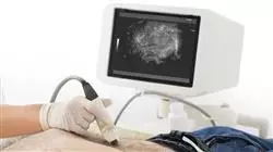
Attend a 3-week intensive internship in a prestigious center and acquire an advanced management of the best ultrasound technologies that are essential for the modern practice of nursing"
In this proposed Professional Master’s Degree, of professionalizing character and hybrid learning modality, the program is aimed at updating nursing professionals who require a high level of qualification. The content is based on the latest scientific evidence and is organized in a didactic way to integrate theoretical knowledge into nursing practice. The theoretical-practical elements allow professionals to update their knowledge and help them to make the right decisions in patient care.
Thanks to its multimedia content elaborated with the latest educational technology, it will allow the nursing professional to obtain a situated and contextual learning, that is to say, a simulated environment that will provide an immersive learning programmed to train in real situations. This program is designed around Problem-Based Learning, whereby the professional must try to solve the different professional practice situations that arise throughout the program. For this purpose, the student will be assisted by an innovative interactive video system created by renowned experts.
This Hybrid professional master’s degree allows you to practice in simulated environments, which provide immersive learning within a hospital facility of the highest prestige in the field of Nursing"

Update your skills and practical procedures in Clinical Ultrasound for Nursing through an innovative learning strategy where you will study in a theoretical and practical way all the advances in the sector"
Teaching Planning
The syllabus of this Hybrid professional master’s degree is composed of a total of 10 didactic modules. Through them, the nurse will learn the most updated criteria on ultrasound-guided intervention procedures for the insertion of drains, probes and punctures. At the same time, it examines the ultrasound approaches to major syndromes such as shock, sepsis, stroke, trauma, among others. Additionally, this syllabus is distinguished from others by its emphasis on pediatric critical care nursing techniques. At the same time, all these contents will be available on a 100% online and interactive platform, without predefined schedules or evaluation chronograms.

Get up to date in a direct way, on TECH's 100% online learning platform, through theoretical materials and multimedia resources such as videos and infographics"
Module 1. Ultrasound imaging
1.1. Physical principles |
1.1.1. Sounds and Ultrasound
1.1.2. Nature of ultrasound
1.1.3. Interaction of ultrasound with matter
1.1.4. Concept of Ultrasound
1.1.5. Ultrasound safety
1.2. Ultrasound Sequence
1.2.1. Ultrasound emission
1.2.2. Tissue interaction
1.2.3. Echo formation
1.2.4. Echo reception
1.2.5. Ultrasound image generation
1.3. Ultrasound Modes
1.3.1. Mode A
1.3.2. M-Mode
1.3.3. Mode B
1.3.4. Color Doppler
1.3.5. Angio-Doppler
1.3.6. Spectral Doppler
1.3.7. Combined Modes
1.3.8. Other modalities and techniques
1.4. Ecography
1.4.1. Console Ecograph Ultrasound Scanners
1.4.2. Portable Ecograph Ultrasound scanners
1.4.3. Specialised Ecograph Ultrasound Scanners
1.4.4. Transducers
1.5. Ultrasound maps and Eco Navigation
1.5.1. Sagittal plane
1.5.2. Transverse plane
1.5.3. Coronal plane
1.5.4. Oblique planes
1.5.5. Ultrasound Marking
1.5.6. Transducer Movements
Module 2. Clinical Cardiac Ultrasound
2.1. Cardiac Anatomy
2.1.1. Basic Three-Dimensional Anatomy
2.1.2. Basic Cardiac Physiology
2.2. Technical Requirements
2.2.1. Probes
2.2.2. Characteristics of the Equipment used in a Cardiac Ultrasound
2.3. Pericardial Windows and Cardiac Ultrasound
2.3.1. Windows and Planes Applied in Emergencies and Intensive Care Situations
2.3.2. Basic Doppler (Color, Pulsating, Continuous and Tissue Doppler)
2.4. Structural Alterations
2.4.1. Basic Measures in Cardiac Ultrasound
2.4.2. Thrombi
2.4.3. Suspected Endocarditis
2.4.4. Valvulopathies
2.4.5. Pericardium
2.4.6. How is an ultrasound reported in emergency and intensive care?
2.5. Structural Alterations I
2.5.1. Left Ventricle
2.5.2. Right Ventricle
2.6. Hemodynamic Ultrasound
2.6.1. Left Ventricular Hemodynamics
2.6.2. Right Ventricular Hemodynamics
2.6.3. Preload Dynamic Tests
2.7. Transesophageal Echocardiogram
2.7.1. Technique
2.7.2. Indications in Emergencies and Intensive Care Cases
2.7.3. Ultrasound-Guided Study of Cardioembolism
Module 3. Clinical Thoracic Ultrasound
3.1. Fundamentals of Thoracic Ultrasound and Anatomical Review
3.1.1. Study of the Normal Thorax
3.1.2. Pulmonary Ultrasound Semiology
3.1.3. Pleural Ultrasound Semiology
3.2. Technical Requirements. Examination Technique
3.2.1. Types of Probes Used
3.2.2. Ultrasound with Contrast in the Thorax
3.3. Ultrasound of the Thoracic Wall and the Mediastinum
3.3.1. Examination of Pulmonary Pathology
3.3.2. Examination of Pleural Pathology
3.3.3. Examination of Mediastinal and Thoracic Wall Pathology
3.4. Ultrasound of the Pleura
3.4.1. Pleural Effusion and Solid Pleural Pathology
3.4.2. Pneumothorax
3.4.3. Pleural Interventionism
3.4.4. Adenopathies and Mediastinal Masses
3.4.5. Adenopathies of the Thoracic Wall
3.4.6. Osteomuscular Pathology of the Thoracic Wall
3.5. Pulmonary Ultrasound Scan
3.5.1. Pneumonia and Atelectasis
3.5.2. Pulmonary Neoplasms
3.5.3. Diffuse Pulmonary Pathology
3.5.4. Pulmonary Infarction
3.6. Diaphragmatic Ultrasound
3.6.1. Ultrasound Approach to the Diaphragmatic Pathology
3.6.2. Usefulness of Ultrasound in the Study of the Diaphragm
Module 4. . Vascular Clinical Ultrasound in Emergencies and Primary Care
4.1. Anatomy Recap.
4.1.1. Venous Vascular Anatomy of the Upper Limbs
4.1.2. Arterial Vascular Anatomy of the Upper Limbs
4.1.3. Venous Vascular Anatomy of the Lower Limbs
4.1.4. Arterial Vascular Anatomy of the Lower Limbs
4.2. Technical Requirements
4.2.1. Ultrasound Scanners and Probes
4.2.2. Curve Analysis
4.2.3. Image-Color Media
4.2.4. Echo Contrasts
4.3. Examination Technique
4.3.1. Positioning
4.3.2. Insonation. Examining Technique
4.3.3. Study of Normal Curves and Speeds
4.4. Large Thoracoabdominal Vessels
4.4.1. Venous Vascular Anatomy of the Abdomen
4.4.2. Arterial Vascular Anatomy of the Abdomen
4.4.3. Abdomino-Pelvic Venous Pathology
4.4.4. Abdomino-Pelvic Arterial Pathology
4.5. Supra-Aortic Trunks
4.5.1. Venous Vascular Anatomy of the Supra-Aortic Trunks
4.5.2. Arterial Vascular Anatomy of the Supra-Aortic Trunks
4.5.3. Venous Pathology of the Supra-Aortic Trunks
4.5.4. Arterial Pathology of the Supra-Aortic Trunks
4.6. Peripheral Arterial and Venous Circulation
4.6.1. Venous Pathology of Lower and Upper Limbs
4.6.2. Arterial Pathology of Lower and Upper Limbs
Module 5. Clinical Cerebral Ultrasound
5.1. Cerebral Hemodynamics
5.1.1. Carotid Circulation
5.1.2. Vertebro-Basilar Circulation
5.1.3. Cerebral Microcirculation
5.2. Ultrasound Modes
5.2.1. Transcraneal Doppler
5.2.2. Cerebral Ultrasound
5.2.3. Special Tests (Vascular Reaction, HITS, etc.)
5.3. Acoustic Windows and Examination Technique
5.3.1. Acoustic Windows
5.3.2. Operator Position
5.3.3. Examination Sequence
5.4. Structural Alterations
5.4.1. Collections and Masses
5.4.2. Vascular Anomalies.
5.4.3. Hydrocephalus
5.4.4. Venous Pathology
5.5. Hemodynamic Alterations
5.5.1. Spectral Analysis
5.5.2. Hyperdynamics
5.5.3. Hypodynamics
5.5.4. Asystole of the Brain
5.6. Ocular Ultrasonography
5.6.1. Pupil Size and Reactivity
5.6.2. Diameter of the Optic Nerve Sheath
5.7. Echo-Doppler in the Diagnosis of Brain Death
5.7.1. Clinical Diagnosis of Encephalic Death
5.7.2. Necessary Conditions Before Transcranial Doppler Examination (TCD) for the Diagnosis of Cerebral Circulatory Arrest
5.7.3. TCD Application Techniques
5.7.4. Advantages of a TCD
5.7.5. Limitations of TCD and Interpretation
5.7.6. TCD Ultrasound for the Diagnosis of Encephalic Death
5.7.7. TCD Ultrasonography in the Diagnosis of Brain Death.
Module 6. Clinical Abdominal Ultrasound
6.1. Anatomy Recap.
6.1.1. Abdominal Cavity
6.1.2. Liver
6.1.3. Gallbladder and Bile Ducts
6.1.4. Retroperitoneum and Great Vessels
6.1.5. Pancreas
6.1.6. Bladder
6.1.7. Kidneys
6.1.8. Bladder
6.1.9. Prostate and Seminal Vesicles
6.1.10. Uterus and Ovaries
6.2. Technical Requirements
6.2.1. Ultrasound Equipment
6.2.2. Types of Transductors for Abdominal Examination
6.2.3. Basic Ultrasound Settings
6.2.4. Patient Preparation
6.3. Examination Technique
6.3.1. Examination Planes
6.3.2. Probe Movements
6.3.3. Visualization of Organs According to Conventional Sectioning
6.3.4. Systematic Examination
6.4. ECO-FAST Methodology
6.4.1. Equipment and Transducers
6.4.2. FAST I
6.4.3. FAST II
6.4.4. FAST III. Perivesical Effusion
6.4.5. FAST IV. Pericardial Effusion
6.4.6. ECO-FAST V. Exclude ABD Aortic Aneurysm
6.5. Ultrasound Scan of the Digestive System
6.5.1. Liver
6.5.2. Gallbladder and Bile Ducts
6.5.3. Pancreas
6.5.4. Bladder
6.6. Genitourinary Ultrasound
6.6.1. Kidney
6.6.2. Urinary Bladder
6.6.3. Male Genital System
6.6.4. Female Genital System
6.7. Usefulness of Ultrasound in the Renal, Hepatic and Pancreatic Transplant Patient.
6.7.1. Normal Ultrasound in the Renal Transplant Patient.
6.7.2. Acute Tubular Necrosis (ATN)
6.7.3. Acute Rejection (AR)
6.7.4. Chronic Transplant Dysfunction
6.7.5. Normal Ultrasound in the Patient with Liver Transplant
6.7.6. Normal Ultrasound in the Patient with Pancreas Transplantation
Module 7. Clinical Musculoskeletal Ultrasound
7.1. Anatomy Recap.
7.1.1. Anatomy of the Shoulder
7.1.2. Anatomy of the Elbow
7.1.3. Anatomy of the Wrist and Hand
7.1.4. Anatomy of the Hip and Thigh
7.1.5. Anatomy of the Knee
7.1.6. Anatomy of the Ankle, Foot, and Leg
7.2. Technical Requirements
7.2.1. Musculoskeletal Ultrasound Equipment
7.2.2. Methodology of Implementation
7.2.3. Ultrasound imaging
7.2.4. Validation, Reliability, and Standardization
7.2.5. Ultrasound-Guided Procedures
7.3. Examination Technique
7.3.1. Basic Concepts in Ultrasound
7.3.2. Rules for Correct Examinations
7.3.3. Examination Technique in Ultrasound Study of the Shoulder
7.3.4. Examination Technique in Ultrasound Study of the Elbow
7.3.5. Examination Technique in Ultrasound Study of the Wrist and Hand
7.3.6. Examination Technique in Ultrasound Study of the Hip
7.3.7. Examination Technique in Ultrasound Study of the Thigh
7.3.8. Examination Technique in Ultrasound Study of the Knee
7.3.9. Examination Technique in Ultrasound Study of the Leg and Ankle
7.4. Sonoanatomy of the Locomotor System: I. Upper Extremities
7.4.1. Shoulder Ultrasound Anatomy
7.4.2. Elbow Ultrasound Anatomy
7.4.3. Wrist and Hand Anatomy Ultrasound
7.5. Sonoanatomy of the Locomotor System: II. Lower Extremities
7.5.1. Hip Ultrasound Anatomy
7.5.2. Thigh Ultrasound Anatomy
7.5.3. Knee Ultrasound Anatomy
7.5.4. Leg and Ankle Ultrasound Anatomy
7.6. Ultrasound in the Most Frequent Acute Locomotor System Injuries
7.6.1. Muscle Injuries
7.6.2. Tendon Injuries
7.6.3. Ligament Injuries
7.6.4. Subcutaneous Tissue Injuries
7.6.5. Bone Injuries
7.6.6. Joint Injuries
7.6.7. Peripheral Nerve Injuries
Module 8. Ultrasonographic Approach to the Major Syndromes.
8.1. Ultrasound in Acute Renal Failure
8.1.1. Introduction
8.1.1.1. Pre-Renal ARF
8.1.1.2. Renal or Intrinsic ARF
8.1.1.3. Post-Renal or Obstructive ARF
8.1.2. Hydronephrosis
8.1.3. Lithiasis
8.1.4. Acute Tubular Necrosis
8.1.5. Doppler Ultrasound in Acute Renal Failure
8.1.6. Bladder Ultrasound in Acute Renal Failure
8.2. Ultrasound in Trauma
8.2.1. FAST and E-FAST (Hemo and Pneumothorax)
8.2.2. Ultrasound Assessment in Special Situations
8.2.3. Hemodynamic Assessment Focused on Trauma
8.3. Ultrasound in Strokes
8.3.1. Introduction
8.3.2. Justification
8.3.3. Initial Assessment
8.3.4. Ultrasound Appraisal
8.3.5. Ultrasound-Guided Management
8.4. Ultrasound in Cardiac Arrest
8.4.1. Cerebral Hemodynamics
8.4.2. Hemodynamics in Cardiac Arrest
8.4.3. Usefulness of Ultrasound in Resuscitation
8.4.4. Usefulness of Ultrasound After Recovery of Spontaneous Circulation
8.5. Ultrasound in Shock
8.5.1. Definition, Types of Shock and Echocardiographic Findings
8.5.1.1. Definition
8.5.1.2. Types of Shock
8.5.1.3. Advantages of Ultrasound in the Recognition and Management of the Different Etiologies of Shock
8.5.1.4. Considerations in ICU
8.5.1.5. Hemodynamic Monitoring by Ultrasound
8.6. Ultrasound in Respiratory Failure
8.6.1. Clinical Ethology of Dyspnea
8.6.2. Approach to the Patient with Dyspnea
8.6.3. Usefulness of Clinical Ultrasound in the Patient with Dyspnea
8.6.4. Pulmonary Ultrasound Scan
8.6.5. Echocardiography
Module 9. Echoguided Procedures in Emergencies and Critical Care
9.1. Airway
9.1.1. Advantages and Disadvantages
9.1.2. Basic Aspects: Ultrasound Specifications and Ultrasound Anatomy
9.1.3. Orotracheal Intubation Technique
9.1.4. Percutaneous Tracheotomy Technique
9.1.5. Common Problems, Complications, and Practical Advice
9.2. Vascular Cannulation
9.2.1. Indications and Advantages of the Anatomical Reference Technique
9.2.2. Current Evidence on Ultrasound-Guided Vascular Cannulation
9.2.3. Basic Aspects: Ultrasound Specifications and Ultrasound Anatomy
9.2.4. Ultrasound-Guided Central Venous Cannulation Technique
9.2.5. Single Peripheral Catheter and Peripherally Inserted Central Catheter (PICC) Cannulation Technique.
9.2.6. Arterial Cannulation Technique
9.2.7. Implementation of an Ultrasound-Guided Vascular Cannulation Protocol
9.2.8. Common Problems, Complications, and Practical Advice
9.3. Thoracentesis and Pericardiocentesis
9.3.1. Indications and Advantages of the Anatomical Reference Technique
9.3.2. Basic Aspects: Ultrasound Esp and Ultrasound Anatomy
9.3.3. Ultrasound Specifications and Pericardial Drainage Technique
9.3.4. Ultrasound Specifications and Thoracic Drainage Technique
9.3.5. Common Problems, Complications, and Practical Advice
9.4. Paracentesis
9.4.1. Indications and Advantages of the Anatomical Reference Technique
9.4.2. Basic Aspects: Ultrasound Specifications and Ultrasound Anatomy
9.4.3. Ultrasound Specifications and Technique
9.4.4. Common Problems, Complications, and Practical Advice
9.5. Lumbar Puncture
9.5.1. Indications and Advantages of the Anatomical Reference Technique
9.5.2. Basic Aspects: Ultrasound Specifications and Ultrasound Anatomy
9.5.3. Technique
9.5.4. Common Problems, Complications, and Practical Advice
9.6. Drainage and Probing
9.6.1. Suprapubic Probing
9.6.2. Collection Drainage
9.6.3. Extraction of Foreign Bodies
Module 10. Clinical Pediatric Ultrasound
10.1. Technical Requirements
10.1.1. Ultrasound at the Patients Bedside
10.1.2. Physical Space
10.1.3. Basic Equipment
10.1.4. Equipment for Interventionalist Ultrasounds
10.1.5. Ultrasound Scanners and Probes
10.2. Examination Technique
10.2.1. Pediatric Patient Preparation
10.2.2. Tests and Probes
10.2.3. Ultrasound Section Planes
10.2.4. Examination System
10.2.5. Ultrasound-Guided Procedures
10.2.6. Images and Documentation
10.2.7. Test Report
10.3. Pediatric Sonoanatomy and Sonophysiology
10.3.1. Normal Anatomy
10.3.2. Sonoanatomy
10.3.3. Sonophysiology of a Child in the Different Stages of Development
10.3.4. Variants of Normality
10.3.5. Dynamic Ultrasound
10.4. Ultrasound of the Major Pediatric Syndromes
10.4.1. Emergency Thorax Ultrasound
10.4.2. Acute Abdomen
10.4.3. Acute Scrotum
10.5. Ultrasound-Guided Procedures in Pediatrics
10.5.1. Vascular Access
10.5.2. Extraction of Superficial Foreign Bodies
10.5.3. Pleural Effusion
10.6. Introduction to Neonatal Clinical Ultrasound
10.6.1. Emergency Transfontanellar Ultrasound
10.6.2. Most Common Examination Indications in Emergencies
10.6.3. Most Common Pathologies in Emergencies

This program employs didactic methods such as Relearning that will help you master state-of-the-art concepts and work protocols"
Hybrid Professional Master's Degree in Clinical Ultrasound in Emergencies and Intensive Care for Nurses
Scientific studies support the need for the healthcare professional to have extensive knowledge about the use of clinical ultrasound, as it reduces care times, improves effectiveness and efficiency in the diagnosis of different pathologies. It is undoubtedly an aspect that favors the patient, as has the improvement of the ultrasound devices themselves thanks to technological development. TECH Global University has already thought about this, that is why it already has its Hybrid Professional Master's Degree available. The Hybrid Professional Master's Degree in Clinical Ultrasound in Emergencies and Intensive Care is an innovative academic program that combines the flexibility of online learning with face-to-face interaction with experts in the field of clinical ultrasound.
Study and take advantage of this blended master's degree in TECH and advance professionally
This intensive one-year seminar is based on the theory of clinical ultrasound and its practical application. The academic program focuses on topics including evaluation and treatment of critical conditions using clinical ultrasound, relevant anatomy, physiology and pathology, understanding ultrasound imaging and interpretation of results. The Hybrid Professional Master's Degree in Clinical Ultrasound in Emergencies and Intensive Care is taught by a team of highly experienced and eminent specialists in the field of clinical ultrasound in emergency and critical care. The Hybrid Professional Master's Degree in Clinical Ultrasound in Emergencies and Intensive Care offers a unique opportunity for students to acquire advanced skills in this field, which will enable them to improve their ability to diagnose and treat patients in critical situations. This academic program is conducted in an interactive and practical learning environment, taught by experts in the field. At the end of the program you will be able to enjoy on-site internships. Make the decision and study at TECH.


