University certificate
The world's largest faculty of veterinary medicine”
Introduction to the Program
Our highly academic Professional Master's Degree will allow you to acquire the specialization you are looking for and achieve a remarkable improvement in your daily practice quickly"

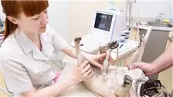
Studying veterinary radiology online is a reality nowadays due to the introduction of more advanced technological tools which enable the possibility of learning this specialty from wherever you happen to be. TECH takes advantage of the this in order to provide students with the most complete educational experience of the moment, employing an innovative methodology that allows for a contextual study of the cases presented. In addition, studies have shown that the veterinarian who has become familiar with radiological images and is able to associate them with different conditions will have a much better diagnostic capacity in the future, therefore, the clarity offered by the new technology allows the learning process to be complete.
For the realization of this Professional Master's Degree, our university's teaching team has made a careful selection of the different radiological diagnostic procedures, together with other diagnostic alternatives for veterinary professionals. In this way, clear clinical guidance is provided by the use of radiology to resolve the diagnosis of small animal diseases diseases without forgetting the wide variety of other diagnostic options that are of great use in veterinary practices.
In short, it is a program based on scientific evidence and daily practice, with all the nuances that each professional can contribute, so that the student can have all this in mind alongside the bibliography enriched by the critical evaluation that every professional must have in mind.
Thus, throughout this training, the students will go through all the current approaches to the different challenges in their profession. A high-level step that will become a process of improvement, not only on a professional level, but also on a personal level. In addition, TECH assumes a social commitment: to help update highly qualified professionals and to develop their personal, social and work skills throughout the duration of the course. And, to do so, it will not only take you through the theoretical knowledge offered, but will show you another more organic, simpler and more efficient way of studying and learning. It works to maintain motivation and to create a passion for learning; it encourages thinking and the development of critical thinking.
The studies in Veterinary Radiology will allow you to use the main diagnostic imaging techniques with total confidence"
This Professional master’s degree in Veterinary Radiology in Small Animals contains the most complete and up-to-date scientific program on the market. The most important features include:
- The development of case studies presented by experts in Veterinary Radiology
- The graphic, schematic, and eminently practical contents with which they are created, provide scientific and practical information on the disciplines that are essential for professional practice.
- Latest developments in Veterinary Radiology
- Practical exercises where the self-assessment process can be carried out to improve learning
- Special emphasis on innovative methodologies in Veterinary Radiology
- Theoretical lessons, questions to the expert, debate forums on controversial topics, and individual reflection work
- Content that is accessible from any fixed or portable device with an Internet connection
This online training course, will give you the possibility to expand your knowledge with a multitude of virtual tools, making your learning faster and more effective"
TECH's teaching staff includes professionals belonging to the veterinary field, who contribute their work experience to this training, as well as renowned specialists from reference societies and prestigious universities.
The multimedia content, developed with the latest educational technology, will provide the professional with situated and contextual learning, i.e., a simulated environment that will provide immersive training programmed to train in real situations.
This program is designed around Problem Based Learning, whereby the specialist must try to solve the different professional practice situations that arise during the academic year. For this purpose, the professional will be assisted by an innovative system of interactive videos made by renowned and experienced experts in Veterinary Radiology.
Our innovative methodology has had a great rate of success amongst our students, due to the benefits it provides for contextual study, a factor which enables them to learn more easily"

Learn efficiently, with the real objective of attaining a valid qualification, on this Professional Master's Degree which is unique for its quality and price, all of which helps it to stand out in the online teaching market"
Why study at TECH?
TECH is the world’s largest online university. With an impressive catalog of more than 14,000 university programs available in 11 languages, it is positioned as a leader in employability, with a 99% job placement rate. In addition, it relies on an enormous faculty of more than 6,000 professors of the highest international renown.

Study at the world's largest online university and guarantee your professional success. The future starts at TECH”
The world’s best online university according to FORBES
The prestigious Forbes magazine, specialized in business and finance, has highlighted TECH as “the world's best online university” This is what they have recently stated in an article in their digital edition in which they echo the success story of this institution, “thanks to the academic offer it provides, the selection of its teaching staff, and an innovative learning method aimed at educating the professionals of the future”
A revolutionary study method, a cutting-edge faculty and a practical focus: the key to TECH's success.
The most complete study plans on the university scene
TECH offers the most complete study plans on the university scene, with syllabuses that cover fundamental concepts and, at the same time, the main scientific advances in their specific scientific areas. In addition, these programs are continuously being updated to guarantee students the academic vanguard and the most in-demand professional skills. In this way, the university's qualifications provide its graduates with a significant advantage to propel their careers to success.
TECH offers the most comprehensive and intensive study plans on the current university scene.
A world-class teaching staff
TECH's teaching staff is made up of more than 6,000 professors with the highest international recognition. Professors, researchers and top executives of multinational companies, including Isaiah Covington, performance coach of the Boston Celtics; Magda Romanska, principal investigator at Harvard MetaLAB; Ignacio Wistumba, chairman of the department of translational molecular pathology at MD Anderson Cancer Center; and D.W. Pine, creative director of TIME magazine, among others.
Internationally renowned experts, specialized in different branches of Health, Technology, Communication and Business, form part of the TECH faculty.
A unique learning method
TECH is the first university to use Relearning in all its programs. It is the best online learning methodology, accredited with international teaching quality certifications, provided by prestigious educational agencies. In addition, this disruptive educational model is complemented with the “Case Method”, thereby setting up a unique online teaching strategy. Innovative teaching resources are also implemented, including detailed videos, infographics and interactive summaries.
TECH combines Relearning and the Case Method in all its university programs to guarantee excellent theoretical and practical learning, studying whenever and wherever you want.
The world's largest online university
TECH is the world’s largest online university. We are the largest educational institution, with the best and widest online educational catalog, one hundred percent online and covering the vast majority of areas of knowledge. We offer a large selection of our own degrees and accredited online undergraduate and postgraduate degrees. In total, more than 14,000 university degrees, in eleven different languages, make us the largest educational largest in the world.
TECH has the world's most extensive catalog of academic and official programs, available in more than 11 languages.
Google Premier Partner
The American technology giant has awarded TECH the Google Google Premier Partner badge. This award, which is only available to 3% of the world's companies, highlights the efficient, flexible and tailored experience that this university provides to students. The recognition as a Google Premier Partner not only accredits the maximum rigor, performance and investment in TECH's digital infrastructures, but also places this university as one of the world's leading technology companies.
Google has positioned TECH in the top 3% of the world's most important technology companies by awarding it its Google Premier Partner badge.
The official online university of the NBA
TECH is the official online university of the NBA. Thanks to our agreement with the biggest league in basketball, we offer our students exclusive university programs, as well as a wide variety of educational resources focused on the business of the league and other areas of the sports industry. Each program is made up of a uniquely designed syllabus and features exceptional guest hosts: professionals with a distinguished sports background who will offer their expertise on the most relevant topics.
TECH has been selected by the NBA, the world's top basketball league, as its official online university.
The top-rated university by its students
Students have positioned TECH as the world's top-rated university on the main review websites, with a highest rating of 4.9 out of 5, obtained from more than 1,000 reviews. These results consolidate TECH as the benchmark university institution at an international level, reflecting the excellence and positive impact of its educational model.” reflecting the excellence and positive impact of its educational model.”
TECH is the world’s top-rated university by its students.
Leaders in employability
TECH has managed to become the leading university in employability. 99% of its students obtain jobs in the academic field they have studied, within one year of completing any of the university's programs. A similar number achieve immediate career enhancement. All this thanks to a study methodology that bases its effectiveness on the acquisition of practical skills, which are absolutely necessary for professional development.
99% of TECH graduates find a job within a year of completing their studies.
Professional Master's Degree in Small Animal Veterinary Radiology
.
Diagnostic imaging is of vital importance for the approach of pathologies, since it facilitates the early detection of complications caused by them. Taking into account that in TECH Global University, one of our main objectives is to promote professional specialization processes, we have created this program focused on the general principles, installation requirements, quality control and the necessary positioning for the performance of ionizing radiation. During the 12 months required by the curriculum, students will develop the key technical skills to carry out neurological or orthopedic radiodiagnostics, as well as those of the cardiovascular system, respiratory system, intrathoracic structures, digestive system and abdominal structures. In addition, you will be able to develop your skills in the application of minimally invasive and interventional techniques. Thanks to the theoretical-practical course, you will be able to identify trauma problems, to correctly interpret radiological images and to adapt to the follow-up and supervision protocols required by current regulations.
Postgraduate course in Small Animal Veterinary Radiology
.
Studying this TECH postgraduate course is an interesting opportunity to fully immerse yourself in diagnostic methods using ultrasound, CT and magnetic resonance imaging (MRI). The contents, designed by our teaching team, provide key tools for the safe handling of the equipment, for the examination of problems or distortions present in the tomography obtained and for the careful positioning of the patient. With this background, the professionals will be qualified for the complementary exploration of the different organs in evaluative studies, which will allow them to safely practice any type of intervention and to be able to operate the technical instruments with ease. In addition, to adopt the respective innovations coined in the field. This Professional Master's Degree in veterinary radiology, then, will empower the future expert in the field of assistance, since he/she will provide effective comprehensive care to injured patients, who are in an emergency situation or require critical care. All this, will enrich their daily work and favor their rapid integration into the labor market.


