University certificate
The world's largest faculty of medicine”
Introduction to the Program
Specialize in Nuclear Medicine and increase your prestige by helping to detect and treat different pathologies thanks to this Master's Degree"

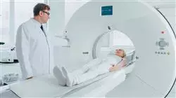
Nuclear Medicine is one of the health areas that is currently experiencing greatest advances. This specialty makes it possible to find and treat different pathologies that, by other means, would be imperceptible or would be detected late. Moreover, its efficiency and precision make it one of the most sought-after fields by the major medical services of the best clinics in the world.
For that reason, going deeper into this subject can make the physicians a prestigious professional who enjoys great opportunities to advance their careers, in addition to updating their knowledge in a field in constant transformation. Thus, this
Master's Degree in Nuclear Medicine is the perfect educational program for all those who wish to deepen their knowledge in this area that will turn them into reputed doctors.
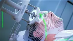
Thus, this degree offers its students highly specialized contents with which they will be able to master issues such as single photon emission applied to Nuclear Medicine, Nuclear Medicine related to pediatrics, nuclear treatments of neuroendocrine tumors or the use of radioguided surgery.
With this knowledge, physicians who complete the program will have become experts in this field and will have updated their skills so that they have mastered the latest techniques in this area. Thus, they will be able to progress professionally, being able to access the Nuclear Medicine services of the major clinics in the country.
This program, moreover, is taught through an innovative 100% online teaching methodology that will allow doctors to combine their professional careers and personal lives with their studies, since it has been designed to adapt to the circumstances of each individual. In this way, the learning process is facilitated while maintaining a high educational level and guaranteeing that students will be true specialists in Nuclear Medicine when they complete this Master's Degree.
Nuclear Medicine offers innovative techniques to treat complex pathologies. Enroll now and offer the best services to your patients with this innovative degree"
This Master's Degree in Nuclear Medicine contains the most complete and up-to-date scientific program on the market. Its most notable features are:
- The development of case studies presented by experts in Nuclear Medicine
- The graphic, schematic, and eminently practical contents with which they are created, provide scientific and practical information on the disciplines that are essential for professional practice
- Practical exercises where self-assessment can be used to improve learning
- Its special emphasis on innovative methodologies
- Theoretical lessons, questions to the expert, debate forums on controversial topics, and individual reflection assignments
- Content that is accessible from any fixed or portable device with an Internet connection
Specialization is the key: with this degree you will enhance your reputation and progress in the exciting field of Nuclear Medicine"
The program’s teaching staff includes professionals from the sector who contribute their work experience to this training program, as well as renowned specialists from leading societies and prestigious universities.
The multimedia content, developed with the latest educational technology, will provide the professional with situated and contextual learning, i.e., a simulated environment that will provide immersive training programmed to train in real situations.
This program is designed around Problem-Based Learning, whereby the professional must try to solve the different professional practice situations that arise during the academic year. This will be done with the help of an innovative system of interactive videos made by renowned experts in the field of Nuclear Medicine with extensive teaching experience.
Update your knowledge in Nuclear Medicine and become a prestigious specialist thanks to this Master's Degree"

Nuclear Medicine services are booming. Specialize and achieve all your professional goals"
Why study at TECH?
TECH is the world’s largest online university. With an impressive catalog of more than 14,000 university programs available in 11 languages, it is positioned as a leader in employability, with a 99% job placement rate. In addition, it relies on an enormous faculty of more than 6,000 professors of the highest international renown.

Study at the world's largest online university and guarantee your professional success. The future starts at TECH”
The world’s best online university according to FORBES
The prestigious Forbes magazine, specialized in business and finance, has highlighted TECH as “the world's best online university” This is what they have recently stated in an article in their digital edition in which they echo the success story of this institution, “thanks to the academic offer it provides, the selection of its teaching staff, and an innovative learning method aimed at educating the professionals of the future”
A revolutionary study method, a cutting-edge faculty and a practical focus: the key to TECH's success.
The most complete study plans on the university scene
TECH offers the most complete study plans on the university scene, with syllabuses that cover fundamental concepts and, at the same time, the main scientific advances in their specific scientific areas. In addition, these programs are continuously being updated to guarantee students the academic vanguard and the most in-demand professional skills. In this way, the university's qualifications provide its graduates with a significant advantage to propel their careers to success.
TECH offers the most comprehensive and intensive study plans on the current university scene.
A world-class teaching staff
TECH's teaching staff is made up of more than 6,000 professors with the highest international recognition. Professors, researchers and top executives of multinational companies, including Isaiah Covington, performance coach of the Boston Celtics; Magda Romanska, principal investigator at Harvard MetaLAB; Ignacio Wistumba, chairman of the department of translational molecular pathology at MD Anderson Cancer Center; and D.W. Pine, creative director of TIME magazine, among others.
Internationally renowned experts, specialized in different branches of Health, Technology, Communication and Business, form part of the TECH faculty.
A unique learning method
TECH is the first university to use Relearning in all its programs. It is the best online learning methodology, accredited with international teaching quality certifications, provided by prestigious educational agencies. In addition, this disruptive educational model is complemented with the “Case Method”, thereby setting up a unique online teaching strategy. Innovative teaching resources are also implemented, including detailed videos, infographics and interactive summaries.
TECH combines Relearning and the Case Method in all its university programs to guarantee excellent theoretical and practical learning, studying whenever and wherever you want.
The world's largest online university
TECH is the world’s largest online university. We are the largest educational institution, with the best and widest online educational catalog, one hundred percent online and covering the vast majority of areas of knowledge. We offer a large selection of our own degrees and accredited online undergraduate and postgraduate degrees. In total, more than 14,000 university degrees, in eleven different languages, make us the largest educational largest in the world.
TECH has the world's most extensive catalog of academic and official programs, available in more than 11 languages.
Google Premier Partner
The American technology giant has awarded TECH the Google Google Premier Partner badge. This award, which is only available to 3% of the world's companies, highlights the efficient, flexible and tailored experience that this university provides to students. The recognition as a Google Premier Partner not only accredits the maximum rigor, performance and investment in TECH's digital infrastructures, but also places this university as one of the world's leading technology companies.
Google has positioned TECH in the top 3% of the world's most important technology companies by awarding it its Google Premier Partner badge.
The official online university of the NBA
TECH is the official online university of the NBA. Thanks to our agreement with the biggest league in basketball, we offer our students exclusive university programs, as well as a wide variety of educational resources focused on the business of the league and other areas of the sports industry. Each program is made up of a uniquely designed syllabus and features exceptional guest hosts: professionals with a distinguished sports background who will offer their expertise on the most relevant topics.
TECH has been selected by the NBA, the world's top basketball league, as its official online university.
The top-rated university by its students
Students have positioned TECH as the world's top-rated university on the main review websites, with a highest rating of 4.9 out of 5, obtained from more than 1,000 reviews. These results consolidate TECH as the benchmark university institution at an international level, reflecting the excellence and positive impact of its educational model.” reflecting the excellence and positive impact of its educational model.”
TECH is the world’s top-rated university by its students.
Leaders in employability
TECH has managed to become the leading university in employability. 99% of its students obtain jobs in the academic field they have studied, within one year of completing any of the university's programs. A similar number achieve immediate career enhancement. All this thanks to a study methodology that bases its effectiveness on the acquisition of practical skills, which are absolutely necessary for professional development.
99% of TECH graduates find a job within a year of completing their studies.
Master's Degree in Nuclear Medicine
Enter the fascinating world of Nuclear Medicine with the Master's Degree in Nuclear Medicine from TECH Global University. Discover our online classes and acquire the necessary knowledge to become a cutting-edge professional in this highly important medical specialty. In a world in constant technological advancement, Nuclear Medicine plays a fundamental role in the diagnosis and treatment of various diseases. With our Master's Degree program, you will be able to learn the most advanced techniques and procedures in nuclear medicine, using radiopharmaceuticals and high-tech equipment to obtain images and perform precise therapies. TECH Global University's online classes give you the flexibility to study from anywhere, anytime. You will be able to access the Master's Degree content, participate in interactive sessions and perform online internships that will allow you to gain practical experience without leaving home.
The best online teaching methodology is at TECH.
Our team of Postgraduate Diplomas in Nuclear Medicine will guide you through the program, providing you with cutting-edge theoretical and practical knowledge. You will learn how to interpret nuclear images, make accurate diagnoses, plan and administer therapeutic treatments, and collaborate with other health professionals in the comprehensive care of patients. By choosing the Master's Degree in Nuclear Medicine from TECH Global University, you will be investing in your professional future. You will open yourself to job opportunities in hospitals, clinics and centers specialized in nuclear medicine, where you will be able to contribute to the early and accurate diagnosis of diseases, as well as to the improvement of patients' quality of life. Take advantage of this unique opportunity to become a Postgraduate Diploma in Nuclear Medicine. Enroll in our Master's Degree and open your way to a successful and rewarding career in the healthcare field - get ready to make a difference in the medicine of the future!


