University certificate
The world's largest faculty of veterinary medicine”
Why study at TECH?
This course in Thoracic Radiology will allow veterinarians to obtain better qualifications providing them with a higher level of skills with which to improve the health of animals"

Improvements in diagnostic imaging techniques in the field of veterinary medicine make it necessary for teaching centers to propose new high level training courses, with fully up to date information which include the latest developments in this field. TECH always wants to be at the forefront in terms of training proposals and, therefore, this time we present this Postgraduate diploma in Thoracic Radiology in Small Animals A program designed by a team specialized in Veterinary Radiology, which aims to provide the keys to success in your practice.
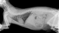
Specifically, this Postgraduate diploma covers ionizing radiation for diagnostic purposes to radiodiagnosis of the cardiovascular and respiratory systems and other intrathoracic structures. As for cardiac radiology, it is necessary to take into account that it is very present in daily clinical practices and it is a challenge when it comes to interpreting it. Therefore, this program deals with the identification of cardiac anatomy in radiological projections, an essential part of cardiac and vascular diagnosis.
In addition, it is necessary to work in the field of Thoracic Radiology with the highest technical quality, because poor patient positioning or poor development technique can greatly affect the interpretation of the images. TECH has set out to teach all the key details that will enable professional growth in this field.
In short, it is a program based on scientific evidence and daily practice, with all the nuances that each professional can contribute, so that the student can keep it all in mind and compare it with the bibliography and feel enriched by the critical evaluation that every professional must have.
Throughout this course, the student will learn about all the current approaches to the different challenges posed by their profession. A high level step that will become a process of improvement, not only on a professional level, but also on a personal level. Additionally, at TECH we have a social commitment: to help highly qualified professionals to update and to develop their personal, social and professional skills throughout the course of their studies. And, to do so, it will not only take you through the theoretical knowledge offered, but will show you another more organic, simpler and more efficient way of learning and studying. It works to maintain motivation and to create passion for learning; it encourages thinking and the development of critical thinking.
Advances in Thoracic Radiology make this Postgraduate diploma a unique opportunity to enhance your training"
This Postgraduate diploma in Thoracic Radiology in Small Animals contains the most complete and up to date educational program on the market. The most important features of the program include:
- The development of practical cases presented by university experts in Veterinary Radiology
- The graphic, schematic, and eminently practical contents with which they are created, provide scientific and practical information on the disciplines that are essential for professional practice
- Latest developments in Veterinary Radiology
- Practical exercises where self-assessment can be used to improve learning.
- Special emphasis on innovative methodologies in Veterinary Radiology
- Theoretical lessons, questions to the university experts, debate forums on controversial topics, and individual reflection assignments
- Content that is accessible from any fixed or portable device with an Internet connection
The application of Thoracic Radiology is a complex process, as any small error can lead to misdiagnosis. If you want to specialize in this field, don't think twice and join TECH"
Its teaching staff includes professionals belonging to the veterinary field, who contribute their work experience to this training, as well as renowned specialists from reference societies and prestigious universities.
Its Multimedia Content, elaborated with the latest Educational Technology, will allow the Professional a situated and contextual learning, that is to say, a Simulated Environment that will provide an immersive specialization programmed to train in real situations.
This program is designed around Problem Based Learning, whereby the specialist must try to solve the different professional practice situations that arise during the academic year. For this purpose, the professional will be assisted by an innovative system of interactive videos made by renowned and experienced university experts in Veterinary Radiology.
We give you all the necessary resources so that you can specialize in a field which offers great employment possibilities"

Our online format will allow you to study in a comfortable way from wherever you choose"
Syllabus
The contents of this Postgraduate diploma in Thoracic Radiology in Small Animals have been designed by a team of university experts, backed by their years of experience. In this way, they have been in charge of programming a totally up to date syllabus aimed at the 21st century professional, who demands high quality training and knowledge of the main innovations in the field.

Module 1. Ionizing Radiation for Diagnostic Purposes
1.1. General Principles
1.1.1. Electron Acceleration
1.1.2. Electrical Current Intensity
1.1.3. The Anode Where the Anions Collide
1.2. Photon Formation with Diagnostic Effects
1.2.1. Types of Photon
1.2.2. Photon Energy
1.2.3. Orientation of Emitted Photons
1.2.4. Scattering of the Energy Generated by Photons
1.3. Scattered Radiation
1.3.1. Anode Scattering
1.3.2. Patient Scattering
1.3.3. Implications for Clinical Imaging
1.3.4. Dispersion of Objects in the Radiodiagnostic Room
1.4. The Formation of Radiological Imaging
1.4.1. Radiological Chassis
1.4.2. Radiological Films
1.4.3. RC Processing
1.4.4. DR Processing
1.5. Radiological Film Processing
1.5.1. Development in Automatic Processors and Development Tanks
1.5.2. Liquid Recycling
1.5.3. Processing with Digital Chassis
1.5.4. Direct Digital Processing
1.6. Factors Affecting Radiological Imaging
1.6.1. Time
1.6.2. Voltage
1.6.3. Amperage
1.7. Alterations in the Perception of the Radiological Image
1.7.1. Pareidolia
1.7.2. Magnification
1.7.3. Distortion
1.8. Radiological Interpretation
1.8.1. Systematization of Interpretation
1.8.2. Validity of the Image Obtained
1.8.3. Differences Between Tissues
1.8.4. Identification of Healthy Organs
1.8.5. Identification of Radiological Alterations
1.8.6. Typical Diseases of the Different Anatomical Regions
1.9. Limiting Factors in Radiological Diagnosis, Time
1.9.1. Regions in Motion
1.9.2. Still Regions
1.9.3. Fuzziness
1.9.4. Anesthesia in Radiology
1.9.5. Radiological Positioners
1.9.6. Anatomical Regions in Which Time Has To Be Taken into Consideration
1.10. Limiting Factors in Radiological Diagnosis, Voltage
1.10.1. Density of the Radiographic Region
1.10.2. Contrast
1.10.3. Sharpness
1.10.4. Anatomical Regions in Which the Energy of Photons Must Be taken into Consideration
Module 2. Radiodiagnosis of the Cardiovascular System
2.1. Positioning in Cardiovascular Radiological Diagnosis
2.1.1. Right Lateral Projection
2.1.2. Dorsoventral Projection
2.1.3. Differences with Other Projections
2.2. Physiological Radiological Imaging of the Cardiovascular System
2.2.1. Cardiac Silhouette
2.2.2. Cardiac Cameras
2.2.3. Large Vessels
2.3. Altered Radiological Image of the Cardiovascular System
2.3.1. Cardiac Size Alteration
2.3.2. Vascular Alteration
2.3.3. Radiographic Signs of Heart Failure
2.4. Acquired Heart Diseases I
2.4.1. Mitral Degenerative Disease
2.4.2. Canine Cardiomyopathy
2.4.3. Pericardial Diseases
2.5. Acquired Heart Diseases II
2.5.1. Feline Cardiomyopathies
2.5.2. Dirofilariasis
2.5.3. Systemic Diseases with Cardiac Implications
2.6. Oncology
2.6.1. Neoplasia of the Right Atrium
2.6.2. Cardiac-based Neoplasm
2.6.3. Congenital Heart Diseases
2.7. Patent Ductus Arteriosus
2.7.1. Introduction
2.7.2. Existing Forms
2.7.3. Radiological Characteristics
2.7.4. CAP with D-I Shunt
2.8. Vascular Ring Anomalies
2.8.1. Introduction
2.8.2. Types
2.8.3. Radiological Characteristics
2.9. Other Congenital Diseases
2.9.1. Pulmonary Stenosis
2.9.2. Atrioventricular Septal Defect
2.9.3. Tetralogy of Fallot
2.9.4. Aortic Stenosis
2.9.5. Interatrial Septal Defect
2.9.6. Mitral Dysplasia
2.9.7. Tricuspid Dysplasia
2.9.8. Microcardia
2.10. Radiological Diagnosis of Pericardial Diseases
2.10.1. Radiological Diagnosis of Pericardial Diseases
2.10.1.1. Pericardial Effusion
2.10.1.2. Introduction
2.10.1.3. Radiological Characteristics
2.10.2. Peritoneopericardial Diaphragmatic Hernia
2.10.2.1. Introduction
2.10.2.2. Radiological Characteristics
Module 3. Radiodiagnostics of the Respiratory System and Other Intrathoracic Structures
3.1. Positioning for Thorax Radiology
3.1.1. Ventrodorsal and Dorsoventral Positioning
3.1.2. Right and Left Laterolateral Positioning
3.2. Physiological Imaging of the Thorax
3.2.1. Trachea Physiological Imaging
3.2.2. Mediastinum Physiological Imaging
3.3. Pathologic Imaging in Thoracic Radiology
3.3.1. Alveolar Pattern
3.3.2. Bronchial Pattern
3.3.3. Interstitial Pattern
3.3.4. Vascular Pattern
3.4. Radiological Diagnosis of Acquired Pulmonary Diseases I
3.4.1. Structural Pathologies
3.4.2. Infectious Pathologies
3.5. Radiological Diagnosis of Acquired Pulmonary Diseases II
3.5.1. Inflammatory Pathology
3.5.2. Neoplasms
3.6. Feline-specific Thoracic Radiology
3.6.1. Radiology of the Heart in the Cat
3.6.1.1. Radiographic Anatomy of the Heart
3.6.1.2. Radiographic Diagnosis of Cardiac Pathologies
3.6.2. Radiology of the Thoracic Wall and Diaphragm of the Cat.
3.6.2.1. Anatomy of the Thoracic Cage
3.6.2.2. Radiographic Diagnosis of Thoracic Wall and Diaphragm Pathologies
3.6.2.2.1. Congenital Skeletal Malformations
3.6.2.2.2. Fractures
3.6.2.2.3. Neoplasms
3.6.2.2.4. Alterations of the Diaphragm
3.6.3. Radiology of the Pleura and Pleural Cavity of the Cat
3.6.3.1. Radiographic Diagnosis of the Pleura and Pleural Cavity Pathologies
3.6.3.1.1. Pleural Effusion
3.6.3.1.2. Pneumothorax
3.6.3.1.3. Hydropneumothorax
3.6.3.1.4. Pleural Masses
3.6.4. Radiology of the Cat Mediastinum
3.6.4.1. Radiographic Anatomy of the Mediastinum
3.6.4.2. Radiographic Diagnosis of Pathologies of the Mediastinum and the Organs it Contains.
3.6.4.2.1. Pneumomediastinum
3.6.4.2.2. Mediastinal Masses
3.6.4.2.3. Esophageal Diseases
3.6.4.2.4. Tracheal Diseases
3.6.5. Pulmonary Radiology of the Cat
3.6.5.1. Normal Pulmonary Radiologic Anatomy
3.6.5.2. Radiographic Diagnosis of Pulmonary Pathologies
3.6.5.2.1. Pulmonary Patterns
3.6.5.2.2. Decreased Pulmonary Opacity
3.7. Radiology of the Mediastinum
3.7.1. Radiographic Anatomy of the Mediastinum
3.7.2. Mediastinal Effusion
3.7.3. Pneumomediastinum
3.7.4. Mediastinal Masses
3.7.5. Mediastinal Deviation
3.8. Congenital Thoracic Diseases
3.8.1. Patent Ductus Arteriosus
3.8.2. Pulmonary Stenosis
3.8.3. Aortic Stenosis
3.8.4. Ventricular Septal Defect
3.8.5. Tetralogy of Fallot
3.9. Oncology
3.9.1. Pleural Masses
3.9.2. Mediastinal Masses
3.9.3. Cardiac Tumors
3.9.4. Pulmonary Tumors
3.10. Radiology of the Thoracic Cage
3.10.1. Anatomy Radiologic of the Thoracic Cage
3.10.2. Radiological Alterations of the Ribs
3.10.3. Radiological Alterations of the Sternum

A comprehensive teaching program, structured into well-developed teaching units, oriented towards a learning experience that is fully compatible with your personal and professional life"
Postgraduate Diploma in Small Animal Thoracic Radiology
.
Learn small animal thoracic radiology and become a veterinary diagnostic specialist with the Postgraduate Diploma in Small Animal Thoracic Radiology Postgraduate Diploma program offered by the College of Veterinary Medicine at TECH Global University, the world's best digital university. Our online classes offer flexibility and convenience, allowing you to access training from anywhere and enjoy the benefits of our recognized institution. With this postgraduate program, you will be able to acquire solid skills that will allow you to perform as a professional trained in the use of radiology as a fundamental diagnostic tool for respiratory, cardiac and thoracic diseases in companion animals. Our program gives you the opportunity to study at your own pace with online classes accessible from any device with an internet connection. You will learn to interpret thoracic radiographs and acquire practical skills in performing specialized radiological studies. You will also master positioning techniques and analysis of common thoracic pathologies. Our practical, case-oriented approach will allow you to apply your theoretical knowledge in clinical situations.
Specialize in small animal thoracic radiology
.
Do you know why TECH is considered one of the best universities in the world? Because we have a catalog of more than ten thousand academic programs, presence in multiple countries, innovative methodologies, unique academic technology and a highly qualified faculty; that's why you can't miss the opportunity to study with us. Work with expert faculty and professionals in the field of veterinary radiology, who will guide you throughout the program. Upon completion, you will receive a recognized certificate to support your skills and knowledge in small animal thoracic radiology. Specialize in thoracic radiology and expand your career opportunities in veterinary clinics, hospitals and diagnostic imaging centers. Enroll in TECH Global University's Postgraduate Diploma in Small Animal Thoracic Radiology and take your veterinary career to the next level.""


