University certificate
The world's largest faculty of veterinary medicine”
Why study at TECH?
This Advanced master’s degree in Anesthesia and Surgery in Small Animals is an incomparable, highly qualified tool for veterinary professionals, which will allow you, in a single training itinerary, to acquire the most up-to-date knowledge and skills in the sector”
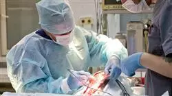
The goal of this complete Advanced master’s degree is to know all the aspects of Anesthesia and Surgery in Small Animals, which we now present to you. With a wide methodological development, throughout this specialization, you will be able to learn each and every one of the fundamental points in this area of work.
In this sense, the Advanced master’s degree will prepare you in everything concerning the phases prior to the application of anesthesia on the patient: knowledge of the equipment, previous management of the patient, drugs and study of drug interactions.
The study of the physiology most closely related to anesthesia, focusing on the involvement of the cardiocirculatory, respiratory, nervous system and endocrine systems, is essential to understand the functioning and consequences on the patient of the application of anesthesia.
However, the success of an anesthetic procedure goes far beyond the administration of the appropriate drugs. It is imperative to master the pre-anesthetic assessment, induction, maintenance and education of the process in order to achieve its success and a return to normality without after-effects. Fluid therapy, and even transfusion, must also be taken into account and, therefore, become the subject of study in our comprehensive Advanced master’s degree in Anesthesia and Surgery in Small Animals.
The anesthesiologist must also take care of pain management. A basic vital sign that, if not adequately controlled, can be one of the main causes of delayed discharge and perioperative complications. Acquiring competence in this part of care is another of our major objectives.
Monitoring, anesthetic complications, management of anesthesia under special conditions and the application of balanced anesthesia and multimodal anesthesia protocols will complete the more extensive review. But the purpose of an anesthetic is usually to allow surgical intervention. For this reason, this Advanced master’s degree also deals comprehensively with the techniques and new developments in this area.
We will review the new surgical materials available and the advances in infection treatment. In addition, we will learn everything we need to know about wound healing. In this unit, the ways of performing the cures and their progress will be part of the agenda.
A complete refresher in Anesthesia and Surgery in Small Animals with this highly effective educational Advanced master’s degree, which opens new paths to your professional advancement”
This Advanced master’s degree in Anesthesia and Surgery in Small Animals, contains the most comprehensive and up-to-date academic course on the university scene. The most important features include:
- Latest technology in online teaching software
- A highly visual teaching system, supported by graphic and schematic contents that are easy to assimilate and understand
- Practical cases presented by practising experts
- State-of-the-art interactive video systems
- Teaching supported by practising remotely
- Continuous updating and recycling systems
- Autonomous learning: full compatibility with other occupations
- Practical exercises for self-evaluation and learning verification
- Support groups and educational synergies: questions to the expert, debate and knowledge forums
- Communication with the teacher and individual reflection work
- Content that is accessible from any , fixed or portable device , with an Internet connection
- Supplementary documentation databases are permanently available, even after the program
This exceptional specialization is the answer to the veterinary professionals need for updating and specialization. A process that you will finish with the solvency of a high-level professional”
Our teaching staff is made up of professionals from different fields related to this specialty. In this way we make sure to offer you the objective of educational updating from all related sectors, with the direct and experienced vision of experts. A multidisciplinary team of doctors with training and experience in different environments, who will develop the theoretical knowledge in an efficient way, but above all, they will bring their practical knowledge from their own experience to the course: one of the differential qualities of this specialization.
This mastery of the subject is complemented by the effectiveness of the methodological design of this Advanced master’s degree in Anesthesia and Surgery in Small Animals developed by a multidisciplinary team of e-learning experts, it integrates the latest advances in educational technology. This way, you will be able to study with a range of easy-to-use and versatile multimedia tools that, will give you the necessary skills you need for your specialization. A new way of learning that transcends physical and temporal barriers, opening the doors to the highest qualification, regardless of place or time.
With a methodological design based on proven teaching techniques, this Advanced master’s degree in Anesthesia and Surgery in Small Animals will take you through different teaching approaches to allow you to learn in a dynamic and effective way"
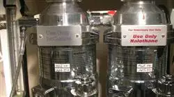
Our innovative remote practise concept will give you the opportunity to learn through an immersive experience, which will provide you with a faster integration and a much more realistic view of the contents: “Learning from an expert”
Syllabus
The contents of this Advanced master’s degree have been developed by the different experts on this course, with a clear purpose: to ensure that our students acquire each and every one of the necessary skills to become true experts in this field.
Throughout the syllabus, the professional will cover the two areas of interest of this Advanced master’s degree; anesthesiology and small animal surgery. Both areas will be developed independently, but in a coordinated manner, covering all possible paradigms of intervention in which the professional may find themselves.
A complete and well-structured program, that will take you to the highest standards of quality and success.
A complete and very well structured syllabus”
Anesthesiology Area
Module 1. Introduction. Anaesthetic Equipment
1.1. Brief History of Anesthesia
1.1.1. Important Facts About Human Anesthesiology
1.1.2. Relevant Historic Facts in Veterinary Anesthesiology
1.2. Optimization of the Surgical Patient. Pre-operative Fasting
1.2.1. Importance of Liquid Fasting
1.2.2. Solid Fasting, Why and How Much?
1.3. Peri-operative Drugs
1.3.1. Precautions in the Polymedicated Patient. General Aspects
1.3.2. Medication Guidelines for Patients with Cardiac Medication
1.3.3. Medication Guidelines in Diabetic Patients
1.3.4. Medication Guidelines for Patient with Epilepsy
1.3.5. Other Chronic Medications
1.4. Anesthetic Machines and Systems
1.4.1. General Aspects
1.4.2. Technical Description and Equipment Care
1.4.3. Anaesthetic Circuits
1.4.3.1. No Reinhalation
1.4.3.2. With Reinhalation
1.5. Mechanical Ventilators
1.5.1. Introduction
1.5.2. Types of Ventilators
1.6. Systems of Administrating Drugs
1.6.1. Systems of Administrating Inhalants
1.6.2. Basic Systems
1.6.3. Volumetric Infusion Pumps
1.6.4. Perfusers
1.7. Patient Classification Systems
1.7.1. Introduction
1.7.2. Conduction Heating Systems
1.7.3. Heating Systems with Hot Air
1.8. Miscellaneous (Endotracheal Tubes and Other Intubation Systems, Laryngoscope)
1.8.1. Endotracheal Tubes
1.8.2. Supraglottic Devices
1.8.3. Laryngoscopy
1.9. Clinical Safety
1.10. Contributions of Current Anesthesiology to Veterinary Medicine and Client Expectations
Module 2. Physiology and Pharmacology Related to Anesthesia
2.1. Ventilatory Physiology
2.1.1. Introduction
2.1.2. Ventilation of the Awake Patient
2.1.3. Ventilation in Anesthesia
2.2. Cardiovascular Physiology
2.2.1. Introduction
2.2.2. Anesthesia-related Characteristics of the Cardiovascular System
2.3. Neurological Physiology. Central and Autonomic Nervous System
2.3.1. Introduction
2.3.2. Anesthesia-related Characteristics of the SNA
2.4. Renal Physiology Acid/ Base Balance
2.4.1. Introduction
2.4.2. Anesthesia-related Characteristics of the Renal System
2.4.3. Mechanism of Regulating the Acid/ Base Balance
2.5. Gastrointestinal and Endocrine Physiology
2.5.1. Introduction
2.5.2. Characteristics of the Digestive System in Anesthesia
2.5.3. Characteristics of the Endocrine System in Anesthesia
2.6. Age Related Physiological Changes
2.6.1. Ventilatory Changes
2.6.2. Cardiovascular Changes
2.6.3. Nervous System Changes
2.6.4. Endocrine Changes
2.6.5. Other Changes Related to Anestesia
2.7. Pharmacology and Anesthesia I. Basic Principles
2.7.1. Pharmacokinetics Applied to Anesthesia
2.7.2. Pharmacodynamics Applied to Anesthesia
2.8. Pharmacology and Anesthesia II. Inhalation Drugs
2.8.1. Main Halogenated Agents
2.8.2. Pharmacology of the Main Agents
2.9. Pharmacology and Anesthesia III. Non-inhaled Drugs
2.9.1. Pharmacology of Inducers
2.9.2. Pharmacology of Sedatives
2.9.3. Pharmacology of Opiodes
2.9.4. Pharmacology of Non-steroid Anti-inflammatory Drugs
2.9.5. Pharmacology of Neuromuscular Blockers
2.10. Physiological Constants Charts, Medication Charts, Dosage Calculation (etc.)
2.10.1. Physiological Constants Charts
2.10.2. Continuous Medical Infusion Charts
2.10.3. Dose Calculation Sheets
Module 3. Anesthetic Timing
3.1. Pre-Anesthetic/Anesthetic Risk Assessment
3.1.1. Anesthetic Risk Vs Procedure Risk
3.1.2. ASA Classification
3.2. Pre-medication Premedication Drugs
3.2.1. Sedatives
3.2.2. Opioids
3.2.3. Alpha-2 Agonists
3.2.4. Benzodiazepines
3.2.5. NSAIDS
3.2.6. Others
3.3. Induction Intubation
3.3.1. Induction Drugs
3.3.1.1. Propofol
3.3.1.2. Alfaxalone
3.3.1.3. Thiopental
3.3.1.4. Etomidate
3.3.1.5. Adjuvants
3.3.2. Intubation Maneuver
3.3.2.1. Sellick Maneuver
3.4. Maintenance. Inhalation Anesthesia
3.4.1. Characteristics of Inhalation Maintenance
3.4.2. Main Anesthetic Agents (Halothane, Isoflurane, Sevoflurane, Desflurane)
3.5. Maintenance. Total Intravenous Anesthesia (TIVA)
3.5.1. Maintenance Characteristics in Total Intravenous Anesthesia
3.5.2. Drugs Used in TIVA (Propofol, Alfaxalone)
3.5.3. Partial Intravenous Anesthesia (PIVA)
3.5.3.1. Characteristics
3.5.3.2. Drugs
3.6. Mechanical Ventilation
3.6.1. Principles of Mechanical Ventilation
3.6.2. Controlled Ventilatory Modes
3.6.1.1. Volume Mode
3.6.1.2. Pressure Mode
3.6.3 Assisted Ventilatory Modes
3.6.3.1. Pressure Support
3.6.3.2. Intermittent Synchronized Ventilation
3.6.4. End-expiratory Pressure (PEEP)
3.6.5. Alveolar Recruitment Maneuvers
3.7. Eduction. Immediate Postoperative
3.7.1. Precautions Before Eduction
3.7.2. Precautions In the Immediate Postoperative Period
3.8. Intraoperative Fluid Therapy
3.8.1. Principles of Fluid Therapy
3.8.2. Types of Fluid
3.8.3. Fluid Choice and Infusion Rate
3.9. Coagulation During the Perioperative Period
3.9.1. Coagulation Physiology
3.9.2. Basic Alterations in Perioperative Coagulation
3.9.3. Disseminated Intravascular Coagulation
3.10. Perioperative Transfusion
3.10.1. Indications
3.10.2. Transfusion Techniques
Module 4. Analgesia
4.1. Pain Physiology
4.1.1. Nociceptive Pathways
4.1.2. Peripheric Sensitization
4.1.3. Central Sensitization
4.2. Chronic Pain I. Osteoarthrosis
4.2.1. Peculiarities of OA Pain
4.2.2. Basic Lines of Pain Treatment Due to OA
4.3. Chronic Pain II. Oncological Pain, Neuropathic Pain
4.3.1. Peculiarities of Oncological Pain
4.3.2. Peculiarities of Neuropathic Pain
4.3.3. Basic Lines of Treatment
4.4. Opioid Analgesics
4.4.1. General Characteristics of Opioids
4.4.2. Opioid Peculiarities in Felines
4.5. Nonsteroidal Anti-Inflammatory Drugs
4.5.1. General Characteristics of NSAIDS
4.5.2. NSAIDS Peculiarities in Felines
4.6. Other Analgesics I: Ketamine, Lidocaine
4.6.1. Ketamine General Characteristics
4.6.2. Lidocaine General Characteristics
4.6.2.1. Precautions with Felines
4.7. Other Analgesics II
4.7.1. Paracetamol
4.7.2. Dipyrone
4.7.3. Gabapentinoids (Gabapentin and Pregabalin)
4.7.4. Amantadine
4.7.5. Grapiprant
4.8. Assessment of Post-Surgical Pain
4.8.1. Implications of Post-Surgical Pain
4.8.2. Perioperative Pain Assessment Scales
4.8.2.1. Canines
4.8.2.2. Felines
4.9. Assessment of Chronic Pain
4.9.1. Implications of Chronic Pain
4.9.2. Chronic Pain Assessment Scales
4.9.2.1. Canines
4.9.2.2. Felines
4.10. Analgesia in the Emergency Department and in the Hospitalized Patient
4.10.1. Peculiarities in Emergency and Hospitalized Patients
4.10.2. Analgesic Protocols for Hospitalized Patients
Module 5. Locoregional Anesthesia/Analgesia
5.1. Pharmacology of Local Anesthetics
5.1.1. General Aspects of Local Aaesthetics
5.1.2. Adjuvants in Locoregional Anesthesia
5.2. Basics of Locoregional Anesthesia: Anatomical Localization, Neurolocalizer, Ultrasound
5.2.1. Basic Aspects of Locoregional Anesthesia
5.2.2. Basic Locoregional Anesthesia: Anatomical Localization
5.2.3. Locoregional Anesthesia With Neurolocalizer
5.2.4. Ultrasound-guided Locoregional Anesthesia
5.3. Complications Associated with Locoregional Anesthesia
5.3.1. Toxicity of Local Anaesthetics
5.3.2. Puncture Injury
5.4. Head Blockages I
5.4.1. Anatomic Introduction
5.4.2. Jaw Nerve Blockade
5.4.3. Mandibular Nerve Block
5.5. Head Blockages II
5.5.1. Ophthalmic Blockages
5.5.2. Blockages Related to the Pinna
5.6. Forelimb Blockages
5.6.1. Anatomic Introduction
5.6.2. Paravertebral Brachial Plexus Blockade
5.6.3. Subscapularis Brachial Plexus Blockade
5.6.4. Axillary Brachial Plexus Blockade
5.6.5. RUMM Blocking
5.7. Trunk Blockages I
5.7.1. Intercostal Blockages
5.7.2. Serratus Blockage
5.7.3. Pleural Instillation
5.8. Trunk Blockages II
5.8.1. Lumbar Square Blockage
5.8.2. Transverse Abdominal Blockage
5.8.3. Peritoneal Instillation
5.9. Rear Limb Blockages
5.9.1. Anatomic Introduction
5.9.2. Sciatic Nerve Block
5.9.3. Femoral Nerve Block
5.10. Epidural
5.10.1. Anatomic Introduction
5.10.2. Location of the Epidural Space
5.10.3. Epidural Drug Administration
5.10.4. Epidural Vs Raquidea
5.10.5. Contraindications and Complications
Module 6. Monitoring
6.1. Basic Monitoring
6.1.1. Palpitation
6.1.2. Observation
6.1.3. Auscultation
6.1.4. Temperature Monitoring
6.2. Electrocardiography
6.2.1. Introduction to Electrocardiography
6.2.2. ECG Interpretation in Anesthesia
6.3. Arterial Pressure
6.3.1. Introduction to Arterial Pressure Physiology
6.3.2. Medication Methods of Arterial Pressure
6.3.3. Non-invasive Arterial Pressure
6.3.4. Invasive Arterial Pressure
6.4. Cardiac Output Monitoring
6.4.1. Introduction to Cardiac Output Physiology
6.4.2. Different Methods of Monitoring Cardiac Output
6.5. Ventilatory Monitoring I. Pulse Oximetry
6.5.1. Physiological Introduction
6.5.2. Plethysmogram Interpretation
6.6. Ventilatory Monitoring II Capnography
6.6.1. Physiological Introduction
6.6.2. Capnogram Interpretation
6.7. Ventilatory Monitoring III
6.7.1. Spirometry
6.7.2. Anesthetic Gases
6.7.3. Arterial Blood Gases
6.8. Hypnosis Monitoring
6.8.1. Introduction to Hypnosis During Anesthesia
6.8.2. Subjective Monitoring of the Hypnosis Plane
6.8.3. BIS Monitoring
6.9. Nociception Monitoring
6.9.1. Physiology Introduction of Intraoperative Nociception
6.9.2. Monitoring of Nociception by ANI
6.9.3. Other Methods of Intraoperative Nociception Monitoring
6.10. Volemia Monitoring Acid/ Base Balance
6.10.1. Introduction to the Physiology of Volemia During Anesthesia
6.10.2. Monitoring Methods
Module 7. Anesthetic Complications
7.1. Regurgitation/ Aspiration
7.1.1. Definition
7.1.2. Management
7.2. Hypotension/ Hypertension
7.2.1. Definition
7.2.2. Management
7.3. Hypocapnia/ Hypercapnia
7.3.1. Definition
7.3.2. Management
7.4. Bradycardia/ Tachycardia
7.4.1. Definition
7.4.2. Management
7.5. Other Alterations in an Electrodiagram
7.5.1. Definition
7.5.2. Management
7.6. Hypothermia/ Hyperthermia
7.6.1. Definition
7.6.2. Management
7.7. Nociception/Intraoperative Awakening
7.7.1. Definition
7.7.2. Management
7.8. Airway Complications/Hypoxia
7.8.1. Definition
7.8.2. Management
7.9. Cardiorespiratory Arrest
7.9.1. Definition
7.9.2. Management
7.10. Various Complications
7.10.1. Post-anesthetic Blindness
7.10.2. Postanesthetic Tracheitis
7.10.3. Post-anesthesia Cognitive Dysfunction
Module 8. Anesthetic Management in Specific Situations I
8.1. Anesthesia in Elderly Patients
8.1.1. Characteristics to Take into Account
8.1.2. Post-Operative Management
8.1.3. Anesthetic Management
8.1.4. Postoperative Management
8.2. Anesthesia in Pediatric Patients
8.2.1. Characteristics to Take into Account
8.2.2. Postoperative Management
8.2.3. Anesthetic Management
8.2.4. Postoperative Management
8.3. Anesthesia in Patients with Cardiac Pathology I (Congenital Heart Disease)
8.3.1. Characteristics to Take into Account
8.3.2. Postoperative Management
8.3.3. Anesthetic Management
8.3.4. Postoperative Management
8.4. Anesthesia in Patients with Cardiac Pathology II (Acquired Heart Disease)
8.4.1. Characteristics to Take into Account
8.4.2. Postoperative Management
8.4.3. Anesthetic Management
8.4.4. Postoperative Management
8.5. Anesthesia for Patients With Thyroid Pathologies
8.5.1. Hypothyroid Patient
8.5.1.1. Characteristics to Take into Account
8.5.1.2. Postoperative Management
8.5.1.3. Anesthetic Management
8.5.1.4. Postoperative Management
8.5.2 Hypothyroid Patient
8.5.2.1. Characteristics to Take into Account
8.5.2.2. Postoperative Management
8.5.2.3. Anesthetic Management
8.5.2.4. Postoperative Management
8.6. Anesthesia for Patients With Adrenal Pathologies
8.6.1. Patient with Hypoadrenocorticism
8.6.1.1. Characteristics to Take into Account
8.6.1.2. Postoperative Management
8.6.1.3. Anesthetic Management
8.6.1.4. Postoperative Management
8.6.2 Patient with Hyperadrenocorticism
8.6.2.1. Characteristics to Take into Account
8.6.2.2. Postoperative Management
8.6.2.3. Anesthetic Management
8.6.2.4. Postoperative Management
8.7. Anesthesia in Diabetic Patients
8.7.1. Characteristics to Take into Account
8.7.2. Postoperative Management
8.7.3. Anesthetic Management
8.7.4. Postoperative Management
8.8. Anesthesia for Patients With Digestive Pathologies I
8.8.1. Characteristics to Take into Account
8.8.2. Postoperative Management
8.8.3. Anesthetic Management
8.8.4. Postoperative Management
8.9. Anesthesia in Patients with Digestive Pathology II (HepatobiliarySystem)
8.9.1. Characteristics to Take into Account
8.9.2. Postoperative Management
8.9.3. Anesthetic Management
8.9.4. Postoperative Management
8.10. Anesthesia for Patients With Neurological Pathologies
8.10.1. Characteristics to Take into Account
8.10.2. Postoperative Management
8.10.3. Anesthetic Management
8.10.4. Postoperative Management
Module 9. Anesthetic Management in Specific Situations II
9.1. Anesthesia for Patients With Respiratory Pathologies
9.1.1. Characteristics to Take into Account
9.1.2. Postoperative Management
9.1.3. Anesthetic Management
9.1.4. Postoperative Management
9.2. Anesthesia for Ophthalmologic Procedures
9.2.1. Characteristics to Take into Account
9.2.2. Postoperative Management
9.2.3. Anesthetic Management
9.2.4. Postoperative Management
9.3. Anesthesia for Endoscopic and Laparoscopic Procedures
9.3.1. Characteristics to Take into Account
9.3.2. Postoperative Management
9.3.3. Anesthetic Management
9.3.4. Postoperative Management
9.4. Anesthesia in Patients with Altered Body conditions (Obesity, Cachexia)
9.4.1. Obese Patient
9.4.1.1. Characteristics to Take into Account
9.4.1.2. Postoperative Management
9.4.1.3. Anesthetic Management
9.4.1.4. Postoperative Management
9.4.2. Cachectic Patient
9.4.2.1. Characteristics to Take into Account
9.4.2.2. Postoperative Management
9.4.2.3. Anesthetic Management
9.4.2.4. Postoperative Management
9.5. Anesthesia in Brachiocephalic Patients
9.5.1. Characteristics to Take into Account
9.5.2. Postoperative Management
9.5.3. Anesthetic Management
9.5.4. Postoperative Management
9.6. Anesthesia in Patients with Extreme Sizes (Miniature vs. Giant patient)
9.6.1. Characteristics to Take into Account
9.6.2. Postoperative Management
9.6.3. Anesthetic Management
9.6.4. Postoperative Management
9.7. Anesthesia for Patients With Genitourinary Pathologies. Pyometra, Urinary Obstruction
9.7.1. Characteristics to Take into Account
9.7.2. Postoperative Management
9.7.3. Anesthetic Management
9.7.4. Postoperative Management
9.8. Anesthesia in Pregnant Patients and for Cesarean Section
9.8.1. Characteristics to Take into Account
9.8.2. Postoperative Management
9.8.3. Anesthetic Management
9.8.4. Postoperative Management
9.9. Anesthesia in Oncology Patients (OFA)
9.9.1. Characteristics to Take into Account
9.9.2. Postoperative Management
9.9.3. Anesthetic Management
9.9.4. Postoperative Management
9.10. Anesthesia in Thoracic Surgery
9.10.1. Characteristics to Take into Account
9.10.2. Postoperative Management
9.10.3. Anesthetic Management
9.10.4. Postoperative Management
Module 10. Anesthetic Management in Specific Situations III
10.1. Hemoabdomen
10.1.1. Characteristics to Take into Account
10.1.2. Postoperative Management
10.1.3. Anesthetic Management
10.1.4. Postoperative Management
10.2. Ovariohysterectomy and Orchiectomy in Healthy Patients
10.2.1. Characteristics to Take into Account
10.2.2. Postoperative Management
10.2.3. Anesthetic Management
10.2.4. Postoperative Management
10.3. Sedation Procedures in the Hospitalized Patient
10.3.1. Characteristics to Take into Account
10.3.2. Postoperative Management
10.3.3. Anesthetic Management
10.3.4. Postoperative Management
10.4. Pulmonary Lobectomy
10.4.1. Characteristics to Take into Account
10.4.2. Postoperative Management
10.4.3. Anesthetic Management
10.4.4. Postoperative Management
10.5. Anesthetic Management With Felines
10.5.1. Characteristics to Take into Account
10.5.2. Postoperative Management
10.5.3. Anesthetic Management
10.5.4. Postoperative Management
10.6. Anesthesia for Imaging Procedures
10.6.1. Characteristics to Take into Account
10.6.2. Postoperative Management
10.6.3. Anesthetic Management
10.6.4. Postoperative Management
10.7. Enterotomy and Enterectomy
10.7.1. Characteristics to Take into Account
10.7.2. Postoperative Management
10.7.3. Anesthetic Management
10.7.4. Postoperative Management
10.8. Perineal Hernia
10.8.1. Characteristics to Take into Account
10.8.2. Postoperative Management
10.8.3. Anesthetic Management
10.8.4. Postoperative Management
10.9. Cutaneous Tumor Excision and Dermatological Surgery (Mastocytoma, for Example)
10.9.1. Characteristics to Take into Account
10.9.2. Postoperative Management
10.9.3. Anesthetic Management
10.9.4. Postoperative Management
10.10. Anesthesia for Dentistry and Maxillofacial Surgery
10.10.1. Characteristics to Take into Account
10.10.2. Postoperative Management
10.10.3. Anesthetic Management
10.10.4. Postoperative Management
Surgery Area
Module 11. Basic Principles of Soft Tissue Surgery. Medical-Surgical Techniques. Exploratory Laparotomy
11.1. Principles of Asepsis and Sterilization
11.1.1. Definition of the Concepts of Asepsis, Antisepsis and Sterilization
11.1.2. Main Methods for Disinfection
11.1.3. Main Methods for Sterilization
11.2. The Operating Room
11.2.1. Preparation of Surgical Personnel
11.2.2. Hand Washing
11.2.3. Clothing
11.2.4. Preparation of the Operating Environment
11.2.5. Sterilization Maintenance
11.3. Instruments
11.3.1. General Materials
11.3.2. Specific Materials
11.4. Hemostasis Sutures Alternative Hemostasis Methods
11.4.1. Hemostasis Physiopathology
11.4.2. Suture Features
11.4.3. Suture Materials
11.4.4. Suture Patterns
11.4.5. Alternative Techniques of Hemostatis
11.5. Surgical Site Infection (SSI)
11.5.1. Nosocomial Infections
11.5.2. Definition of SSI Types of ISQ
11.5.3. Types of Surgery
11.5.4. Risk Factors
11.5.6. Treatment of SSI
11.5.7. Use of Antimicrobials
11.5.8. Precautions to Avoid SSI
11.6. Surgical Defects. Bandages and Drainage
11.6.1. Use of Cutting Instruments
11.6.2. Use of Gripping Instruments
11.6.3. Use of Retractors
11.6.4. Aspiration
11.6.5. Bandages
11.6.6. Drainages
11.7. Electrosurgery and Lasers
11.7.1. Physical Fundamentals
11.7.2. Monopolar
11.7.3. Bipolar
11.7.4. Sealants
11.7.5. Basic Rules of Use
11.7.6. Main Techniques
11.7.7. Laser
11.7.7.1. CO2 Laser
11.7.7.2. Diode Laser
11.8. Postsurgical Monitoring and Care
11.8.1. Nutrition
11.8.2. Pain Management
11.8.3. Decubitus Patients
11.8.4. Renal Monitoring
11.8.5. Hemostasis
11.8.6. Hyperthermia and Hypothermia
11.8.7. Anorexia
11.9. Medical-surgical Procedures
11.9.1. Feeding Tubes
11.9.2. Nasoesophageal
11.9.3. Esophagostomy
11.9.4. Gastronomy
11.9.5. Thoracostomy Tubes
11.9.6. Temporary Tracheostomy
11.9.7. Other Procedures
11.9.8. Abdominocentesis
11.9.9. Jejunostomy Tubes
11.10. Exploratory Laparotomy. Abdominal Cavity Closure
11.10.1. Abdominal Opening and Closure
11.10.2. Topographic Anatomy
Module 12. Skin. Treatment of Wounds and Reconstructive Surgery
12.1. Skin: Anatomy, Vascularization and Tension
12.1.1. Skin Anatomy
12.1.2. Vascular Contribution
12.1.3. Correct Treatment of the Skin
12.1.4. Tension Lines
12.1.5. Ways to Manage Tension
12.1.6. Sutures
12.1.7. Local Techniques
12.1.8. Flap Types
12.2. Pathophysiology of Healing
12.2.1. Inflammatory Phase
12.2.2. Types of Debridement
12.2.3. Proliferative Phase
12.2.4. Maturation Phase
12.2.5. Local Factors Which Affect Healing
12.2.6. Systemic Factors Which Affect Healing
12.3. Wounds: Types and How to Treat Them
12.3.1. Types of Wounds (Etiology)
12.3.2. Wound Assessment
12.3.3. Wound Infection
12.3.4. Surgical Site Infection (SSI)
12.3.5. Wound Management
12.3.6. Preparation and Cleaning
12.3.7. Dressings
12.3.8. Bandages
12.3.9. Antibiotics: Yes or No
12.3.10. Other Medication
12.4. New Techniques to Aid Healing
12.4.1. Laser Therapy
12.4.2. Vacuum Systems
12.4.3. Others
12.5. Plasties and Subdermal Plexus Flaps
12.5.1. Z-plasty, V-Y Plasty
12.5.2. Bow-tie Technique
12.5.3. Advance Flaps
12.5.4. U
12.5.5. H
12.5.6. Rotation Flaps
12.5.7. Transposition Flaps
12.5.8. Interpolation Flaps
12.6. Other Flaps. Grafts
12.6.1. Pedicle Flaps
12.6.2. What They Are and Why Do They Work?
12.6.3. Most Common Pedicle Flaps
12.6.4. Muscle and Myocutaneous Flaps
12.6.5. Grafts
12.6.6. Indications
12.6.7. Types
12.6.8. Bedding Requirements
12.6.9. Collection and Preparation Technique
12.6.10. Postoperative Care
12.7. Common Head Injuries
12.7.1. Eyelids
12.7.2. Techniques for Eyelid Reconstruction
12.7.3. Advance Flaps
12.7.4. Rotation
12.7.5. Transposition
12.7.6. Superficial Temporalis Axial Flap
12.7.7. Nose
12.7.8. Rotation Flaps
12.7.9. Lip to Nose Plasty
12.7.10. Lips
12.7.11. Direct Closure
12.7.12. Advance Flaps
12.7.13. Rotation Flaps. Lip to Eye
12.7.14. Ears
12.8. Neck and Torso Techniques
12.8.1. Advance Flaps
12.8.2. Myocutaneous Flap of the Latissimus Dorsi
12.8.3. Axillary Crease and Inguinal Crease
12.8.4. Cranial Epigastric Axial Flap
12.8.5. Episioplasty
12.9. Techniques for Wounds and Defects in the Extremities (I)
12.9.1. Problems Related to Compression and Tension
12.9.2. Alternative Closure Methods
12.9.3. Thoracodorsal Axial Flap
12.9.4. Lateral Thoracic Axial Flap
12.9.5. Superficial Brachial Axial Flap
12.9.6. Caudal Epigastric Axial Flap
12.10. Techniques for Wounds and Defects in the Extremities (II)
12.10.1. Problems Related to Compression and Tension
12.10.2. Axial Flap of the Deep Iliac Circumflex (Dorsal and Ventral Branches)
12.10.3. Genicular Axial Flap
12.10.4. Reverse Saphenous Flap
12.10.5. Pads and Interdigital Pads
Module 13. Gastrointestinal Surgery
13.1. Anatomy of the Gastrointestinal Tract
13.1.1. Stomach
13.1.2. Small Intestine
13.1.3. Large Intestine
13.2. General Aspects
13.2.1. Sutures and Materials
13.2.2. Laboratory and Imaging Tests
13.3. Stomach
13.3.1. Surgical Principles
13.3.2. Clinical Stomach Pathologies
13.3.3. Foreign Bodies
13.3.4. Gastric Dilatation-Volvulus Syndrome
13.3.5. Gastropexy
13.3.6. Gastric Retention and Obstruction
13.3.7. Gastroesophageal Intussusception
13.3.8. Hiatal Hernia
13.3.9. Neoplasty
13.4. Surgical Techniques
13.4.1. Biopsy Sampling
13.4.2. Gastronomy
13.4.3. Gastrectomy
13.4.3.1. Simple Gastrectomy
13.4.3.2. Billroth I
13.4.3.3. Billroth II
13.5. Small Intestine
13.5.1. Surgical Principles
13.5.2. Clinical Pathologies of the Small Intestine
13.5.2.1. Foreign Bodies
13.5.2.1. Non-linear
13.5.2.2. Linear
13.5.2.3. Duplication of the Intestinal Wall
13.5.2.4. Intestinal Perforation
13.5.2.5. Intestinal Incarceration
13.5.2.6. Intestinal Intussusception
13.5.2.7. Mesenteric Volvulus
13.5.2.8. Neoplasty
13.6. Surgical Techniques
13.6.1. Biopsy Sampling
13.6.2. Enterotomy
13.6.3. Enterectomy
13.6.4. Enteroplication
13.7. Large Intestine
13.7.1. Surgical Principles
13.7.2. Clinical Pathologies
13.7.2.1. Ileocolic Intussusception or Cecal Inversion
13.7.2.2. Megacolon
13.7.2.3. Transmural Migration
13.7.2.4. Neoplasty
13.8. Surgical Techniques
13.8.1. Biopsy Sampling
13.8.2. Typhlectomy
13.8.3. Colopexy
13.8.4. Colotomy
13.8.5. Colectomy
13.9. Rectum
13.9.1. Surgical Principles
13.9.2. Clinical Pathologies and Rectum Surgical Techniques
13.9.2.1. Rectal Prolapse
13.9.2.3. Anal Atresia
13.9.2.4. Neoplasty
13.10. Perianal Zone and Anal Sacs
13.10.1. Pathology and Perianal Area Surgical Technique
13.10.1.1. Perianal Fistulas
13.10.1.2. Neoplasms
13.10.2. Pathologies and Anal Sacs Surgical Techniques
Module 14. Genitourinary Surgery. Mammary Surgery
14.1. Introduction to Urogenital Surgical Pathology
14.1.1. Surgical Principles Applied in Urogenital Surgery
14.1.2. Surgical Material Used
14.1.3. Suture Materials
14.1.4. Pathophysiology of Urinary Surgical Problems: Introduction
14.1.5. Urinary Obstruction
14.1.6. Urinary Trauma
14.2. Kidney
14.2.1. Anatomy Recap
14.2.2. Techniques (I)
14.2.2.1. Renal Biopsy
14.2.2.2. Nephrotomy. Pyelolithotomy
14.2.3. Techniques (II)
14.2.3.1. Nephrectomy
14.2.3.2. Nephropexy
14.2.3.3. Nephrostomy
14.2.4. Congenital Diseases
14.2.5. Renal Trauma
14.2.6. Infection. Abscesses
14.3. Urether
14.3.1. Anatomy Recap
14.3.2. Techniques (I)
14.3.2.1. Ureterotomy
14.3.2.2. Anastomosis
14.3.3. Techniques (II)
14.3.3.1. Ureteroneocystostomy
14.3.3.2. Neoureterostomy
14.3.4. Congenital Diseases
14.3.5. Urethral Trauma
14.3.6. Ureteral Obstruction
14.3.6.1. New Techniques
14.4. Bladder
14.4.1. Anatomy Recap
14.4.2. Techniques (I)
14.4.2.1. Cystostomy
14.4.2.2. Cystectomy
14.4.3. Techniques (II)
14.4.3.1. Cystopexy Serosal Patch
14.4.3.2. Cystostomy
14.4.3.3. Boari Flap
14.4.4. Congenital Diseases
14.4.5. Bladder Trauma
14.4.6. Bladder Lithiasis
14.4.7. Bladder Torsion
14.4.8. Neoplasms
14.5. Urethra
14.5.1. Anatomy Recap
14.5.2. Techniques (I)
14.5.2.1. Urethrotomy
14.5.2.2. Anastomosis
14.5.3. Techniques (II): Urethrostomy
14.5.3.1. Introduction
14.5.3.2. Feline Perineal Urethrostomy
14.5.3.3. Canine Pre-scrotal Urethrostomy
14.5.3.4. Other Urethrostomies
14.5.4. Congenital Diseases
14.5.5. Urethral Trauma
14.5.6. Urethral Obstruction
14.5.7. Urethral Prolapse
14.5.8. Sphincter Incompetence
14.6. Ovaries, Uterus, Vagina
14.6.1. Anatomy Recap
14.6.2. Techniques (I)
14.6.2.1. Ovariectomy
14.6.2.2. Ovariohysterectomy
14.6.3. Techniques (II)
14.6.3.1. Cesarean Section
14.6.3.2. Episiotomy
14.6.4. Congenital Diseases
14.6.4.1. Ovaries and Uterus
14.6.4.2. Vagina and Vestibule
14.6.5. Ovarian Remnant Syndrome
14.6.5.1. Effects of Gonadectomy
14.6.6. Pyometra
14.6.6.1. Stump Pyometra
14.6.7. Uterine Prolapse and Vaginal Prolapse
14.6.8. Neoplasms
14.7. Penis, Testicles and Scrotum
14.7.1. Anatomy Recap
14.7.2. Techniques (I)
14.7.2.1. Pre-scrotal Orchiectomy
14.7.2.2. Feline Scrotal Orchiectomy
14.7.2.3. Abdominal Orchiectomy
14.7.3. Techniques (II)
14.7.3.1. Scrotum Ablation
14.7.3.2. Penis Amputation
14.7.4. Techniques (III)
14.7.4.1. Preputial Plasties
14.7.4.2. Phallopexy
14.7.5 Congenital Alterations of the Penis and Foreskin
14.7.5.1. Hypospadias
14.7.5.2. Phimosis vs Paraphimosis
14.7.6. Congenital Alterations to the Testicles
14.7.6.1. Anorchia/Monorchidism
14.7.6.2. Cryptorchidism
14.7.7. Neoplasms in the Penis
14.7.8. Testicular Neoplasms
14.8. Prostate Ancillary Techniques in Urogenital Surgery
14.8.1. Anatomy Recap
14.8.2. Techniques
14.8.2.1. Omentalization
14.8.2.2. Marsupialization
14.8.3. Prostatic Hyperplasia
14.8.4. Prostatic Cysts
14.8.5. Prostatitis and Prostatic Abscesses
14.8.6. Neoplasms
14.8.7. Auxiliary Techniques Catheterization and Cystopuncture
14.8.8. Abdomen Drainage
14.9. Complementary Tests in Urogenital Surgical Pathology
14.9.1. Diagnostic Imaging Techniques(I)
14.9.1.1. Simple Radiography
14.9.1.2. Contrast Radiography
14.9.2. Diagnostic Imaging Techniques (II)
14.9.2.1. Ultrasound
14.9.3. Diagnostic Imaging Techniques (III)
14.9.4. Importance of Laboratory Diagnosis
14.10. Breast
14.10.1. Anatomy Recap
14.10.2. Techniques (I)
14.10.2.1. Nodulectomy
14.10.2.2. Lymphadenectomy
14.10.3. Techniques (II)
14.10.3.1. Simple Mastectomy
14.10.3.2. Regional Mastectomy
14.10.3.3. Radical Mastectomy
14.10.4. Postoperative Care
14.10.4.1. Analgesic Catheters
14.10.5. Hyperplasia and Pseudo-gestation
14.10.6. Canine Mammary Tumors
14.10.7. Feline Mammary Tumors
Module 15. Surgical Oncology. Basic Principles. Cutaneous and Subcutaneous Tumors
15.1. Principles of Surgical Oncology (I)
15.1.1. Pre-operative Considerations
15.1.2. Surgical Approach
15.1.3. Biopsies and Sample Collecting
15.2. Principles of Surgical Oncology (II)
15.2.1. Surgical Considerations
15.2.2. Definition of Surgical Margins
15.2.3. Cytoreductive and Palliative Surgeries
15.3. Principles of Surgical Oncology (III)
15.3.1. Post-operative Considerations
15.3.2. Adjuvant Therapy
15.3.3. Multimodal Therapy
15.4. Cutaneous and Subcutaneous Tumors Soft Tissue Sarcomas (I)
15.4.1. Clinical presentation
15.4.2. Diagnosis
15.4.3. Staging
15.4.4. Surgical Aspects
15.5. Cutaneous and Subcutaneous Tumors Soft Tissue Sarcomas (II)
15.5.1. Reconstructive Surgery
15.5.2. Adjuvant Therapies
15.5.3. Paliative Procedures
15.5.4. Prognosis
15.6. Cutaneous and Subcutaneous Tumors Mastocytoma (I)
15.6.1. Clinical presentation
15.6.2. Diagnosis
15.6.3. Staging
15.6.4. Surgery (I)
15.7. Cutaneous and Subcutaneous Tumors Mastocytoma (II)
15.7.1. Surgery (II)
15.7.2. Post-operative Recommendations
15.7.3. Prognosis
15.8. Cutaneous and Subcutaneous Tumors Other Cutaneous and Subcutaneous Tumors (I)
15.8.1. Melanoma
15.8.2. Epitheliotropic Lymphoma
15.8.3. Hemangiosarcoma
15.9. Cutaneous and Subcutaneous Tumors Other Cutaneous and Subcutaneous Tumors (II)
15.9.1. Cutaneous and Subcutaneous Benign Tumors
15.9.2. Feline Injection Site Sarcoma
15.10. Interventional Oncology
15.10.1. Material
15.10.2. Vascular Interventions
15.10.3. Non-Vascular Interventions
Module 16. Liver and Biliary System Surgery. Spleen Surgery. Endocrine System Surgery
16.1. Liver Surgery Basic Principles
16.1.1. Liver Anatomy
16.1.2. Liver Pathophysiology
16.1.3. General Principles of Liver Surgery
16.1.4. Hemostasis Techniques
16.2. Liver Surgery (II). Techniques
16.2.1. Hepatic Biopsy
16.2.2. Partial Hepatectomy
16.2.3. Hepatic Lobectomy
16.3. Hepatic Surgery (III) Liver Cysts and Abscesses
16.3.1. Liver Tumors
16.3.2. Hepatic Abscesses
16.4. Liver Surgery (IV)
16.4.1. Portosystemic Shunt
16.5. Extrahepatic Biliary Tree Surgery
16.5.1. Anatomy
16.5.2. Techniques Cholecystectomy
16.5.3. Cholecystitis (Biliary Mucocele)
16.5.4. Bladder Stones
16.6. Spleen Surgery (I)
16.6.1. Spleen Anatomy
16.6.2. Techniques
16.6.3. Splenorrhaphy
16.6.4. Partial Splenectomy
16.6.5. Complete Splenectomy
16.6.6. Three Clamp Technique Approach
16.7. Spleen Surgery (II)
16.7.1. Splenic Mass Approach
16.7.2. Hemoabdomen
16.8. Thyroid Gland Surgery
16.8.1. Anatomy Recap
16.8.2. Surgical Techniques
16.8.3. Thyroidectomy
16.8.4. Parathyroidectomy
16.8.5. Diseases
16.8.6. Thyroid Tumors in Dogs
16.8.7. Hyperthyroidism in Cats
16.8.8. Hyperparathyroidism
16.9. Adrenal Gland Surgery
16.9.1. Anatomy Recap
16.9.2. Surgical Technique
16.9.3. Adrenalectomy
16.9.4. Hypophysectomy
16.9.5. Diseases
16.9.6. Adrenal Adenomas/Adenocarcinomas
16.9.7. Pheochromocytomas
16.10. Endocrine Pancreatic Surgery
16.10.1. Anatomy Recap
16.10.2. Surgical Technique
16.10.3. Pancreatic Biopsy
16.10.4. Pancreatectomy
16.10.5. Diseases
16.10.6. Insulinoma
Module 17. Head and Neck Surgery
17.1. Salivary Glands
17.1.1. Anatomy
17.1.2. Surgical Technique
17.1.3. Sialocele
17.2. Laryngeal Paralysis
17.2.1. Anatomy
17.2.2. Diagnosis
17.2.3. Pre-operative Considerations
17.2.4. Surgical Techniques
17.2.5. Post-operative Considerations
17.3. Brachycephalic Syndrome (I)
17.3.1 Description
17.3.2 Syndrome Components
17.3.3 Anatomy and Physiopathology
17.3.4 Diagnosis
17.4. Brachycephalic Syndrome (II)
17.4.1 Pre-operative Considerations
17.4.2 Surgical Techniques
17.4.3 Post-operative Considerations
17.5. Tracheal Collapse
17.5.1 Anatomy
17.5.2 Diagnosis
17.5.3 Medical Management
17.5.4 Surgical Management
17.6. Ears (I)
17.6.1 Anatomy
17.6.2 Techniques
17.6.3 Technique for Treating Otohematoma
17.6.4 Aurectomy
17.6.5 External Auditory Canal Ablation with Trephination of the Bulla
17.6.6 Ventral Osteotomy of the Tympanic Bulla
17.7. Ears (II)
17.7.1 Diseases
17.7.2 Otohematomas
17.7.3 External Auricular Pavilion Tumors
17.7.4 Chronic Otitis
17.7.5 Nasopharyngeal Polyps
17.8. Oral and Nasal Cavity (I)
17.8.1 Anatomy
17.8.2 Techniques
17.8.3 Maxillectomy
17.8.4 Mandibulectomy
17.8.5 Techniques for Oral Cavity Reconstruction
17.8.6 Rhinotomy
17.9. Oral and Nasal Cavity (II)
17.9.1 Diseases
17.9.2 Oral and Lip Tumors
17.9.3 Nasal Cavity Tumors
17.9.4 Aspergillosis
17.9.5 Cleft Palate
17.9.6 Oronasal Fistulas
17.10. Other Head and Neck Diseases
17.10.1 Nasopharyngeal Stenosis
17.10.2 Laryngeal Tumors
17.10.3 Tracheal Tumors
17.10.4 Cricopharyngeal Achalasia
Module 18. Thoracic Cavity Surgery
18.1. Pleural Cavity Surgery (I)
18.1.1. Basic Principles and Anatomy
18.1.2. Pleural Effusions
18.1.2.1. Pleural Drainage Techniques
18.2. Pleural Cavity Surgery (II)
18.2.1. Clinical Pathologies
18.2.1.1. Trauma
18.2.1.2. Pneumothorax
18.2.1.3. Chylothorax
18.2.1.3.1. Thoracic Duct Ligation
18.2.1.3.2. Cisterna Chyli Ablation
18.2.1.4. Pyothorax
18.2.1.5. Hemothorax
18.2.1.6. Malignant Pleural Effusion
18.2.1.7. Benign Cysts
18.2.1.8. Neoplasty
18.3. Rib Wall Surgery
18.3.1. Basic Principles and Anatomy
18.3.2. Clinical Pathologies
18.3.2.1. Floating Thorax
18.3.2.2. Pectus Excavatum
18.3.2.3. Neoplasty
18.4. Diagnostic Methods
18.4.1. Laboratory Tests
18.4.2. Imaging Tests
18.5. Thorax Surgery Approaches
18.5.1. Instruments and Material
18.5.2. Types of Thorax Approach
18.5.2.1. Intercostal Thoracotomy
18.5.2.2. Thoracotomy for Costal Resection
18.5.2.3. Median Sternotomy
18.5.2.4. Transsternal Thoracotomy
18.5.2.5. Transdiaphragmatic Thoracotomy
18.5.3. Restoration of Negative Pressure
18.6. Lung Surgery
18.6.1. Basic Principles and Anatomy
18.6.2. Surgical Techniques
18.6.2.1. Partial Lobectomy
18.6.2.2. Total Lobectomy
18.6.2.3. Pneumonectomy
18.6.3. Clinical Pathologies
18.6.3.1. Trauma
18.6.3.2. Pulmonary Abscess
18.6.3.3. Pulmonary Torsion
18.6.3.4. Neoplasty
18.7. Heart Surgery (I)
18.7.1. Basic Principles and Anatomy
18.7.2. Surgical Techniques
18.7.2.1. Pericardiocentesis
18.7.2.2. Partial Pericardiectomy
18.7.2.3. Partial Auriculectomy
18.7.2.4. Pacemaker Insertion
18.8. Heart Surgery (II)
18.8.1. Clinical Pathologies
18.8.1.1. Septal Defects
18.8.1.2. Pulmonary Stenosis
18.8.1.3. Subaortic Stenosis
18.8.1.4. Tetralogy of Fallot
18.8.1.5. Pericardial Effusion
18.8.1.6. Neoplasty
18.9. Vascular Anomalies and Vascular Rings
18.9.1. Basic Principles and Anatomy
18.9.2. Clinical Pathologies
18.9.2.1. Persistent Ductus Arteriosus
18.9.2.2. Persistent Right Aortic Arch
18.10. Thoracic Esophageal Surgery
18.10.1. Basic Principles and Anatomy
18.10.2. Surgical Techniques
18.10.2.1. Esophagotomy
18.10.2.2. Esophagectomy
18.10.3. Clinical Pathologies
18.10.3.1. Foreign Bodies
18.10.3.2. Idiopathic Megaesophagus
18.10.3.3. Neoplasty
Module 19 Amputations: Thoracic Limb, Pelvic Limb, Caudectomy, Phalanges. Umbilical, Inguinal, Scrotal, Traumatic, Perineal, Diagrammatic and Peritoneopericardial Diaphragmatic Hernias
19.1. Thoracic Limb Amputation
19.1.1. Indications
19.1.2. Pre-operative Considerations Patient Selection and Owner Aesthetic Considerations
19.1.3. Surgical Techniques
19.1.4. With Scapulectomy
19.1.5. Humeral Osteotomy
19.1.6. Post-operative Considerations
19.1.7. Short and Long-Term Complications
19.2. Pelvic Limb Amputation
19.2.1. Indications
19.2.2. Patient Selection Aesthetic Considerations
19.2.3. Pre-operative Considerations
19.2.4. Surgical Techniques
19.2.5. Coxofemoral Disarticulation
19.2.6. Femoral and Tibial Osteotomy
19.2.7. Hemipelvectomy
19.2.8. Post-operative Considerations
19.2.9 Complications
19.3. Diseases
19.3.1. Osteosarcoma
19.3.2. Other Bone Tumors
19.3.4. Trauma, Old Articular Fractures, Osteomyelitis
19.4. Other Amputations
19.4.1. Phalange Amputation
19.4.2. Caudectomy
19.4.3. Tumors that Affect the Phalanges
19.5. Umbilical, Inguinal, Scrotal and Traumatic Hernias
19.5.1. Umbilical Hernia
19.5.2. Inguinal Hernia
19.5.3. Scrotal Hernia
19.5.4. Traumatic Hernias
19.6. Traumatic Hernias
19.6.1. Polytraumatized Patient Care
19.6.2. Pre-operative Considerations
19.6.3. Surgical Techniques
19.6.4. Post-operative Considerations
19.7. Perineal Hernia (I)
19.7.1. Anatomy
19.7.2. Pathophysiology
19.7.3. Types of Perineal Hernias
19.7.4. Diagnosis
19.8. Perineal Hernia (II)
19.8.1. Preoperative Considerations
19.8.2. Surgical Techniques
19.8.3. Postoperative Considerations
19.8.4. Complications
19.9. Diaphragmatic Hernia
19.9.1. Diaphragmatic Hernia
19.9.2. Anatomy
19.9.3. Diagnosis
19.9.4. Preoperative Considerations
19.9.5. Surgical Techniques
19.9.6. Postoperative Considerations
19.10. Peritoneopericardial Diaphragmatic Hernia
19.10.1. Anatomy
19.10.2. Diagnosis
19.10.3. Preoperative Considerations
19.10.4. Surgical Techniques
19.10.5. Postoperative Considerations
Module 20. Minimally Invasive Surgery. Laparoscopy. Thoracoscopy. Interventional Radiology
20.1. History and Advantages/ Disadvantages of Minimally Invasive Surgery
20.1.1. History of Laparoscopy and Thoracoscopy
20.1.2. Advantages and Disadvantages
20.1.3. New Perspectives
20.2. Equipment and Instruments
20.2.1. Equipment
20.2.2. Instruments
20.3. Laparoscopy Techniques Training Program
20.3.1. Laparoscopy Sutures
20.3.2. Conventional Sutures
20.3.3. Mechanical Sutures
20.3.4. Laparoscopy Training Program
20.4. Laparoscopy (I) Approaches
20.4.1. Techniques for Performing Pneumoperitoneum Surgery
20.4.2. Port Placement
20.4.3. Ergonomics
20.5. Laparoscopy (II) Most Common Techniques
20.5.1. Ovariectomy
20.5.2. Abdominal Cryptorchidism
20.5.3. Preventive Gastropexy
20.5.4. Hepatic Biopsy
20.6. Laparoscopy (III) Less Common Techniques
20.6.1. Cholecystectomy
20.6.2. Assisted Cystoscopy
20.6.3. Digestive Examination
20.6.4. Splenectomy
20.6.5. Biopsy
20.6.6. Renal
20.6.7. Pancreatic
20.6.8. Lymph Nodes
20.7. Thoracoscopy (I) Approaches. Specific Materials
20.7.1. Specific Materials
20.7.2. Most Common Approaches Port Placement
20.8. Thoracoscopy (II) Most Common Techniques Pericardiectomy
20.8.1. Indications and Techniques for Pericardiectomy
20.8.2. Pericardial Examination Subtotal Pericardiectomy Versus Pericardial Window
20.9. Thoracoscopy (II) Less Common Techniques
20.9.1. Pulmonary Biopsy
20.9.2. Pulmonary Lobectomy
20.9.3. Chylothorax
20.9.4. Vascular Rings
20.10. Interventional Radiology
20.10.1. Equipment
20.10.2. More Common Techniques
A very complete teaching program, structured in very well-developed didactic units, oriented towards learning that is compatible with your personal and professional life”
Advanced Master's Degree in Small Animal Anesthesia and Surgery
.
Among the various advances that the veterinary field has undergone in recent years is the development of techniques and protocols to efficiently address pathologies and conditions that jeopardize the welfare of the animal. In addition to an advance in instrumental technology, the methods of surgical intervention and anesthesiology have evolved radically, paying attention to multiple factors such as physiological differences between patients. This, however, requires professionals to remain abreast of permanent updates in order to offer a safe and quality service. At TECH Global University we developed the Advanced Master's Degree in Small Animal Anesthesia and Surgery, a program with which you can prepare yourself and acquire the necessary knowledge, skills and competencies to incorporate these tools into your daily practice.
Specialize in Veterinary Anesthesia and Surgery
.
If you want to become one of the most reputable veterinary anesthesiologists and surgeons in the industry, this program is for you. With our Advanced Master's Degree you will expand your knowledge and intervention capacity through the mastery of the most innovative surgical and anesthetic techniques. Together with the guidance of experts in the field, the most complete theoretical and practical content and the study of real cases, you will manage all types of situations by understanding the protocol to be used, monitoring, detection of complications and their resolution. Thus, you will define the essential surgical principles to take into account in the pre and postoperative processes; you will understand the mechanics of anesthesia and its effects on the organic systems of the animals; and you will know the main diseases of surgical resolution and their intervention protocols. From this, you will strengthen your competencies and develop new techniques that will highlight and revalue your professional profile, boosting your career growth.