University certificate
The world's largest faculty of veterinary medicine”
Why study at TECH?
Specialize in Orthopedic and Neurological Radiological Diagnosis and take advantage of the latest tools on offer to this field"

Diagnostic imaging tests are indispensable in the veterinary practice, becoming a common tool in the professional practice, as they allow them to make early diagnoses that can potentially save the lives of animals with certain conditions. Therefore, on this occasion, TECH offers an academic program prepared by a team of experts in the field that focuses on Orthopedic and Neurological Radiological Diagnosis in Small Animals .
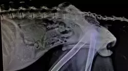
In general, definitive diagnoses of central nervous system diseases require advanced imaging tests (CT and MRI), cerebrospinal fluid analysis and histopathological study (among others). However, in the case of some conditions it is possible to make an approximation to the diagnosis and, sometimes, a definitive diagnosis in the clinic through the use of simple radiography and myelography, as a complement to the rest of the diagnostic tests. Therefore, its study is of great value to any veterinarians looking to improve their level of training.
This program focuses on the field of orthopedics and traumatology, taking into account that the bone is a complex tissue and requires specialized knowledge to understand the fundamental functions. However, through the radiological method, specialized knowledge of the different conditions that may affect it can be developed.
In short, it is a program based on scientific evidence and daily practice, with all the nuances that each professional can contribute, enriched by the critical assessment that every professional must keep in mind.
Throughout this course, the student will learn about all the current approaches to the different challenges posed by their profession. A high-level step that will become a process of improvement, not only on a professional level, but also on a personal level. In addition, TECH assumes a social commitment: to help the updating of highly qualified professionals and to develop their personal, social and labor skills during the development of the same. And, to do so, it will not only take you through the theoretical knowledge offered, but will show you another way of studying and learning which is more organic, simpler and more efficient. It works to maintain motivation and to create a passion for learning; it encourages thinking and the development of critical thinking.
With this Postgraduate diploma we give you the opportunity to acquire superior training in Radiological Diagnosis so that it can be applied to the veterinary clinic"
This Postgraduate diploma in Orthopedic and Neurological Radiological Diagnosis in Small Animals contains the most complete and up-to-date educational program on the market. The most important features of the program include:
- The development of case studies presented by experts in Veterinary Radiology
- The graphic, schematic, and eminently practical contents with which they are created, provide scientific and practical information on the disciplines that are essential for professional practice
- Latest developments in Veterinary Radiology
- Practical exercises where self-assessment can be used to improve learning
- Special emphasis on innovative methodologies in Veterinary Radiology
- Theoretical lessons, questions to the expert, debate forums on controversial topics, and individual reflection assignments
- Content that is accessible from any fixed or portable device with an Internet connection
At TECH we help you to specialize in Orthopedic and Neurological Radiological Diagnosis in a simple way, thanks to our innovative methodology"
TECH's teaching staff includes professionals belonging to the veterinary field, who contribute their work experience to this training, as well as renowned specialists from reference societies and prestigious universities.
The Multimedia Content, elaborated with the latest Educational Technology, will allow professionals a situated and contextual learning, that is to say, a simulated environment that will provide an immersive specialization programmed to train in real situations.
This program is designed around Problem Based Learning, whereby the specialist must try to solve the different professional practice situations that arise during the academic year. For this purpose, the professional will be assisted by an innovative system of interactive videos made by renowned and experienced experts in Veterinary Radiology.
Our specialization, of high academic level, will allow you to achieve superior training in a short period of time"

Our online format will allow you to study in a comfortable way from wherever you choose"
Syllabus
The contents of this Postgraduate diploma in Orthopedic and Neurological Radiological Diagnosis in Small Animals have been designed by a team of university experts, backed by their years of experience. In this way, they have been in charge of programming a totally up-to-date syllabus aimed at the 21st century professional, who demands high educational quality and knowledge of the main innovations in the field.

Our syllabus has been created following a criteria of excellence set by our own institution and demanded by today's society"
Module 1. Radiological Diagnosis in Neurology
1.1. Radiological Anatomy
1.1.1. Structures Assessable by Radiology
1.1.2. Normal Radiological Anatomy of the Spine
1.1.3. Normal Radiological Anatomy of the Skull and its Structures
1.2. Radiological Examination of the Spine
1.2.1. C1-C6
1.2.2. T1-T13
1.2.3. L1-L7
1.2.4. S1-Cd
1.3. Contrast Examination
1.3.1. Cisternal Myelography
1.3.2. Lumbar Myelography
1.3.3. Pathological Alterations Observed by Myelography
1.4. Diagnosis of Vascular Pathologies
1.4.1. Vascular Pathologies: How Far Can We Go with Conventional Radiology
1.4.2. Assessment of Vascular Pathologies by Contrast Techniques
1.4.3. Assessment of Vascular Pathologies by Other Imaging Techniques
1.5. Cerebral and Meningeal Malformations
1.5.1. Hydrocephalus
1.5.2. Meningocele
1.6. Inflammatory Pathology
1.6.1. Infectious
1.6.2. Non-infectious
1.6.3. Disc Spondylitis
1.7. Degenerative Pathologies
1.7.1. Degenerative Disc Disease
1.7.2. Wobbler Syndrome
1.7.3. Lumbosacral Instability, Cauda Equina Syndrome
1.8. Spiral Trauma
1.8.1. Pathophysiology
1.8.2. Fractures
1.9. Oncology
1.9.1. Primary Neoplastic Diseases
1.9.2. Secondary Metastatic Diseases
1.10. Other Neurological Diseases
1.10.3. Metabolic
1.10.4. Nutritional
1.10.5. Congenital
Module 2. Orthopedic Radiological Diagnosis I
2.1. The Growth Plate
2.1.1. Organization of the Growth Plate and its Impact on Radiological Imaging
2.1.2. Blood Supply of the Growth Plate
2.1.3. Structure and Function of the Growth Plate Cartilaginous Components
2.1.3.1. Reserve Zone
2.1.3.2. Proliferative Zone
2.1.3.3. Hypertrophic Zone
2.1.4. Bone Components (Metaphysis)
2.1.5. Fibrous and Fibrocartilaginous Components
2.1.6. Radiological Imaging of the Growth Plate at Different Stages of Growth
2.1.6.1. Epiphysiolysis
2.1.6.2. Other Growth Disorders
2.2. Fracture Repair
2.2.1. Radiological Response of Traumatized Bone
2.2.2. Phased Fracture Repair
2.2.2.1. Inflammatory Phase
2.2.2.2. Repair Phase
2.2.2.3. Remodelling Phase
2.2.2.4. Callus formation
2.2.2.5. Fracture Healing
2.2.2.6. First Intention Repair
2.2.2.7. Second Intention Repair
2.2.2.8. Clinical Union
2.2.2.9. Clinical Union Ranges
2.3. Fracture Complications
2.3.1. Delayed Union
2.3.2. Non-union
2.3.3. Bad Union
2.3.4. Osteomyelitis
2.4. Radiologic Imaging of Arthritis and Polyarthritis
2.4.1. Types of Arthritis and Polyarthritis
2.4.2. Clinical Diagnosis
2.4.3. Differential Diagnosis Radiology
2.5. Radiological Imaging of Osteoarthritis
2.5.1. Etiology
2.5.2. Radiological Diagnosis
2.5.3. Prognosis According to Radiological Imaging
2.6. Decision-making in Traumatology and Orthopedics Based on Radiologic Diagnosis
2.6.1. Fulfilled Clinical Function
2.6.2. Implant Ruptures
2.6.3. Implant Bends
2.6.4. Implant Migrates
2.6.5. Rejection
2.6.6. Infections
2.6.7. Thermal Interference
2.7. Radiology of Orthopedic Diseases
2.7.1. Radiology of Osteochondritis Dissecans
2.7.2. Panosteitis
2.7.3. Retained Cartilaginous Nucleus
2.7.4. Hypertrophic Osteodystrophy
2.7.5. Craniomandibular Osteopathy
2.7.6. Bone Tumors
2.7.7. Other Bone Diseases
2.8. Radiology of Hip Dysplasia
2.8.1. Physiological Hip Radiology
2.8.2. Pathological Hip Radiology
2.8.3. Gradation of Hip Dysplasia
2.8.4. Surgical Treatments for Hip Dysplasia
2.8.5. Clinical/Radiographic Progression of Hip Dysplasia
2.9. Radiology of Elbow Dysplasia
2.9.1. Physiological Elbow Radiology
2.9.2. Pathological Elbow Radiology
2.9.3. Types of Elbow Dysplasia
2.9.4. Surgical Treatments for Elbow Dysplasia
2.9.5. Clinical/Radiographic Progression of Elbow Dysplasia
2.10. Radiology of the Knee
2.10.1. Radiology of Anterior Cruciate Ligament Rupture
2.10.1.1. Surgical Treatment of Anterior Cruciate Ligament Rupture
2.10.2. Radiology of Patellar Dislocation
2.10.2.1. Gradation of Patellar Dislocation
2.10.2.2. Surgical Treatment of Patellar Dislocation
Module 3. Orthopedic Radiological Diagnosis II
3.1. Anatomy Radiology of the Pelvis
3.1.1. General Considerations
3.1.2. Radiologic Assessment of Stable Hip Fractures
3.1.3. Surgical Radiological Indication
3.1.3.1. Intra-articular Fracture
3.1.3.2. Closure of the Pelvic Canal
3.1.3.3. Joint Instability of a Hemipelvis
3.1.4. Fracture Separation of the Sacro-Iliac Joint
3.1.5. Fractures of the Acetabulum
3.1.6. Fracture of the Ilium
3.1.7. Ischial Fractures
3.1.8. Pubic Symphysis Fractures
3.1.9. Fractures of the Ischial Tuberosity
3.2. Radiological Imaging of Femur Fractures
3.2.1. Proximal Femoral Fractures
3.2.2. Fractures of the Medium Third of the Femur
3.2.3. Fractures of the Distal Third of the Femur
3.3. Radiological Imaging of Tibial Fractures
3.3.1. Fractures of the Proximal Third
3.3.2. Fractures of the Middle Third of the Tibia
3.3.3. Fractures of the Distal Third of the Tibia
3.3.4. Fractures of the Tibial Malleoli
3.4. Anterior Member
3.4.1. Radiological Imaging of the Scapula Fractures
3.4.2. Radiological Imaging of the Humerus Fractures
3.4.3. Radiological Imaging of the Radius and Ulnar Fractures
3.5. Fractures of the Maxilla and Mandible, Radiological Imaging of the Skull
3.5.1. Jaw Radiology
3.5.1.1. Rostral Jaw
3.5.1.2. Dental Radiology
3.5.1.3. Temporomandibular Joint (TMJ)
3.5.2. Radiology of the Maxilla
3.5.2.1. Dental Radiology
3.5.2.2. Radiology of the Maxilla
3.5.3. Radiology to the Paranasal Sinus
3.5.4. Radiology of the Skull
3.5.5. Oncology
3.6. Radiology of Fractures and Other Alterations Resulting in Incongruence of the Articular Surface
3.6.1. Fractures Affecting the Growth Nucleus
3.6.2. Classification of the Epiphysis Based on its Type
3.6.3. Classification of Slipped or Split Fractures Involving the Growth Nucleus and Adjacent Epiphyseal Metaphysis
3.6.4. Clinical Assessment and Treatment of Damage to Nucleus Growth
3.6.5. Radiology of Joint Fractures in Adult Animals
3.7. Joint Dislocations, Radiology
3.7.1. Radiological Positioning
3.7.2. Nomenclature
3.7.3. Traumatic Dislocations
3.7.4. Scapulohumeral Instability
3.8. Interventional Radiology in Traumatology
3.8.1. Radiology of the Fractures Affecting the Growth Nucleus
3.8.2. Radiology of Fractures Involving the Epiphysis based on Their Type
3.8.3. Radiology of Slipped or Split Fractures Involving the Growth Nucleus, Epiphysis and Adjacent Metaphysis
3.8.4. Radiology of Joint Fractures in Adult Animals
3.9. Radiology of Muscular, Tendinous and Ligamentous Diseases
3.9.1. Radiology of Muscular Diseases
3.9.2. Radiology of Tendinous and Ligamentous Diseases
3.9.3. Other Alternatives for Diagnostic Imaging of these Pathologies
3.10. Radiology of Metabolic and Nutritional Disorders
3.10.1. Introduction
3.10.2. Radiologic Imaging in Secondary Nutritional Hyperparathyroidism
3.10.3. Radiologic Imaging in Secondary Renal Hyperparathyroidism
3.10.4. Radiological Imaging in Hypervitaminosis A
3.10.5. Radiologic Imaging in Pituitary Dwarfism

Our syllabus has been created following the criteria of excellence set by our own institution and demanded by today's society"
Postgraduate Diploma in Orthopedic and Neurological Radiological Diagnosis in Small Animals
.
Veterinary professionals need to have effective tools to make accurate and early diagnoses in their animal patients, and Diagnostic Imaging tests are a fundamental part of their daily clinical practice. For this reason, a Postgraduate Diploma in Orthopedic and Neurological Radiological Diagnosis in Small Animals focused on radiological diagnosis in the field of Orthopedics, Traumatology and Neurology in small pets is presented.
Get up to date on the latest advances in this field by the hand of TECH
.
The Postgraduate Diploma in Orthopedic and Neurological Radiological Diagnosis in Small Animals is designed on the basis of scientific evidence and daily clinical practice, and has a team of experts in the field to ensure the quality of the preparation. Thus, participants will have access to high quality multimedia content, analysis of clinical cases, master classes and video techniques, which will allow them to get up to date with all the guarantees in Diagnostic Imaging in the veterinary field. In addition, the program is fully online, making it easy for them to organize their time and pace of study according to their schedules and needs.


