University certificate
The world's largest faculty of veterinary medicine”
Introduction to the Program
An intensive education in the surgery of corneal, crystalline, uveal and retinal lesions and conditions in small animal ophthalmology"

The cornea is one of the most exposed and visible areas, and any alteration can be detected very quickly. Each corneal component heals to a different degree, at a different speed and by completely different mechanisms. Understanding these differences will help us to identify if the repair is occurring abnormally, in order to intervene early and improve the prognosis of our patients.
This Postgraduate diploma develops specialized knowledge about the different diagnostic methods and their indications and includes learning the basic and necessary instruments for a complete ophthalmological examination. The complete ophthalmological examination will be approached starting with the anamnesis, the clinical history of the patient up to the different procedures that can be used to reach a correct diagnosis. We examine the most important procedures, tests and devices that facilitate accurate diagnosis.
In addition, the keys will be presented so that the student can approach one of the most complex phases of the ophthalmological examination: the identification of changes in color, edges and visual "texture", and their association to each corneal pathology and clinical relevance.
For all these reasons, this is the most complete Postgraduate diploma that students will find in the market, and it also includes an online methodology that will allow them to learn from the comfort of the place of their choice, without schedules and without time limits. Through state-of-the-art audiovisual content, this revolutionary methodology will turn the graduate into an Expert in Eye Surgery in Small Animals.
A world-renowned expert will join this educational itinerary. TECH has invited a renowned veterinary ophthalmology researcher to serve as the International Guest Director of this program. Likewise, this specialist is responsible for teaching a series of comprehensive Masterclasses where the main therapeutic and surgical advances in animal eye diseases are covered.
The International Guest Director of this program will update you on the most cutting-edge corneal healing technique in animals”
This Postgraduate diploma in Corneal, Crystalline Lens, Uveal and Retinal Surgery in Small Animals contains the most complete and up-to-date scientific program on the market. The most important features include:
- The development of case studies presented by experts in Veterinary Ophthalmology
- The graphic, schematic, and practical contents with which they are created, provide scientific and practical information on the disciplines that are essential for professional practice
- Practical exercises where self-assessment can be used to improve learning
- Its special emphasis on innovative methodologies
- Theoretical lessons, questions to the expert, debate forums on controversial topics, and individual reflection assignments
- Content that is accessible from any fixed or portable device with an Internet connection
Stand out from other professionals with the ability to care for small animal eye diseases”
The program’s teaching staff includes professionals from the industry who contribute their work experience to this program, as well as renowned specialists from leading societies and prestigious universities.
The multimedia content, developed with the latest educational technology, will provide the professional with situated and contextual learning, i.e., a simulated environment that will provide immersive education programmed to prepare for real situations.
This program is designed around Problem-Based Learning, whereby the professional must try to solve the different professional practice situations that arise during the course. For this purpose, the students will be assisted by an innovative interactive video system created by renowned and experienced experts.
A process of total quality growth that will allow you to specialize in a field of great interest and demand"

With an intensive and efficient process, this Postgraduate diploma will lead students to acquire theoretical and practical knowledge quickly and in a way that is compatible with other obligations"
Why study at TECH?
TECH is the world’s largest online university. With an impressive catalog of more than 14,000 university programs available in 11 languages, it is positioned as a leader in employability, with a 99% job placement rate. In addition, it relies on an enormous faculty of more than 6,000 professors of the highest international renown.

Study at the world's largest online university and guarantee your professional success. The future starts at TECH”
The world’s best online university according to FORBES
The prestigious Forbes magazine, specialized in business and finance, has highlighted TECH as “the world's best online university” This is what they have recently stated in an article in their digital edition in which they echo the success story of this institution, “thanks to the academic offer it provides, the selection of its teaching staff, and an innovative learning method aimed at educating the professionals of the future”
A revolutionary study method, a cutting-edge faculty and a practical focus: the key to TECH's success.
The most complete study plans on the university scene
TECH offers the most complete study plans on the university scene, with syllabuses that cover fundamental concepts and, at the same time, the main scientific advances in their specific scientific areas. In addition, these programs are continuously being updated to guarantee students the academic vanguard and the most in-demand professional skills. In this way, the university's qualifications provide its graduates with a significant advantage to propel their careers to success.
TECH offers the most comprehensive and intensive study plans on the current university scene.
A world-class teaching staff
TECH's teaching staff is made up of more than 6,000 professors with the highest international recognition. Professors, researchers and top executives of multinational companies, including Isaiah Covington, performance coach of the Boston Celtics; Magda Romanska, principal investigator at Harvard MetaLAB; Ignacio Wistumba, chairman of the department of translational molecular pathology at MD Anderson Cancer Center; and D.W. Pine, creative director of TIME magazine, among others.
Internationally renowned experts, specialized in different branches of Health, Technology, Communication and Business, form part of the TECH faculty.
A unique learning method
TECH is the first university to use Relearning in all its programs. It is the best online learning methodology, accredited with international teaching quality certifications, provided by prestigious educational agencies. In addition, this disruptive educational model is complemented with the “Case Method”, thereby setting up a unique online teaching strategy. Innovative teaching resources are also implemented, including detailed videos, infographics and interactive summaries.
TECH combines Relearning and the Case Method in all its university programs to guarantee excellent theoretical and practical learning, studying whenever and wherever you want.
The world's largest online university
TECH is the world’s largest online university. We are the largest educational institution, with the best and widest online educational catalog, one hundred percent online and covering the vast majority of areas of knowledge. We offer a large selection of our own degrees and accredited online undergraduate and postgraduate degrees. In total, more than 14,000 university degrees, in eleven different languages, make us the largest educational largest in the world.
TECH has the world's most extensive catalog of academic and official programs, available in more than 11 languages.
Google Premier Partner
The American technology giant has awarded TECH the Google Google Premier Partner badge. This award, which is only available to 3% of the world's companies, highlights the efficient, flexible and tailored experience that this university provides to students. The recognition as a Google Premier Partner not only accredits the maximum rigor, performance and investment in TECH's digital infrastructures, but also places this university as one of the world's leading technology companies.
Google has positioned TECH in the top 3% of the world's most important technology companies by awarding it its Google Premier Partner badge.
The official online university of the NBA
TECH is the official online university of the NBA. Thanks to our agreement with the biggest league in basketball, we offer our students exclusive university programs, as well as a wide variety of educational resources focused on the business of the league and other areas of the sports industry. Each program is made up of a uniquely designed syllabus and features exceptional guest hosts: professionals with a distinguished sports background who will offer their expertise on the most relevant topics.
TECH has been selected by the NBA, the world's top basketball league, as its official online university.
The top-rated university by its students
Students have positioned TECH as the world's top-rated university on the main review websites, with a highest rating of 4.9 out of 5, obtained from more than 1,000 reviews. These results consolidate TECH as the benchmark university institution at an international level, reflecting the excellence and positive impact of its educational model.” reflecting the excellence and positive impact of its educational model.”
TECH is the world’s top-rated university by its students.
Leaders in employability
TECH has managed to become the leading university in employability. 99% of its students obtain jobs in the academic field they have studied, within one year of completing any of the university's programs. A similar number achieve immediate career enhancement. All this thanks to a study methodology that bases its effectiveness on the acquisition of practical skills, which are absolutely necessary for professional development.
99% of TECH graduates find a job within a year of completing their studies.
Postgraduate Diploma in Cornea, Lens, Uvea and Retina Surgery in Small Animals
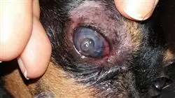
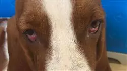
Corneal, lens, uveal and retinal surgery in small animals refers to a series of surgical procedures performed on the ocular structures of pets, such as dogs, cats or other small animals. Corneal surgery is performed to correct a variety of conditions, such as corneal ulcers, wounds, congenital defects, scarring or deformities. Corneal surgery procedures may include suturing or wetting the cornea to repair an injury, or removing diseased corneal tissue to treat diseases such as keratoconus. Crystalline lens surgery is used to correct cataracts, an opacity of the lens that can affect the animal's vision. The surgical procedure usually involves removing the diseased lens and replacing it with an intraocular lens to restore the animal's vision. Uveal surgery is performed to treat diseases of the uvea, the vascular layer that lies between the retina and the sclera of the eye. Uveal surgery procedures may include correction of retinal detachments or treatment of inflammation or tumors in the region of the uvea. Retinal surgery is performed to treat a variety of conditions, such as retinal detachments, macular holes, tumors or inflammation. Surgical procedures on the retina may include removal of diseased tissue or insertion of a prosthetic device to correct eye diseases or defects.
TECH, the world's largest online university, has the Postgraduate Diploma in Cornea, Lens, Uvea and Retina Surgery in Small Animals academic program, delivered completely online and designed to provide students with a thorough and comprehensive understanding of ocular anatomy in small animals, as well as specialized surgical techniques for the treatment of ocular diseases in the areas of the cornea, lens, uvea and retina. Students learn to diagnose and treat a wide variety of ocular conditions, and develop specialized clinical skills in small animal ophthalmic surgery.


