University certificate
The world's largest faculty of dentistry”
Why study at TECH?
If you want to update your knowledge in the field of dentistry, don't hesitate any longer. At TECH we offer you the most complete specialization in the market to reach a higher level of professionalism"
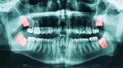
Gingival and periodontal diseases are among the most common human diseases. Gingivitis affects approximately 50% of school-age children and more than 70% of the adult population has suffered from gingivitis, periodontitis or both. In addition, it is estimated that periodontitis is responsible for 30-35% of all tooth extractions, while caries and its sequelae account for 50%.
With these data, it is not surprising how important it is for dental professionals to have extensive knowledge in this field, since all surgery, no matter how small, must be carried out following certain protocols that are fundamental for the good short and long term results of the surgery.
It should also be noted that, in recent years, dentistry, and periodontics and osseointegration in particular, have undergone enormous changes, with an increase in the number of patients coming to dental clinics seeking treatments that restore optimal oral health conditions, not only from a functional but also from an esthetic point of view.
Throughout this specialization, the student will learn all of the current approaches to the different challenges posed by their profession. A high-level step that will become a process of improvement, not only on a professional level, but also on a personal level. We will not only take you through the theoretical knowledge, but we will show you another way of studying and learning, more organic, simpler and more efficient.
This Advanced master’s degree is designed to give you access to the specific knowledge of this discipline in an intensive and practical way. A great value for any professional. Furthermore, as it is a 100% online specialization, the student decides where and when to study. Without the restrictions of fixed timetables or having to move between classrooms, this course can be combined with work and family life.
A high level scientific training program, supported by advanced technological development and the teaching experience of the best professionals"
This Advanced master’s degree in Periodontics, Implantology and Oral Surgery contains the most comprehensive and up-to-date academic course on the university scene. The most important features of the program include:
- The latest technology in online teaching software
- A highly visual teaching system, supported by graphic and schematic contents that are easy to assimilate and understand
- Practical cases presented by practising experts
- State-of-the-art interactive video systems
- Teaching supported by remote training
- Continuous updating and retraining systems
- Self organised learning which makes the course completely compatible with other commitments
- Practical exercises for self-evaluation and learning verification.
- Support groups and educational synergies: Questions to the expert, discussion forums and knowledge
- Communication with the teacher and individual reflection work
- Content that is accessible from any, fixed or portable device with an Internet connection.
- The banks of complementary documentation are permanently available, even after the program has been completed.
This Advanced master’s degree may be the best investment you can make when choosing a refresher program for two reasons: in addition to updating your Dentistry knowledge, you will obtain a diploma from TECH Global University"
Our teaching staff is made up of working professionals. In this way, we ensure that we provide you with the up-to-date training we are aiming for. A multidisciplinary team of doctors with training and experience in different environments, who will develop the theoretical knowledge in an efficient way, but above all, they will bring their practical knowledge from their own experience to the course.
The efficiency of the methodological design of this Advanced master’s degree enhances the student's understanding of the Advanced master’s degreee. Developed by a multidisciplinary team of e-learning experts, it integrates the latest advances in educational technology. In this way, you will be able to study with a range of easy-to-use and versatile multimedia tools that will give you the necessary skills you need for your specialization.
The design of this program is based on Problem-Based Learning, an approach that conceives learning as a highly practical process. To achieve this remotely, we will use telepractice learning. With the help of an innovative interactive video system, and learning from an expert, you will be able to acquire the knowledge as if you were actually dealing with the scenario you are learning about. A concept that will allow you to integrate and fix learning in a more realistic and permanent way.
A training program created for professionals who aspire to excellence that will allow you to acquire new skills and strategies in a smooth and effective way"
We offer you the best specialization of the moment so that you can carry out a deep study in this field, in such a way that you will be able to develop your profession with total guarantees of success"
Syllabus
The contents of this Periodontics, Implantology and Oral Surgery have been developed by the different experts on this course, with a clear purpose: to ensure that our students acquire each and every one of the necessary skills to become true experts in this field. The content of this course enables you to learn all aspects of the different disciplines involved in this field. A complete and well-structured program that will take you to the highest standards of quality and success.
Through a very well compartmentalized development, you will be able to access the most advanced knowledge of the moment to achieve professional success"
Module 1. Basic Periodontics
1.1. Anatomy of the Periodontium
1.1.1. Gingiva: Keratinized, Free, Inserted, Interdental
1.1.2. Alveolar Mucosa
1.1.3. Periodontal Ligament
1.1.4. Root Cement
1.1.5. Alveolar Bone
1.1.6. Blood, Lymphatic and Nervous System of the Periodontium
1.1.7. Periodontal Biotypes
1.1.8. Biological Space
1.2. Epidemiology of Periodontal Disease
1.2.1. Prevalence of Periodontal Diseases
1.2.2. Risk Factors for Periodontitis
1.2.3. Periodontal Diseases and Their Relation to Systemic Diseases
1.3. Microbiology of Periodontal Disease
1.3.1. Biofilm and Dental Calculus Microbiological and Clinical Aspects
1.3.2. Periodontal Infections
1.3.3. Periodontal Pathogens
1.3.4. Bacterial Plaque and Biofilm Disease Onset and Progression
1.4. Host-Parasite Interaction
1.4.1. Disease Onset and Progression
1.4.2. Pathogenesis of Periodontitis
1.4.3. Host-Parasite Interaction
1.5. Factors Associated with Periodontal Disease
1.5.1. Diabetes Mellitus
1.5.2. Puberty, Pregnancy, Menopause
1.5.3. Tobacco use
Module 2. Periodontal Diseases
2.1. Non-Plaque-Induced Inflammatory Gingival Lesions
2.1.1. Gingival Diseases of Bacterial Origin
2.1.2. Gingival Injuries of Viral Origin
2.1.3. Gingival Diseases of Mycotic Origin
2.1.4. Gingival Diseases of Genetic Origin
2.1.5. Gingival Diseases of Systemic Origin
2.1.6. Trauma Lesions
2.2. Plaque-Induced Gingival Lesions
2.2.1. Classification of Gingival Diseases
2.2.2. Plaque-Induced Gingivitis
2.2.3. Gingival Diseases Associated with Medications
2.2.4. Gingival Diseases Associated with Systemic Diseases
2.3. Chronic Periodontitis
2.3.1. General and Clinical Characteristics
2.3.2. Susceptibility and Progression
2.3.3. Risk factors
2.4. Aggressive Periodontitis
2.4.1. Classification
2.4.2. Etiology and Pathogenesis
2.4.3. Microbiological
2.4.4. Therapeutic Principles
2.5. Necrotizing Ulcerative Periodontal Disease
2.5.1. General and Clinical Characteristics Classification
2.5.2. Etiology and Pathogenesis
2.5.3. Microbiological
2.5.4. Therapeutic Principles
2.6. Periodontal Abscess
2.6.1. Introduction
2.6.2. Classification
2.6.3. Etiology, Pathogenesis, Histopathology and Microbiology
2.6.4. Microbiological
2.6.5. Pediatric Dentistry
2.7. Lesion of Endodontic Origin
2.7.1. Introduction
2.7.2. Classification
2.7.3. Etiology, Pulp Pathogenesis and Microbiology
2.7.4. Microbiological
2.7.5. Effects of Periodontal Treatment on the Pulp
2.7.6. Pediatric Dentistry
2.8. Halitosis
Module 3. Examination, Diagnosis and Treatment Plan
3.1. Anamnesis of the Patient with Periodontal Disease
3.1.1. Dental History, Social, Family, Smoking, Hygiene Habits, etc.
3.1.2. Oral Hygiene Status
3.1.3. Signs and Symptoms of Periodontal Disease: Gingiva, Periodontal Ligament and Alveolar Bone
3.2. Intraoral and Radiographic Examination
3.2.1. Intraoral Examination: Periodontogram
3.2.2. X-ray Examination: Periapical Radiographic Series
3.2.3. Screening for Periodontal Disease
3.3. Microbiological
3.3.1. Diagnosis of Periodontal Lesions
3.3.2. Gingivitis
3.3.3. Mild Periodontitis
3.3.4. Moderate or Advanced Periodontitis
3.4. Treatment Plan
3.4.1. Initial Treatment Plan
3.4.2. Pretherapeutic Prognosis
3.4.3. Re-evaluation
3.4.4. Corrective or Reconstructive Therapy
3.4.5. Maintenance Therapy
Module 4. Basic Non-Surgical Periodontal Treatment Initial Phase
4.1. Mechanical Control of Supragingival Plaque
4.1.1. Plaque Control: Brushing and Interdental Cleaning. Techniques
4.1.2. Instruction and Motivation in Plaque Control
4.2. Chemical Control of Supragingival Plaque Use of Antiseptics in Periodontics
4.2.1. Chemical Control Concept, Agents, Mechanisms of Action and Drivers
4.2.2. Chemical Plaque Control Agent Classification
4.2.3. Chlorhexidine: Toxicity, Pigmentation, Mechanism of Action, Clinical Use
4.3. Non-Surgical Treatment of Periodontal Disease
4.3.1. Calculus Detection and Removal
4.3.2. Debridement Techniques. Mechanical and Manual
4.3.3. Postoperative Care and Control of Tooth Sensitivity
4.4. Pharmacological Treatment. Use of Antibiotics in Periodontics
4.4.1. Principles of Antibiotics Therapy Specific Characteristics and Limitations
4.4.2. Evaluation of Antimicrobials for Periodontal Therapy
4.5. Re-evaluation
4.5.1. Interpretation of Results Treatment Evaluation
4.6. Periodontal Maintenance
4.6.1. Risk Assessment: Patient, Tooth, Progression
4.6.2. Objectives of Maintenance in Gingivitis and Periodontitis
4.6.3. Continuous Review and Reassessment
4.6.4. Motivation
Module 5. Surgical Periodontal Treatment Periodontal Surgery Access Therapy
5.1. Periodontal Pocket Reduction Techniques
5.1.1. Gingivectomy
5.1.2. Widman’s Flap
5.1.3. Modified Widman’s Flap
5.1.4. Neumann’s Flap
5.1.5. Apical Repositioning Flap
5.1.6. Papilla Preservation Flap
5.1.7. Distal Wedge Flap
5.1.8. Bone Resective Surgery: Osteoplasty and Ostectomy
5.2. General Guidelines in Periodontal Surgery
5.2.1. Objectives of Surgical Treatment
5.2.2. Indications for Surgical Treatment
5.2.3. Contraindications for Surgical Treatment
5.2.4. Anesthesia in Periodontal Surgery
5.2.5. Instruments in Periodontal Surgery
5.2.6. Root Surface Treatment
5.2.7. Suture in Periodontal Access Surgery
5.2.8. Periodontal Dressings
5.2.9. Pain Control and Postoperative Care
Module 6. Periodontal Restorative Treatment I: Periodontal Regeneration GTR
6.1. Basic Principles of Regeneration
6.1.1. Introduction: Reintegration, New Insertion, Regeneration
6.1.2. Indications for Regenerative Periodontal Surgery
6.1.3. Assessment of Periodontal Regeneration: Probing, Radiographic and Histological
6.1.4. Periodontal Wound Healing Regenerative Capabilities
6.1.4.1. Bone Cells
6.1.4.2. Gingival Connective Tissue
6.1.4.3. Periodontal Ligament
6.1.4.4. Epithelium
6.2. Regenerative Procedures
6.2.1. Scaling and Root Smoothing and-Needle Flap Surgeries
6.2.2. Grafting - Regeneration Procedures
6.2.2.1. Autogenous Grafts
6.2.2.2. Allografts
6.2.2.3. Xenografts
6.2.2.4. Alloplastic Materials
6.2.3. Root Surface Biomodification
6.2.4. Membranes in Periodontal Regeneration Barrier Function
6.2.5. Amelogenins in Periodontal Regeneration
6.3. Guided Tissue Regeneration (GTR)
6.3.1. Clinical Application of GTR Infraosseous Defects
6.3.2. GTR Technique Guidelines
6.3.2.1. Design of the Flap
6.3.2.2. Characteristics of the Defect to be Treated
6.3.2.3. Preparation of the Defect
6.3.2.4. Suture of the Membranes
6.3.2.5. Flap Closure
6.3.2.6. Postoperative Indications
6.3.3. Influencing Factors: Patient, Defect, Technique and Healing
6.3.4. Barrier Materials in GTR
6.3.5. Resorbable Membranes
Module 7. Reconstructive Periodontal Treatment II: Periodontal Surgery Treatment of Furcation Lesions
7.1. Furcations Concept and Anatomy
7.1.1. Upper Molars
7.1.2. Upper Premolars
7.1.3. Lower Molars
7.2. Microbiological
7.2.1. Periodontogram
7.2.2. Radiographic Tests
7.3. Pediatric Dentistry
7.3.1. Grade I Furcation Lesions
7.3.2. Grade II Furcation Lesions
7.3.3. Grade III Furcation Lesions
7.3.4. Plastics of Furcation
7.3.5. Furcation Tunneling
7.3.6. Radectomy
7.3.7. Regeneration of Furcation Lesions
7.3.8. Extraction
7.4. Prognosis of Furcation Lesions
Module 8. Periodontal Reconstructive Treatment III: Periodontal and Mucogingival Plastic Surgery Basic Principles
8.1. Etiopathogenesis and Prevalence of Mucogingival Disorders
8.1.1. Eruption Pattern
8.1.2. Fenestration and Dehiscence
8.1.3. Precipitating and Predisposing Factors
8.1.4. Prevalence of Gingival Recession
8.2. Diagnosis and Indications in Mucogingival Surgery
8.2.1. Diagnosing a Mucogingival Problem
8.2.2. Performance Criteria in Pediatric, Young and Adult Patients
8.3. Gingival Recession
8.3.1. Classification
8.4. Prognosis and Predetermination in Root Canal Veneering
8.5. Surgical Technique Selection
8.5.1. Criteria for Choosing a Surgical Technique
8.5.2. Anatomical Factors that Affect Prognosis
8.5.3. Scientific Evidence
8.5.4. Variables to be Taken Into Account Depending on the Technique
8.6. Root Surface Treatment
8.7. Amelogenins in Mucogingival Surgery
8.8. Surgical Principles in Periodontal Plastic Surgery
8.8.1. Incisions and Bevels
8.8.2. Flaps
8.9. Sutures, Surgical Instruments and Postoperative Care
8.9.1. Sutures, Materials, Characteristics, Knots and Suturing Techniques
8.9.2. Surgical Instruments in Mucogingival Surgery
8.9.3. Post-Operative Care
Module 9. Periodontal Reconstructive Treatment IV: Periodontal and Mucogingival Plastic Surgery Autografts and Displaced Flaps for Root Resurfacing
9.1. Epithelialized Free Autograft
9.1.1. Basic Principles
9.1.1.1. Indications and Contraindications
9.1.1.2. Advantages and Disadvantages
9.1.1.3. Phases when Performing Epithelialized Autografts
9.1.1.4. Donor Site Treatment
9.1.1.5. Nourishment and Healing of the Graft and Donor Site
9.1.1.6. Immediate postoperative complications
9.1.2. Step-by-Step Technique
9.1.2.1. Prophylactic Autograft
9.1.2.2. Therapeutic Autograft
9.1.2.3. Technique for Obtaining an Epithelialized Graft
9.1.2.4. Creeping Attachment
9.2. Displaced Flaps Indications, Advantages and Disadvantages and Technique
9.2.1. Coronal Displaced Flap (Single or Multiple)
9.2.2. Multiple Coronal Displaced Flap with No Offloading
9.2.3. Laterally Displaced and Coronally Advanced Flap
9.2.4. Semilunar Flap
9.2.5. Bipediculated Flap
Module 10. Periodontal Reconstructive Treatment V: Periodontal and Mucogingival Plastic Surgery Bilaminar Techniques for Root Canal Veneering
10.1. Introduction to Bilaminar Techniques
10.1.1. Indications, Contraindications, Advantages, Disadvantages, Classification, Total-Partial Thicknesses
10.2. Surgical Techniques for Obtaining Connective Tissue Grafts
10.2.1. Characteristics of the Palatal Fibromucosa
10.2.2. Trap-door Technique (Three Incisions)
10.2.3. “I" Technique (Two Incisions)
10.2.4. Envelope Technique (One Incision)
10.2.5. De-Epithelialized Epithelial-Connective Tissue Grafting Technique
10.3. Connective Tissue Grafts Associated with Displaced Flaps
10.3.1. Coronal Displaced Flap Associated with Subepithelial Connective Tissue Grafting
10.3.2. Multiple Coronal Non-Discharged Displaced Flap Associated with Subepithelial Connective Tissue Grafting
10.3.3. Lateral Displaced Flap Associated with Subepithelial Connective Tissue Grafting
10.3.4. Bipedicled Flap Associated with Subepithelial Connective Tissue Grafting
10.4. Pocket or Envelope Connective Tissue Grafting and Tunneling
10.4.1. Indications, Contraindications, Advantages and Disadvantages
10.4.2. Techniques
10.5. Biomaterial Substitutes for Autologous Grafts
10.5.1. Soft Tissue Allografts and Xenografts
10.5.2. Indications, Contraindications, Advantages and Disadvantages
10.5.3. Types, Characteristics and Handling
Module 11. Periodontal Reconstructive Treatment VI: Periodontal and Mucogingival Plastic Surgery Corrective Plastic Surgery
11.1. Surgical Lengthening of the Dental Crown
11.1.1. Coronary Lengthening for Prosthodontic Reasons
11.1.2. Multiple Coronary Elongation for the Treatment of APE
11.1.2.1. Altered Passive Eruption
11.1.2.2. APE Treatment
11.1.2.3. Apically Displaced Flap with Vestibular Osteoplasty
11.1.2.4. Apically Displaced Flap with Vestibular Osteoplasty
11.2. Frenulum Surgery
11.2.1. Upper Labial Frenulum Surgery
11.2.2. Lower Labial Frenulum Surgery
11.3. Vestibular Plastic Surgery Vestibuloplasty
11.3.1. Vestibuloplasty
11.3.2. Vestibuloplasty Associated with Grafting
11.4. Treatment of Cervical Abrasions and Caries Associated with Gingival Recession
11.5. Treatment of Gingival Clefts
11.6. Composite Restorative Treatment in Conjunction with Surgical Root Canal Veneering
11.7. Treatment of Alveolar Ridge Defects Using Soft Tissue Augmentation
11.7.1. Etiology and Classification of Alveolar Ridge Defects
11.7.2. Surgical Techniques for Volume and Keratinized Gingival Augmentation
Module 12. Mucogingival Surgery in Implant Dentistry
12.1. Morphologic Differences Between Periodontal and Peri-Implant Soft Tissues
12.1.1. Morfoligical
12.1.2. Vascularization
12.2. Influence of Gingival Biotype and Keratinized Gingiva in Implant Dentistry
12.2.1. Fine Biotype in Implant Dentistry
12.2.2. Coarse Biotype in Implant Dentistry
12.2.3. Risk Areas Implant-Soft Tissue Junction
12.2.4. Keratinized Gingiva Vs. Mucosa
12.3. Tissue Reconstruction Simultaneous to Implant Placement
12.3.1. Tissue Reconstruction Simultaneous to Implant Placement immediately After an Extraction
12.3.1.1. Clinical Benefits Vs. Biological Limitations
12.3.2. Tissue Reconstruction Simultaneous to Implant Placement Delayed After an Extraction
12.4. Delayed Tissue Reconstruction is After Placing an Implant
12.4.1. Delayed Tissue Reconstruction After an Implant Placement During Surgical Reopening - Second Phase
12.4.2. Delayed Tissue Reconstruction After Placing an Implant Approach to Esthetic Implant Failure
12.5. Surgical Techniques
12.5.1. Alveolar Ridge Preservation Techniques
12.5.1.1. Collagen Matrix
12.5.1.2. Alveolar Sealing by Free Grafting
12.5.1.3. Alveolar Sealing by Pedicle Grafting of the Palate
12.5.1.4. Temporary Alveolar Sealing (Bio-Col)
12.5.1.5. Combined Soft-Tissue-Bone Graft Tuber-Trephine Technique
12.5.2. Surgical Techniques for Obtaining Keratinized Gingiva Over Implants
12.5.2.1. Palatal to Vestibular Fibromucosa Displacement
12.5.2.2. Interproximal Pedicles
12.5.2.3. Vestibular Pocket Pedicles
12.5.2.4. Free Grafting on Implants
12.5.3. Surgical Techniques to Obtain Connective Tissue Volume
12.5.3.1. Envelope Connective Tissue Grafting
12.5.3.2. Pedicle Graft of the Palate
Module 13. Periimplantitis
13.1. Structural Differences Between Peri-Implant and Periodontal Tissues
13.1.1. Tooth-Gum Vs. Implant-Gum Interface
13.1.2. Connective Tissue
13.1.3. Vascularization
13.1.4. Biological Space
13.1.5. Microbiology
13.2. Mucositis
13.3. Mucositis Vs. Periimplantitis
13.4. Peri-Implantitis
13.4.1. Risk factors
13.5. Treatment of Peri-Implant Diseases
13.5.1. Mucositis Treatment
13.5.2. Peri-Implantitis Treatment
13.5.3. Non-surgical Treatment
13.5.4. Surgical Management
13.6. Maintenance of Peri-Implant Diseases
Module 14. Periodontics and Endodontics
14.1. Interactions Between Pulpal Disease and Periodontal Disease
14.2. Anatomic Considerations
14.2.1. Dentinal Tubules
14.2.2. Apical Foramen
14.2.3. Periodontium
14.2.4. Interactions of the Disease
14.3. Etiology
14.3.1. Bacteria
14.3.2. Fungi
14.3.3. Virus
14.3.4. Other Pathogens: Intrinsic and Extrinsic
14.4. Contributing Factors
14.4.1. Incorrect Endodontic Treatment
14.4.2. Incorrect Restorations
14.4.3. Trauma
14.4.3.1. Enamel Fracture
14.4.3.2. Crown Fractures without Pulp Exposure
14.4.3.3. Crown Fractures with Pulp Exposure
14.4.3.4. Coronoradicular Fracture
14.4.3.5. Root Fracture
14.4.3.6. Dislocation
14.4.3.7. Avulsion
14.4.4. Perforation
14.4.5. Dental Malformation
14.5. Differential Diagnosis
14.5.1. Endodontic Lesions
14.5.2. Periodontal Injuries
14.5.3. Combined Injuries
14.5.3.1. Primary Endodontic Lesions with Secondary Periodontal Involvement
14.5.3.2. Primary Periodontal Lesions with Secondary Periodontal Involvement
14.5.3.3. Concomitant Lesion: Independent or Communicated
14.6. Prognosis
Module 15. Periodontics, Orthodontics and Occlusion
15.1. Indications and Contraindications for Orthodontic Treatment in the Periodontal Patient
15.1.1. Indications
15.1.2. Contraindications
15.1.3. Orthodontic Planning in the Periodontal Patient
15.2. Advantages and Disadvantages of Orthodontic Forces in the Patient with Controlled Periodontitis
15.3. Biological Considerations
15.3.1. Periodontal and Bone Response to Normal Function
15.3.2. Structure and Function of the Periodontal Ligament
15.3.3. Response of the Periodontal Ligament and Alveolar Bone to Maintained Orthodontic Forces
15.3.4. Biological Control of Tooth Movement - Bioelectrical and Pressure-Voltage Theory
15.3.5. Orthodontic Basics: Center of Resistance, Center of Rotation, Controlled Forces, Force-Transfer, Anchorage
15.4. Orthodontic Tooth Movement in Patients with Periodontal Tissue Destruction
15.4.1. Considerations
15.4.2. Tooth Movement into Infraosseous Pockets
15.4.3. Types of Orthodontic Movements and Their Influence on Periodontal Teeth
15.5. Symptomatology of Trauma due to Occlusion
15.5.1. Angular Bone Defects
15.5.2. Increased Tooth Mobility
15.6. Treatment of Increased Tooth Mobility
15.6.1. Classification According to the Degree of Mobility, Periodontal Ligament Status and Alveolar Bone Status
15.6.2. Treatment of Tooth Mobility
Module 16. Laser in Periodontics
16.1. Introduction to the Laser
16.1.1. History of the Laser
16.1.2. Low-Power Laser
16.1.3. High-Power of Surgical Laser
16.1.4. Laser Safety
16.2. Types of Laser Features
16.2.1. Diode Laser
16.2.2. Erbium Laser
16.3. Indications and Applications of Lasers in Periodontics
16.3.1. As a Stand-Alone Treatment
16.3.2. As a Complement to Conventional Treatment
16.4. Laser Therapy - Photobiomodulation
Module 17. Maintenance of Periodontal and Implant Dentistry Patients
17.1. Maintenance of Periodontal Patients
17.1.1. Periodontal Maintenance in Patients with Gingivitis
17.1.2. Periodontal Maintenance in Patients with Periodontitis
17.1.3. Objectives of Periodontal Maintenance Therapy
17.1.4. Risk Assessment
17.1.5. Periodontal Maintenance Therapy in the Clinic
17.1.5.1. Examination, Reassessment and Diagnosis
17.1.5.2. Motivation, Reinstruction and Instrumentation
17.1.5.3. Site-Specific Treatment
17.1.5.4. Establishing Periodic Maintenance Intervals
17.2. Maintenance of Implant Patients
17.2.1. Maintenance of Patients with Dental Implants
17.2.2. Objectives of Implant Dentistry Maintenance Therapy
17.2.3. Diagnosis of the Peri-Implant Problem
17.2.3.1. Bleeding, Suppuration, Probing Depth, Radiographic Interpretation, Mobility
17.2.4. Preventive and Therapeutic Strategies
Module 18. Microbiological
18.1. Clinical History: First Visit, Anamnesis and Patient's Expectations
18.2. Medical Assessment of the Surgical Patient
18.2.1. Complementary Tests in Implantology and Oral Surgery
18.3. Patient With Diseases of Risk in Implant Dentistry and Surgery: Medical Considerations and Dental Management
18.3.1. Diabetic Patients
18.3.2. Immunosuppressed Patients
18.3.3. Patients Taking Anticoagulants
18.3.4. The Medically Compromised Patient: Bisphosphonates
18.4. Anaesthetic Techniques in Surgery and Implantology
18.4.1. Drugs:
18.4.2. Loco-regional Anaesthesia Techniques in Surgery and Implantology
18.5. Sedation and General Anaesthesia
Module 19. Oral Surgery Pathology
19.1. Tooth Retention
19.1.1. Concept, Etiology and Possible Treatment
19.2. Third Molar Included
19.2.1. Pathology and Clinical Manifestations
19.2.2. Diagnosis and Treatment
19.3. Pathology and Treatment of Included Canines
19.3.1. Microbiological
19.3.2. Surgical Management
19.3.3. Surgical-Orthodontic Treatment
19.4. Pre-prosthetic Surgery Techniques on Soft and Hard Tissue
19.4.1. Laser in Oral Surgery
19.4.2. Types of Laser in Oral Surgery
19.5. Periapical Surgery
19.5.1. Materials
19.5.2. Techniques
Module 20. Implant Planning
20.1. Extraoral and Intraoral Examination
20.1.1. Extraoral Examination: Symmetry, Facial Thirds, Extraoral Aesthetic Parameters
20.1.2. Intraoral Examination: Hard Tissue, Soft Tissue, Occlusion and TMJ
20.2. Impression Taking and Study Models in Implantology
20.2.1. Materials and Impression Techniques in Implant Diagnosis
20.2.2. Facebow and Mounting on a Semi-Adjustable Articulator
20.3. Diagnostic Wax-Up and Radiological Splints
20.3.1. Waxing Techniques and Clinical Considerations
20.3.2. Radiological Splints: Classification and Laboratory Manufacturing
20.4. Radiological Diagnosis in Implantology
20.4.1. Classification of Techniques
20.4.2. Planning in 2D
20.4.3. Cone Beam Computed Tomography (CBCT): Planning Software
20.5. Photographic Records in Implantology
20.6. Presentation of a Treatment Plan Strategies
Module 21. Implantology and Osseointegration
21.1. Historical Review and Generic Terminology of Dental Implants
21.1.1. Evolution of Implantology up to the 21st Century
21.1.2. Generic Terminology of Dental Implants: Components and Nomenclature
21.2. Biology of Osseointegration
21.2.1. Inflammatory Phase
21.2.2. Proliferative Phase
21.2.3. Maturation Phase
21.2.4. Contact and Remote Osteogenesis
21.3. Anatomy in Implantology
21.3.1. Anatomy of the Upper Jaw
21.3.2. Anatomy of the Mandible
21.4. Histology of Bone Tissue, Periodontium and Peri-implant Tissue
21.5. Bone Availability in Implantology
21.6. Preparation of the Surgical Field, Sterilization and Premedication Protocols
21.6.1. Table Preparation
21.6.2. Surgical Asepsis of the Patient: Premedication
21.6.3. Surgical Asepsis of the Surgeon and Assistants
Module 22. Basic Surgical Technique and Implantology
22.1. Incision Techniques in Implantology
22.1.1. Incisions in a Total Edentulous Patient
22.1.2. Incisions in a Partial Edentulous Patient
22.1.3. Incisions in the Aesthetic Sector
22.1.4. Incisions in Bone Guided Regeneration Techniques
22.1.5. Flapless
22.2. Surgical Instruments Detachment, Separation and Bone Regulation
22.3. Drilling Techniques in Implantology
22.3.1. Drills and Components of the Surgical Trays
22.3.2. Sequential Drilling
22.3.3. Biological Drilling
22.4. Single-stage Implants and Two-stage Implants
22.5. Sutures in Implantology
22.5.1. Suture Instruments and Materials
22.5.2. Suture Techniques
Module 23. Biomaterials and Bone Guided Regeneration
23.1. Types of Bone Grafts and Biological Mechanisms of Bone Formation
23.1.1. Classification, Advantages and Disadvantages
23.1.2. Osteogenesis, Osteoconduction and Osteoinduction
23.2. Autologous Bone Grafts: Chin and Mandibular Ramus
23.3. Other Biomaterials in Bone Regeneration
23.3.1. Homologous Grafts
23.3.2. Heterologous Grafts
23.3.3. Alloplastic Grafts
23.3.4. Plasma Which Is Rich in Growth Factors
23.4. Membranes and Bone Guided Regeneration
23.4.1. Non-resorbable Membranes
23.4.2. Resorbable Membranes
Module 24. Maxillary Sinus Lift
24.1. Diagnosis and Anatomical Recall of the Sinus Lift
24.2. Sinus Lift Technique Via the Crestal Approach
24.2.1. Sinus Lift with Osteotome Technique
24.2.2. Minimally Invasive Crestal Sinus Lift
24.2.2.1. Atraumatic Drilling Kits
24.2.2.2. Balloon Technique
24.3. Sinus Lift Technique Via the Lateral Approach
24.3.1. Step - by - Step Description of the Technique
24.3.2. Piezoelectric Systems
24.3.3. Biomaterials in Maxillary Sinus Elevation
Module 25. Immediate Implantology
25.1. Post-extraction Implants
25.1.1. Surgical Aspects of Immediate Implants
25.1.1.1. Immediate Implant
25.1.1.2. Early Implant Placement
25.2. Immediate Implants in Posterior Sectors
25.3. Immediate Aesthetic
25.3.1. Emergency Profile Transmission
25.3.2. Immediate Provisional
Module 26. Advanced Surgical Techniques in Implantology
26.1. Crest Expansion
26.1.1. Crest Expansion with Manual Instruments
26.1.2. Crest Expansion with Motorized Instruments
26.2. Pterygoid Implants
26.3. Zygomatic Implants
26.4. Treatment with Dental Implants without Grafts
26.4.1. Short Implants
26.4.2. Narrow Implants
26.4.3. Angled Implants
Module 27. Periodontics Applied to the Treatment of the Implantology Patient
27.1. Basic Concepts of Periodontics Applied to a Patient With Implants
27.1.1. Peridontal Diagnosis
27.1.2. Prognosis and Treatment Plan
27.2. Mucogingival Procedures to Increase Keratinized Tissue
27.2.1. Free Gingival Grafting
27.2.2. Bilaminar Grafts
27.3. Mucogingival Procedures to Increase the Volume of Connective Tissue
27.3.1. Subepithelial Free Grafts
27.3.2. Pedicled Grafts
27.4. Alveolar Ridge Preservation Techniques
27.5. Implant Maintenance
27.5.1. Hygiene Techniques
27.5.2. Revisions and Maintenance in Implantology
Module 28. Implant Prosthesis
28.1. Restoration as a Guide to Global Implantology Treatment
28.1.1. Nomenclature
28.2. Impression Taking in Implantology Work Models
28.2.1. Impression Materials in Implantology
28.2.2. Impression Techniques: Open or Closed Cuvette Impressions
28.2.3. Pouring Impressions and Obtaining the Working Model
28.3. Selection of Abutments in Implantology
28.3.1. Preformed Abutments
28.3.2. Calcinable Abutments
28.3.3. Cad-Cam Abutments
28.3.4. Direct Prosthesis to Implant or on Transepithelials
28.4. Materials for Implant Prosthesis
28.4.1. Porcelain Metal Prostheses
28.4.2. Resin Metal Prostheses
28.4.3. Zirconium Prosthesis
28.5. Screw-Retained Versus Cemented Prostheses
28.5.1. Indications
28.5.2. Advantages and Disadvantages
28.6. Color Acquisition
28.6.1. Color Map, Color Guides and Colorimeters
28.6.2. Color Acquisition Technique
28.7. Clinical Sequence for Implant Prosthetics on Single Crowns and Partial Bridges
Module 29. Implant Prosthesis in a Totally Edentulous Patient
29.1. Treatment Options for a Totally Edentulous Patient
29.1.1. Key Positions of Implants
29.2. Removable Complete Restorations
29.2.1. Concept
29.2.2. Overdenture with Single Anchors
29.2.3. Overdentures on Bars
29.2.4. Clinical Sequence of Implant Prostheses in Totally Edentulous Patients Treated with Overdentures
29.3. Complete Fixed Restorations with Hybrid Prosthesis
29.3.1. Concept
29.3.2. Materials: Metal– Composite and Metal– Resin Fixed Prosthesis
29.3.3. Clinical Sequence of Implant Prostheses in Totally Edentulous Patients Treated with Hybrid Prosthesis
29.4. Complete Fixed Restorations with Fixed Prosthesis
29.4.1. Concept
29.4.2. Metal-Porcelain– Zirconium
29.4.3. Clinical Sequence of Implant Prostheses in Totally Edentulous Patients Treated with Fixed Prosthesis
Module 30. Implant Prosthesis in the Anterior Aesthetic Sector
30.1. Problems of the Anterior Single Tooth
30.2. Aesthetics in Oral Restoration with Dental Implants
30.2.1. Pink Aesthetic
30.2.2. White Aesthetic
30.3. Aesthetic Parameters in Implantology
30.3.1. Shape, Color, Dental Size
30.3.2. Gingival Symmetry
30.4. Prosthodontic Management of the Immediate Postextraction Implant
30.4.1. Indications and Contraindications
30.4.2. Management of Temporaries in the Anterior Aesthetic Sector
30.4.3. Prosthodontic Aspects of Immediate Provisionalization in Single Teeth: Immediate Aesthetics
Module 31. Computer Guided Surgery and Immediate Loading
31.1. Introduction and General Considerations in Immediate Loading
31.1.1. Parameters and Selection of Patient with Immediate Loading
31.2. Computer-Guided Surgery
31.2.1. Guided Surgery Software
31.2.2. Guided Surgery Splints: Mucosal, Dental and Bone Support
31.2.3. Surgical Components Adapted to Computer-Guided Surgery
31.2.4. Surgical Techniques in Computer-Guided Surgery
Module 32. Occlusion in Implantology
32.1. Occlusal Patterns in Implant Dentistry
32.1.1. Occlusion in a Totally Edentulous Patient
32.1.2. Occlusion in a Partially Edentulous Patient
32.2. Occlusal Splints
32.3. Occlusal Adjustment and Selective Grinding
Module 33. Complications in Implantology
33.1. Emergencies and Complications in Implant Surgery: How They Are and How to Solve Them
33.1.1. Immediate Complications
33.1.2. Late Complications
33.2. Prosthesis Complications in Implantology
33.3. Biological Complications: Peri-implantitis
33.3.1. Concept
33.3.2. Microbiological
33.3.3. Non-Surgical and Surgical Treatment
33.3.4. Informed Consent and Legal Consequences
Take the step to catch up on the latest developments in Periodontics, Implantology and Oral Surgery"
Advanced Master's Degree in Periodontics, Implant Dentistry and Oral Surgery
Gingival and periodontal diseases are among the most common conditions in the dental office. Since more and more people require medical assistance in order to treat these conditions, it is essential to have specialized dental personnel. At TECH Global University we have developed this Advanced Master's Degree in Periodontics, Implantology and Oral Surgery, a program designed to offer you a complete training on the latest methods and technologies in this field. In this way, you will acquire the conceptual background and the most specialized technical skills and you will offer more specific treatments to each patient.
Specialize in the largest Faculty of Dentistry
Our Advanced Master's Degree will enable you to assist in the different areas of periodontics, implant dentistry and oral surgery. You will be able to review the macroscopic and microscopic anatomy of the periodontium, as well as the jaws and related tissues in order to make a diagnosis and determine the optimal treatment in each case. You will also study in detail the pathologies that can affect this area and the most appropriate form of intervention, always using the laser as an ally in these procedures. Likewise, you will be accompanied by experts in the area who will guide your studies and promote the exploration of your capabilities based on the best didactic material and the study of real clinical cases. This training will promote the growth of your professional career and will guide you towards a better working future.