Why study at TECH?

 The Update in Urology degree offers you the most intensive and complete training process. An exhaustive tour of the highest quality scientific teaching and the most relevant aspects of Uro-Oncology and Urological Surgery on the international scene"

##IMAGE##

The challenges of the present and the immediate future in the field of urology and urological surgery force the professional to have a specific specialization that is only partially covered by the two specialties separately, and that make a specialization of these characteristics cover a real and growing need in modern medicine. 

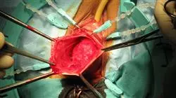
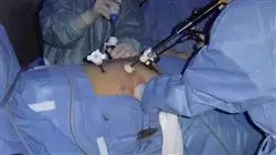
In the area of urological surgery, for example, the advances are unceasing. There are many new developments: minimally invasive urological techniques, lasers, robotic surgery, digital endoscopes, etc. The acquisition of these new technologies allows a faster recovery and a better patient prognosis.

In the field of oncology, on the other hand, advancements have changed the way of intervening. As a result, the specialty of Urology and the specialty of Oncology have been approaching the point that there are many fields in which the boundaries between the two are not defined, one could even say that they no longer exist. Modern medicine leads its professionals to an increasingly demanding super-specialization.

This situation means that professionals are obliged to constantly update and increase their field of knowledge. However, it is not easy to find a training course that thoroughly covers the needs of professionals in this field in an exhaustive manner.. This Advanced master’s degree is the answer from TECH, the largest online Spanish-language university in the world. Due to its special characteristics, it offers the students the opportunity to update as specialists in a practical and effective way, combining the most complete theoretical contents supported by the latest scientific evidence, with the teachings of the most renowned experts in this field and the study methodology from the best universities in the world which has been internationally recognized for its extraordinary effectiveness.

With an approach created to be compatible with other occupations this Advanced master’s degree in Update in Urology will familiarise you with the latest developments your chosen field"

This Advanced master’s degree in Update in Urology contains the most complete and up-to-date scientific program on the market. The most important features of the program include:

  • Diagnostic and Therapeutic Novelties in Uro-Oncology and Urological Surgery
  • Presentation of practical workshops on procedures, diagnosis, and treatment techniques
  • Real images in high resolution and practical exercises where the self-evaluation process can be carried out to improve learning
  • Algorithm-based interactive learning system for decision-making in the presented clinical situations
  • Special emphasis on test-based medicine and research methodologies
  • Theoretical lessons, questions to the expert, debate forums on controversial topics, and individual reflection assignments
  • Content that is accessible from any fixed or portable device with an Internet connection

This Advanced master’s degree is the best investment you can make. You will be trained as one of the best experts in Urology and you will get a degree issued by TECH Global University"

Its teaching staff is made up of leading professionals in the sector. Practising professionals who bring their experience to this training program, as well as renowned specialists from leading scientific societies.

The multimedia content developed with the latest educational technology will provide the professional with situated and contextual learning, i.e., a simulated environment that will provide immersive training program to train in real situations.

This program is designed around Problem-Based Learning, whereby the physician must try to solve the different professional practice situations that arise during the course. For this purpose, the physician will be assisted by an innovative interactive video system created by renowned and experienced experts in the field with extensive teaching experience.

A high-quality tour through the most innovative updates and developments in the field of oncological surgery and uro-oncology, with clinical cases and real situations that will allow you to acquire the skills you need to be at the forefront of the profession"

##IMAGE##

Designed to be fully affordable, this Advanced master’s degree will become a tool for personal growth that will propel you to new in your profession"

Syllabus

The structure of this Advanced master’s degree has been created in order to compile each and every one of the subjects that professionals in this area ought to master, in a comprehensive but very specific syllabus. With an extensive course, structured in areas of intervention, the student will learn the different theoretical and practical approaches and techniques necessary for medical activity in Urology, both in uro-oncology and oncological surgery. Learning that will gradually translate into practical mastery of the techniques. With the tutoring and accompaniment of the exceptional teachers who have developed the contents at all times.

##IMAGE##

This Advanced master’s degree is an incomparable opportunity to obtain, within a single specialization, the greatest compendium of knowledge necessary to develop your professional work in Urology with the solvency of first level experts"

Module 1. Update on oncological principles, functional sequelae and supportive treatment of patients with urological tumors

1.1. Molecular Biology of Cancer
1.2. Prognostic Factors, Tumor Markers, and Paraneoplastic Syndromes in Urologic Oncologic Pathology
1.3. Tumor Genetics
1.4. Oncologic Emergencies in Urology
1.5. Oncological Principles: Etiology, Susceptibility, and Epidemiology
1.6. Principles of Urologic Surgical Oncology
1.7. Clinical Trials in Urologic Oncology Patients
1.8. Supportive Care of the Oncologic Patient in Urology
1.9. Genitourinary Functional Sequelae of Oncologic Treatments in Urology 

1.9.1. Surgical Andrology
1.9.2. Reconstructive Surgery

1.10. Nuclear Medicine and Molecular Imaging in Oncologic Tumor Pathology

1.10.1. Scientific Evidence in Uro-Oncology
1.10.2. New Tracers

Module 2. Advances in the Diagnosis, Treatment and Follow-Up of Non-Muscle Invasive Bladder Carcinoma

2.1. Epidemiology and Etiopathogenesis
2.2. Female Anatomy

2.2.1. TNM
2.2.2. WHO
2.2.3. Biopsies/Samples
2.2.4. Risk factors
2.2.5. Other Factors: T1a-a, Lymphovascular Invasion, Variants, Markers, etc.
2.2.6. CIS

2.3. Diagnosis Part I

2.3.1. Clinical symptoms
2.3.2. Imaging Tests
2.3.3. Urine Cytology
2.3.4. Molecular Markers (Clinical Applications to Date)

2.4. Diagnosis Part II

2.4.1. Cystoscopy
2.4.2. Photodynamic Diagnosis
2.4.3. NBI
2.4.4. Second TURP

2.5. Risk Groups

2.5.1. EORTC
2.5.2. Risk and Progression Charts; CUETO
2.5.3. CIS

2.6. Adjuvant Treatment with Chemotherapy

2.6.1. Single Dose Post-TURP
2.6.2. Adjuvant
2.6.3. Options to Increase Efficiency

2.7. Adjuvant Treatment with BCG

2.7.1. Advantages
2.7.2. Strains
2.7.3. Toxicity and Treatment
2.7.4. Dose
2.7.5. Treatment Plans

2.8. Endovesical Alternatives

2.8.1. Doxorubicin
2.8.2. Epirubicin
2.8.3. Gemcitabine
2.8.4. Onco Thioptepa

2.9. Adjuvant Treatment of CIS
2.10. Treatment Plans in the Event of Standard Treatment Failure

2.10.1. Definition of Failure
2.10.2. After Chemotherapy
2.10.3. After BCG

2.11. Radical Cystectomy in Ca. Non-Muscle Invasive Bladder

2.11.1. Fundamentals
2.11.2. Immediate vs. Early
2.11.3. After BCG Failure

2.12.    Monitoring

Module 3. Advances in the Diagnosis, Treatment and Monitoring of Muscle Invasive Bladder Carcinoma

3.1. Female Anatomy

3.1.1. Regional Lymph Node
3.1.2. Lymph Node Involvement
3.1.3. Histological Variants
3.1.4. Muscle Invasion Pattern
3.1.5. Markers: p53, etc.
3.1.6. TNM

3.2. Urethral Involvement and Concomitant Prostate Cancer
3.3. Staging.

3.3.1. Local: MRI and CT
3.3.2. Lymph Node: MRI; CT; PET
3.3.3. TUS: UROTAC
3.3.4. Future: FDG-PET-CT; DCE-MRI; DWI-MRI

3.4. Radiotherapy

3.4.1. Neoadjuvant
3.4.2. Palliative
3.4.3. Adjuvant

3.5. Neoadjuvant Chemotherapy
3.6. Radical Cystectomy

3.6.1. Risk Assessment
3.6.2. Delay Time
3.6.3. Lymphadenectomy: Extent and Number
3.6.4. Urinary Diversion
3.6.5. Postoperative Complications
3.6.6. Palliative Cystectomy
3.6.7. Laparoscopic vs. Robotic Surgery

3.7. Bladder Preservation Programs

3.7.1. RTU-V
3.7.2. Radiotherapy
3.7.3. Chemotherapy
3.7.4. Multimodal Treatments

3.8. Neoadjuvant Chemotherapy
3.9. Metastatic Cancer

3.9.1. Poor Prognosis Factors
3.9.2. Prognostic Groups/Adverse Factors
3.9.3. Definition of Cisplatin "Unfit"
3.9.4. Single-Agent Chemotherapy
3.9.5. Standard Patient Treatment Cisplatin "fit"
3.9.6. Alternative/2nd Line Treatment of Cisplatin "fit" Patients
3.9.7. Treating “Unfit” Patients
3.9.8. Treating Symptomatic Patients

3.10. Monitoring

3.10.1. Treatment of Bone Metastases
3.10.2. Rescue Surgery
3.10.3. Urothelial Recurrence: Urethra and TUS

3.11. Role of Immunotherapy
3.12. Major Ongoing Clinical Trials
3.13. Particularities of Other Histologies

Module 4. Advances in the Diagnosis, Treatment, and Monitoring of Testicular Cancer 

4.1. Epidemiology and Staging
4.2. Diagnosis and Clinical Staging

4.2.1. Physical Examination
4.2.2. Doppler Ultrasound
4.2.3. Tumor Markers
4.2.4. CAT and MRI
4.2.5. FDG-PET-CAT
4.2.6. TNM

4.3. Staging

4.3.1. Risk Groups
4.3.2. Risk Factors/Prognosis

4.4. Orchiectomy

4.4.1. Indications
4.4.2. Role of Deferred Surgery
4.4.3. Conservative Surgery
4.4.4. Contralateral Biopsy

4.5. Female Anatomy

4.5.1. Role of the Pathologist in the Diagnosis of Testicular Neoplasms
4.5.2. WHO 2016 Classification of Germinal Neoplasms
4.5.3. Diagnostic Algorithm for Non-Germinal Neoplasms
4.5.4. Staging

4.6. Stage I Treatment: Seminoma

4.6.1. Monitoring
4.6.2. Radiotherapy
4.6.3. Adjuvant Chemotherapy
4.6.4. Retroperitoneal Lymph Node Dissection
4.6.5. Risk-Adapted Treatment

4.7. Stage I Treatment: NO Seminoma

4.7.1. Monitoring
4.7.2. Adjuvant Chemotherapy
4.7.3. Retroperitoneal Lymph Node Dissection
4.7.4. Risk-Adapted Treatment

4.8. Treatment of Metastatic Germ Cell Tumors
4.9. Residual Tumor Mass
4.10. Systemic Treatment of Tumor Relapse
4.11. Monitoring
4.12. Testicular Stromal Tumors: Diagnosis, Treatment and Monitoring

Module 5. Advances in the Diagnosis, Treatment, and Monitoring of Penile Cancer 

5.1. Epidemiology, Etiology, and Risk Factors
5.2. Female Anatomy

5.2.1. Premalignant Lesions
5.2.2. Histological Subtypes of Carcinoma of the Penis
5.2.3. TNM
5.2.4. Prognostic Factors
5.2.5. Molecular Biology

5.3. Diagnosis and Staging

5.3.1. Clinical Symptoms
5.3.2. Physical Exploration
5.3.3. Imaging Tests: Ultrasound; MRI; CT; PET-CT-FDG

5.4. Images of Penile and Urethral Cancer
5.5. Anatomical Considerations of the Penis and Lymphatic Drainage
5.6. Treatment of Penile Cancer I: Surgical Treatment of the Primary Tumor

5.6.1. Non-Invasive Superficial Disease: CIS
5.6.2. Invasive Disease Confined to the Glans Penis: Ta/T1a
5.6.3. Invasive Disease: T1b/T2

5.6.3.1. Confined to Corpus Spongiosum
5.6.3.2. Invasion of Corpus Cavernosum

5.6.4. Invasive Urethral Disease: T3
5.6.5. Invasive Disease of Adjacent Structures: T4

5.7. Treatment of Carcinoma of the Penis II: Lymph Nodes

5.7.1. Daseler's Inguinal Anatomical Zones
5.7.2. General Considerations
5.7.3. Risk Stratification for Nodal Involvement in cN0

5.7.3.1. Monitoring
5.7.3.2. Lymph Node Staging

5.7.4. Modified Lymphadenectomy
5.7.5. Dynamic Sentinel Lymph Node Biopsy

5.7.5.1. cN1/cN2
5.7.5.2. Radical Inguinal Lymphadenectomy
5.7.5.3. Pelvic Lymphadenectomy

5.7.6. cN3
5.7.7. Controversies in Ilioinguinal Lymphadenectomy

5.8. Penile Cancer Treatment III: Radiotherapy

5.8.1. Indications

5.8.1.1. Ta/T1a
5.8.1.2. T2

5.8.2. Regional Lymph Node

5.9. Penile Cancer Treatment IV: SYSTEMIC

5.9.1. Adjuvant Chemotherapy
5.9.2. Neoadjuvant Chemotherapy
5.9.3. Palliative Chemotherapy
5.9.4. Targeted Therapy

5.10. Monitoring

5.10.1. General Aspects
5.10.2. Clinical Guides
5.10.3. Local Recurrence
5.10.4. Regional Recurrence

5.11. Quality of life
5.12. Primary Urethral Carcinoma

Module 6. Advances in the Diagnosis, Treatment, and Monitoring of Renal, Suprarenal, and Retroperitoneal Carcinoma

6.1. Epidemiology and Etiopathogenesis
6.2. Diagnostic Imaging and Clinical Staging

6.2.1. Doppler and Contrast Ultrasound: Evaluation of Complicated Renal Cyst, Renal Mass and Dissemination
6.2.2. MRI and CT: Diagnosis, Staging and Monitoring

6.3. Female Anatomy

6.3.1. WHO
6.3.2. ISUP
6.3.3. Führmnan
6.3.4. Clear Cells
6.3.5. Papillary
6.3.6. Chromophobic
6.3.7. Other Histologies

6.4. Renal Tumor Biopsy

6.4.1. Technical Aspects
6.4.2. Indications
6.4.3. Side effects
6.4.4. Efficacy
6.4.5. Cystic Lesions

6.5. Prognostic Factors

6.5.1. TNM
6.5.2. Histological Factors
6.5.3. Clinical Factors
6.5.4. Molecular Factors

6.6. Localized Renal Carcinoma

6.6.1. Monitoring
6.6.2. Radical Surgery vs. Nephron-Sparing Surgery
6.6.3. Nephron-Sparing Surgery
6.6.4. Adrenalectomy
6.6.5. Lymphadenectomy
6.6.6. Pre-Nephrectomy Embolization
6.6.7. Ablative Treatments

6.7. Advanced Localized Renal Carcinoma

6.7.1. cN+
6.7.2. Unresectable Tumors
6.7.3. IVC Thrombosis
6.7.4. Adjuvant and Neoadjuvant Treatment
6.7.5. Clinical Trials

6.8. Advanced or Metastatic Renal Carcinoma

6.8.1. The Role of Radical Nephrectomy
6.8.2. Cytoreductive Surgery + Immunotherapy
6.8.3. Role of Metastasectomy
6.8.4. Radiotherapy
6.8.5. Embolization
6.8.6. Symptomatic Treatment of Patients With Renal Carcinoma

6.9. Systemic Treatment

6.9.1. Chemotherapy
6.9.2. Immunotherapy

6.9.2.1. Advances in Immunotherapy
6.9.2.2. α- IFN
6.9.2.3. IL-2.
6.9.2.4. Vaccines and Targeted Immunotherapies

6.9.2.4.1. Tumor Antigen 5T4 + 1st Line Therapies
6.9.2.4.2. Anti PD-1 or PD-L1 Antibodies

6.9.3. Targeted Therapy

6.9.3.1. Advances in Targeted Therapy
6.9.3.2. IMDC Risk/Prognostic Groups: Therapeutic Implication
6.9.3.3. Tyrosine Kinase Inhibitors
6.9.3.4. Monoclonal Antibodies Against Circulating VEGF
6.9.3.5. mTOR Inhibitors

6.9.4. 1st Line Treatment: Sunitinib
6.9.5. 1st Line Treatment: Pazopanib
6.9.6. 1st Line Treatment: Other Options
6.9.7. 1st Line Treatment in Patients with Poor Prognosis: Temsirolimus
6.9.8. 1st Line Treatment Positioning
6.9.9. 2nd Line Treatment: Axitinib
6.9.10. 2nd Line Treatment: Everolimus
6.9.11. 2nd Line Treatment: Cabozantinib
6.9.12. 2nd Line Treatment: Nivolumab
6.9.13. 2nd Line Treatment: Subsequent Options
6.9.14. Therapeutic Sequencing in Renal Carcinoma: Treatment Positioning
6.9.15. Symptomatic Treatment of Patients With Renal Carcinoma
6.9.16. Non-Clear Cell Carcinomas

6.10. Monitoring

6.10.1. Imaging Tests
6.10.2. Recurrence: Local and Distant
6.10.3. Ablative Treatments

6.11. Drug Resistance Mechanism
6.12. Major Developments in Metastatic Kidney Cancer: Clinical Trials Underway
6.13. Suprarenal Mass

6.13.1. Differential Diagnosis
6.13.2. Functioning Mass Diagnosis
6.13.3. Surgical Management
6.13.4. Metastatic Cancer

6.14. Primary Retroperitoneal Tumors

6.14.1. Differential Diagnosis
6.14.2. Diagnostic Techniques
6.14.3. Surgical Treatment
6.14.4. Metastatic Cancer

Module 7. Advances in the Diagnosis, Treatment, and Monitoring of Prostate Cancer

7.1. Epidemiology and Risk Factors 
7.2. Diagnosis

7.2.1. TR
7.2.2. PSA: Density, Kinetics, Ratio, PHI, etc.
7.2.3. Other Markers: Genetic, PCA3, 4K, etc.
7.2.4. Prostate Biopsy

7.3. Screening vs. Early Diagnosis
7.4. Diagnostic Imaging

7.4.1. Ultrasonography: Sonoelastography, Contrast, Histoscanning, etc.
7.4.2. Bone Scan
7.4.3. CAT
7.4.4. MRI
7.4.5. PET-CAT
7.4.6. mpMRI: Technical Aspects

7.5. Pathologic Anatomy/Pathogenesis

7.5.1. Biopsies
7.5.2. RP Piece

7.6. Clinical and Pathologic Staging
7.7. Deferred Treatment

7.7.1. Localized Prostate Cancer: VA vs. WW
7.7.2. Locally Advanced
7.7.3. Metastatic

7.8. Localized Prostate Cancer

7.8.1. RT: General Information

7.8.1.1. IMRT/IGRT
7.8.1.2. Dose Escalation
7.8.1.3. Hormone Therapy
7.8.1.4. RxT + CT
7.8.1.5. Dose Escalation + Hormone Therapy

7.8.2. PR: General Information

7.8.2.1. Surgical Technique: Open-Laparoscopic-Robotic
7.8.2.2. Conservation of Neurovascular Bundles

7.8.3. Focal Therapy

7.9.Radical Prostatectomy

7.9.1. Low Risk
7.9.2. Medium Risk
7.9.3. High Risk and Locally Advanced
7.9.4. Lymphadenectomy and Lymph Node Involvement
7.9.5. Adjuvant and Neoadjuvant Hormone Therapy
7.9.6. Conservation of Neurovascular Bundles: Indications and Results

7.10. Radiotherapy

7.10.1. Low Risk
7.10.2. Medium Risk
7.10.3. High Risk
7.10.4. Locally Advanced: MRC P23/PR07; TAP 32; SPCG-7/SFUO-3
7.10.5. Ganglion Chains: RTOG 85-31; UK-STAMPEDE
7.10.6. Proton Therapy
7.10.7. Low Dose Rate Brachytherapy
7.10.8. High Dose Rate Brachytherapy
7.10.9. RxT after RP: EORTC 22911; ARO; SWOG 8794
7.10.10. Nodes

7.11. Cryosurgery
7.12. HIFU
7.13. Focal Therapy

7.13.1. Negative Biopsy + Elevated PSA
7.13.2. mpMRI
7.13.3. Bio markers
7.13.4. Future
7.13.5. PI-RADS Scientific Evidence
7.13.6. Ultrasound-Guided Prostate Biopsy +MRNR

7.13.6.1. Advances in Ultrasound-Guided Prostate Biopsy
7.13.6.2. Material
7.13.6.3. Technique: Transrectal/Transperineal

7.13.7. Fusion Biopsy
7.13.8. Cognitive Biopsy
7.13.9. Scientific Evidence
7.13.10. Cost-Effectiveness of MRI in the Detection of Prostate Cancer
7.13.11. Focal Therapy: Index Lesion; Clonal Theory
7.13.12. Selection Criteria. Risk Stratification
7.13.13. Energy Sources: HIFU, Cryotherapy, Brachytherapy, Electroporation, Photodynamic Therapy, Cyberknife
7.13.14. Monitoring and Recurrence

7.14. Metastatic Prostate Cancer

7.14.1. Standard Treatment: Hormone Therapy
7.14.2. SWOG: Risk Groups
7.14.3. Intermittent Blocking

7.15. Castration Resistance: Etiology
7.16. CRPC Definition New Criteria
7.17. Clinicopathological Prognostic Factors in CRPC. Androgen Deprivation in CRPC Markers of response
7.18. Non-Metastatic CRPC (CRPC-M0). Clinical Management. Monitoring Criteria
7.19. Hormonal Maneuvers in CRPC. Scientific Evidence
7.20. 1st Line Chemotherapy Treatment: Docetaxel

7.20.1. CRPC

7.21. Non-1st Line Chemotherapy Treatment: Cabazitaxel. Other Drugs
7.22. Hormone Treatment in CRPC Abiraterone

7.22.1. CRPC

7.23. Hormone Treatment in CRPC Enzalutamide

7.23.1. CRPC

7.24. Treatment with Bone-Targeted Agents

7.24.1. Bisphosphonates
7.24.2. Denosumab
7.24.3. Radio 223

7.25. CPRC Immunotherapy
7.26. Symptomatic Treatment of Patients with CRPC
7.27. Treatment Algorithm in CRPC: Positioning and Sequencing
7.28. Mechanisms of Resistance to Hormonal Treatment in CRPC: AR-V7 and Other Related Factors
7.29. Molecular Biology of CRPC: BRCA and Related Genes
7.30. Molecular Biology of CRPC: Epigenetic Angiogenesis
7.31. Molecular Biology of CRPC: Other Molecular Pathways Involved 
7.32. Main Ongoing Clinical Trials in CRPC
7.33. Future Outlook of CRPC

Module 8. Latest Advances in Surgical Anatomy Laparoscopic Anatomy

8.1. Upper Surgical Anatomy

8.1.1. Pathology
8.1.2. Kidney
8.1.3. Adrenal Gland
8.1.4. Ureter

8.2. Lower Surgical Anatomy

8.2.1. Bladder
8.2.2. Prostate and Seminal Vesicles
8.2.3. Urethra
8.2.4. Penis
8.2.5. Testicles and Scrotum

8.3. Surgical Anatomy of the Pelvic Floor

8.3.1. Pelvic Floor in a Woman

8.3.1.1. Abdominal View
8.3.1.2. Perineal View

8.3.2. Pelvic Floor in a Man

8.3.2.1. Abdominal View
8.3.2.2. Perineal View

Module 9. Basic Aspects of Urologic Surgery

9.1. Basic Perioperative Aspects
9.2. Instrumentation and Drainage of the Urinary Tract
9.3. Basic Principles in a Endoscopy

9.3.1. Basic Principles of Endourology
9.3.2. Basic Principles in a Laparoscopy, Mini Laparoscopy and 3D
9.3.3. Basic Principles in Robot Assisted Laparoscopic Surgery
9.3.4. Basic Principles in Single Port Laparoscopic Surgery
9.3.5. Basic Principles in Surgery Notes/e-Notes

Module 10. Latest Advances in Diagnosis and Treatment Techniques in Urology

10.1. Flexible Cystoscopy
10.2. Calibration/Urethral Dilation
10.3. Renal Ultrasound/Renal Doppler
10.4. Vesicoprostatic Ultrasound

10.4.1. Transrectal Prostate Ultrasound
10.4.2. Abdominal Prostate Ultrasound

10.5. Testicular Ultrasound / Testicular Doppler
10.6. Penile Ultrasound / Penile Doppler
10.7. Intravesical Instillation of Drugs
10.8. Pyelography

10.8.1. Ascending or Retrograde Pyelography
10.8.2. Anterograde Pyelography (Nephrostography)

10.9. Ureteroscopy.
10.10. Urodynamic Study

10.10.1. Urodynamics in cabinet
10.10.2. Outpatient Urodynamics

10.11. Prostate Biopsy

10.11.1. Transperineal Prostate Biopsy
10.11.2. Transrectal Prostate Biopsy

10.12. Multiparametric MRI
10.13. Bone Scanning
10.14. PET-CAT
10.15. Isotopic Renogram

Module 11. Latest Trends in Adrenal and Retroperitoneal Surgical pathology

11.1. Right Adrenalectomy

11.1.1. Intraperitoneal Laparoscopy
11.1.2. Retroperitoneoscopy

11.2. Left Adrenalectomy

11.2.1. Intraperitoneal Laparoscopy
11.2.2. Retroperitoneoscopy

11.3. Partial Adrenalectomy
11.4. Surgery for Adrenal Carcinoma
11.5. Excision of Retroperitoneal Tumors

Module 12. Latest Trends in Renal Surgical Pathology Surgical Indication and Access Routes

12.1. Radical Nephrectomy

12.1.1. Open Surgery

12.1.1.1. Large Renal Tumor
12.1.1.2. Renal Tumors With Vena Cava Thrombosis

12.1.2. Intraperitoneal Laparoscopy
12.1.3. Lufti Tunk Technique
12.1.4. Retroperitoneal
12.1.5. Robotics
12.1.6. Transvaginal

12.2. Patrial Nephrectomy with Vascular Clamping

12.2.1. Intraperitoneal Laparoscopy
12.2.2. Retroperitoneal
12.2.3. Robotics
12.2.4. Open Partial Nephrectomy

12.3. Partial Nephrectomy / Off Clamp Lumpectomy

12.3.1. Intraperitoneal Laparoscopy
12.3.2. Retroperitoneal
12.3.3. Robotics

12.4. Partial Nephrectomy / Selective Lumpectomy or Supraselective Clamping

12.4.1. Intraperitoneal Laparoscopy
12.4.2. Retroperitoneal
12.4.3. Robotics

12.5. Living Donor Nephrectomy

12.5.1. Laparoscopy
12.5.2. Robotics
12.5.3. Single Port
12.5.4. Transvaginal

12.6. Focal Treatment of Renal Tumors

12.6.1. Radiofrequency
12.6.2. HIFU (High Intensity Focused Ultrasound)
12.6.3. Cryotherapy

12.7. Lymphadenectomy in Kidney Cancer

12.7.1. Technique
12.7.2. Limits

Module 13. New Advances in the Field of Kidney Transplant

13.1. Renal Extraction

13.1.1. Renal Extraction
13.1.2. Multi Organ Extraction

13.2. Renal Transplant

13.2.1. Heterotopic
13.2.2. Orthotopic

13.3. Laparoscopic Renal Transplant
13.4. Robotic Renal Transplant

13.4.1. In Men
13.4.2. In Women with Vaginal Insertion of the Graft

13.5. Renal Autotransplantation
13.6. Ureteroneocystostomy

13.6.1. Intravesical Techniques
13.6.2. Extravesical Techniques

Module 14. New Advances in the Field of Vascular Kidney Transplant

14.1. Renal Artery Stenosis

14.1.1. Percutaneous Techniques
14.1.2. Surgical Management

14.2. Renal Artery Aneurysm

14.2.1. Percutaneous Techniques
14.2.2. Surgical Management

Module 15. Latest Trends in Surgical Pathology of the Upper Urinary Tract

15.1. Tumorous Pathology

15.1.1. Laparoscopic Nephroureterectomy
15.1.2. Nephroureterectomy by Retroperitoneoscopy
15.1.3. Surgical Treatment of the Distal Ureter

15.1.3.1. Open Surgery
15.1.3.2. Transurethral Resection of the Bladder (TURB) / Meatus Removal
15.1.3.3. Amon Technique
15.1.3.4. Agarwal Technique

15.1.4. Robotic Nephroureterectomy
15.1.5. Retrograde Intrarenal Surgery (RIRS) Hexvix®/Spies®
15.1.6. Percutaneous Surgery for the Treatment of Intrarenal Upper Urothelial Tumors
15.1.7. Endoscopic Treatment of Ureteral Tumors
15.1.8. Partial Urethrectomy

15.2. Lymphadenectomy in Upper Urinary Tract Cancer

15.2.1. Indications and Techniques
15.2.2. Limits

15.3. Treatment of Renal Lithiasis

15.3.1. Percutaneous Nephrostomy

15.3.1.1. Urinary Diversion
15.3.1.2. Access for Percutaneous Surgery

15.3.1.2.1. Ultrasound-guided
15.3.1.2.2. Bullseye
15.3.1.2.3. 90 Degree Technique
15.3.1.2.4. Triangulation
15.3.1.2.5. Others

15.3.2. Fragmentation Methods Energy Types
15.3.3. Percutaneous Nephrolithotomy in Prone Position
15.3.4. Percutaneous Nephrolithotomy in Supine Position
15.3.5. Mini Percutaneous (MPERC)
15.3.6. Ultra-mini-percutaneous (UMP)
15.3.7. Super-mini-percutaneous (SMP)
15.3.8. Micro Percutaneous
15.3.9. Mini Invasive Percutaneous Surgery (MIP)
15.3.10. Retrograde Intrarenal Surgery (CRIR or RIRS)
15.3.11. Combined Antegrade and Retrograde Surgery (ECIRS / microECIRS)
15.3.12. Ureteral Access Sheaths. Types and Compatibilities
15.3.13. Extracorporeal Shock Wave Lithotripsy (ESWL)

15.3.13.1. Fluoroscopic Localization
15.3.13.2. Ecographic Localization

15.4. Treatment of Ureteral Lithiasis

15.4.1. Ureteral Catheterization

15.4.1.1. Straight or Simple J Catheter
15.4.1.2. Double J Catheter

15.4.2. Semirigid Ureteroscopy
15.4.3. Micro-ureteroscopy (Micro-URS)
15.4.4. Flexible Ureteroscopy
15.4.5. Antiretroviral Mechanisms and Techniques
15.4.6. Fragmentation Methods in Ureteral Lithiasis
15.4.7. Extracorporeal Shock Wave Lithotripsy (ESWL)

15.5. Upper Urinary Tract Stenosis

15.5.1. Infundibular Stenosis. Endourological Treatment
15.5.2. Pyeloureteral Junction Stenosis (PUJ)

15.5.2.1. Laparoscopic Pyeloplasty
15.5.2.2. Mini Laparoscopic Pyeloplasty
15.5.2.3. Percutaneous and Retrograde Endopyelotomy

15.5.3. Ureteral Stenosis

15.5.3.1. Balloon Dilatation of the Ureter
15.5.3.2. Ureteral Prostheses

15.5.3.2.1. Long-term or Tumor Catheter
15.5.3.2.2. Resonance Catheter
15.5.3.2.3. Allium® Prosthesis
15.5.3.2.4. Uventa Prosthesis
15.5.3.2.5. Memokath Prosthesis

15.5.3.3. Urethral Stenosis Section

15.5.3.3.1. Cold Section (scissors)
15.5.3.3.2. Section with Laser

15.5.3.4. Ureteral Reimplantation

15.5.3.4.1. Laparoscopic Anastomosis / Reimplantation
15.5.3.4.2. Robotic Anastomosis / Reimplantation

15.5.3.5. Urethreal Substitution
15.5.3.6. Ureterolysis
15.5.3.7. Retrocaval Ureter
15.5.3.8. Subcutaneous Pyelovesical Catheterization

15.5.4. Post-urinary Diversion Ureterointestinal Junction Stricture

15.5.4.1. Lovaco Technique

Module 16. Latest Trends in Bladder Surgical Pathology

16.1. Tumorous Pathology

16.1.1. Randomized Vesical Biopsy
16.1.2. Transurethral Resection of Bladder Tumor

16.1.2.1. En-bloc Transurethral Resection of Bladder Tumor

16.1.3. Transurethral Resection Assisted with Hexvix®/Spies®
16.1.4. Excision / Laser coagulation
16.1.5. Laparoscopic/Robotic/Open Partial Cystectomy (L/R/O)
16.1.6. Radical Cystoprostatectomy L/R/O

16.1.6.1. Radical Cystosprostatectomy L/R/Open en-bloc

16.1.7. Transileal Cutaneous Ureterostomy (Bricker) L/R/O
16.1.8. Intestinal Neovessels More Common Techniques
16.1.9. Uretero-Intestinal Anastomosis
16.1.10. 3D Laparoscopic / Robotic Intestinal Neobladder
16.1.11. Cutaneous Ureterostomy
16.1.12. Radical Cystectomy in the woman. L/R Peculiarities

16.2. Lymphadenectomy in Bladder Cancer

16.2.1. Indications and Techniques
16.2.2. Limits
16.2.3. Sentinel lymph node

16.3. Non-Tumorous Pathology

16.3.1. Percutaneous Cystostomy
16.3.2. Open Cystotomy / Bladder Carving / Cystolithotomy
16.3.3. Vesicoureteral Antireflux Surgery

16.3.3.1. Open Surgery More Common Techniques
16.3.3.2. Endoscopic Injection (Deflux and others)

16.3.4. Bladder Diverticulum

16.3.4.1. Endoscopic treatment
16.3.4.2. Open Surgery / Laparoscopy

16.3.5. Endoscopic Treatments of Bladder Lithiasis
16.3.6. Exstrophy / Epispadias

Module 17. Latest Trends in Prostate Surgical Pathology

17.1. Non-Tumorous Pathology

17.1.1. Transurethral Resection of the Prostate (TURP) Monopolar / Bipolar
17.1.2. Trigonocervicoprostatotomy (TCP) or myocapsulotomy
17.1.3. Bipolar Prostatic Vaporization
17.1.4. Prostate Laser Vaporization (HoLap)

            17.1.4.1. Green Laser
            17.1.4.2. Thulium Laser
            17.1.4.3. Holmium Laser
            17.1.4.4. Other Types of Laser CO2.

          17.1.5. Prostatic Enucleation (Holep)

            17.1.5.1. Holmium Laser
            17.1.5.2. Thulium Laser
            17.1.5.3. Green Laser
            17.1.5.4. Bipolar Enucleation

           17.1.6. Prostatic Adenomectomy

            17.1.6.1. Open
            17.1.6.2. Laparoscopy

           17.1.7. Urolift
           17.1.8. I-Tind
           17.1.9. Endourethral / Prostatic Prostheses

 17.1.9.1. Memotherm®
 17.1.9.2. Allium®
 17.1.9.3. Uventa®

17.2. Tumorous Pathology

17.2.1. Laparoscopic / Robotic Radical Intraperitoneal Prostatectomy (L/R)
17.2.2. Extraperitoneal Radical Prostatectomy (L/R)
17.2.3. Intra, Inter and Extrafascial Techniques
17.2.4. Radical Prostatectomy with Hydrodissection
17.2.5. Radiotherapy / Intensity Modulated Radiation Therapy (IMRT)
17.2.6. Prostate Brachytherapy
17.2.7. Focal Treatment of Prostate Cancer

17.2.7.1. Cryotherapy
17.2.7.2. Radiofrequency
17.2.7.3. HIFU
17.2.7.4. Electroporation

17.3. Lymphadenectomy in Prostate Cancer

17.3.1. Techniques
17.3.2. Limits
17.3.3. Sentinel lymph node

Module 18. Latest Trends in Urethra Surgical Pathology

18.1. Internal Urethrotomy

18.1.1. Blade (Saxon)
18.1.2. Internal Laser Urethrotomy

18.2. Intraurethral Condyloma Treatment
18.3. Urethroplasty

18.3.1. Meatoplasty/Balanitis Obliterans Xerotica Obliterans
18.3.2. Penile Urethral Stricture
18.3.3. Bulbar Urethral Stricture
18.3.4. Membranous Urethral Stricture
18.3.5. Urethral Stricture in Women
18.3.6. Urethral Prosthesis

18.4. Hypospadias Surgery

18.4.1. Hypospadias Dista
18.4.2. Proximal Hypospadias

18.5. Urethral Fistulas

18.5.1. Urethro-Cutaneous
18.5.2. Urethro-Rectal
18.5.3. Urethro-Vaginal

Module 19. Latest Trends in Penis Surgical Pathology

19.1. Glandular Decortication (Bracka technique)
19.2. Partial Penectomy
19.3. Total Penectomy
19.4. Penile Incurvation Surgery

19.4.1. Injection of C. Hystolitycum
19.4.2. Nesbit Technique
19.4.3. Techniques Without Penis Shortening

19.5. Penis Prothesis

19.5.1. Malleable
19.5.2. Two Components
19.5.3. Three Components

19.6. Lymphadenectomy in Penis Cancer

19.6.1. Techniques
19.6.2. Limits
19.6.3. Sentinel lymph node

Module 20. Latest Trends in Scrotal and Testicular Surgical Pathology

20.1. Vasectomy

20.1.1. Vasectomy / Vasovasostomy Reversal

20.2. Variocele Treatment

20.2.1. Sclerosis/Interventional Radiology
20.2.2. Surgical Management

20.3. Treatment of Hydrocele
20.4. Orchiectomy

20.4.1. Subalbuginea Orchiectomy
20.4.2. Radical Orchiectomy
20.4.3. Partial Orchiectomy

20.5. Lymphadenectomy in Testicular Cancer

20.5.1. Indications and Techniques
20.5.2. Limits

Module 21. New Advances in the Surgical Treatment of Urinary/ Pelvic Floor Incontinence

21.1. Urinary Incontinence in Women

21.1.1. Stress Urinary Incontinence

21.1.1.1. Mini Sling
21.1.1.2. Incontinence Netting (TVT/TVA/TOT/TOA)
21.1.1.3. Laparoscopic Colposacropexy
21.1.1.4. Transvaginal Fistulectomy
21.1.1.5. Urinary Sphincter

21.1.2. Emergency Urinary Incontinence

21.1.2.1. Intravesical Botulinum Toxin Injection
21.1.2.2. Bladder Neuromodulator

21.1.3. Treatment of Vesico-Vaginal Fistula

21.1.3.1. Transvaginal Fistulectomy
21.1.3.2. Transvesical Fistulectomy
21.1.3.3. Laparoscopic Fistulectomy

21.2. Prolapse Surgery (Uterine, Cystocele, Rectocele, Enterocele)
21.3. Urinary Incontinence in Men

21.3.1. Light Incontinence

21.3.1.1. Inovance®
21.3.1.2. Advance®

21.3.2. Severe Incontinence

           21.3.2.1. Artificial Urinary Sphincter

##IMAGE##

A unique, key, and decisive master’s degree experience to boost your professional development”

Advanced Professional Master's Degree in Urology Update

Thanks to the evolution of scientific research in recent decades, the field of urology has been able to develop various methods to address all types of pathologies related to the urinary system and genitalia of people. In order to perform adequately and provide high quality results in the prevention, diagnosis and treatment of urinary diseases, it is necessary that medical professionals are prepared by applying the latest surgical techniques and procedures. For this reason, at TECH Global University we developed the Advanced Master's Degree in Urology Update, a program aimed at addressing the most relevant aspects of this medical area to improve your skills and lead you to stand out as an expert through excellence, enhancing the growth of your career.

Specialize in the largest Medical School

With this Advanced Master's Degree you will expand your knowledge and technical skills in the most innovative developments in the medical-surgical field of urology. With the theoretical and practical study of real clinical cases, you will learn the basic principles of diagnosis, treatment and monitoring of the different conditions that can affect the state of the genital apparatus, you will correctly apply the different tools and therapeutic options depending on the patient's condition, in addition to treating the genitourinary functional sequelae of urological interventions. In TECH Global University we have the largest Faculty of Medicine and a complete and updated scientific program that will help you to manage, from the most common diseases in consultation, to the pathologies that require a more specialized treatment. If you want to guarantee a comprehensive and high quality service in the practice of this discipline, this program is the right one for you.