University certificate
The world's largest faculty of medicine”
Why study at TECH?
This Advanced master’s degree in Ophthalmologywill allow you to acquire advanced knowledge and will be of great use in improving the visual health of your patients”

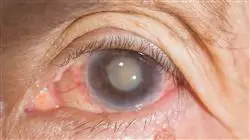
Sciences related to Vision such as Optics, Optometry, Ocular Pharmacology, and Ophthalmology have experienced spectacular progress over the last ten years, together with significant technological development in the field. There have been noteworthy advances in treatment of pathologies that, until recently, were major causes of blindness, such as cataracts, glaucoma, and alterations and degeneration of the retina and the macula.
Therefore, the subspecialization of professionals in this field is of great relevance to improving the health of people suffering from any visual pathology. To increase the training of ophthalmologists, TECH offers an educational proposal of high academic value: the Advanced master’s degree in Ophthalmology. A program that, due to its size, has been distributed intov two large modules. In this way, the student will firstly enter into the study of clinical ophthalmology, to continue with a specific program on the pathology and surgery of the macula, retina and vitreous.
Thus, the program includes everything from cataract surgery, oculoplasty and tear ducts to glaucoma and ophthalmopediatrics. It also deals in depth with all the subspecialties of the retina, delving deeply into other major issues, such as AMD (Age-Related Macular Degeneration). In this case, the specific topics on surgery provide an additional value to this entire educational project, whose main objective is to offer a higher specialization and high academic level to promote the need of these professionals to study and increase their professional training.
This Advanced master’s degree offers the possibility to deepen and update knowledge of this subject, with the use of the latest educational technology. It offers a global vision of Ophthalmology while focusing on the most important and innovative aspects of the macula, retina and vitreous. All of this in a 100% online specialization course, which allows students to expand their knowledge and, therefore, their professional skills and competencies in a simple way, adapting the study time to fit in with the rest of their daily commitments.
Our Advanced master’s degree is a unique opportunity to study the most relevant aspects of Ophthalmology in one single program, providing you with the best training to boost your career"
This Advanced master’s degree in Ophthalmology contains the most scientific and up-to-date program on the market. The most important features include:
- The development of clinical practical case studies presented by experts in Ophthalmology
- The graphic, schematic, and eminently practical contents with which they are created, provide scientific and practical information on the disciplines that are essential for professional practice
- Latests diagnostic and therapeutic developments in Ophthalmology
- The presentation of hands-on workshops on procedures, diagnostic and therapeutic techniques
- Real images in high resolution and practical exercises where the self-evaluation process can be carried out to improve learning
- An algorithm-based interactive learning system for decision-making in the clinical situations presented throughout the course
- Special emphasis on test-based medicine and research methodologies
- Theoretical lessons, questions to the expert, debate forums on controversial topics, and individual reflection assignments
- Content that is accessible from any fixed or portable device with an Internet connection
This Advanced master’s degree is the best investment you can make when choosing a refresher program for two reasons: in addition to expanding your knowledge in Ophthalmology, you will obtain a degree endorsed by TECH"
Forming part of the teaching staff is a group of professionals in the world of psychiatry, who bring their work experience to this course, as well as a group of renowned specialists recognized by esteemed scientific communities.
The multimedia content, developed with the latest educational technology, will provide the professional with situated and contextual learning, i.e., a simulated environment that will provide an immersive training program designed to train in real situations.
This program is designed around Problem Based Learning, whereby the doctor must try to solve different professional practice situations that arise during the program. For this purpose, the physician will be assisted by an innovative interactive video system developed by renowned experts in the field of ophthalmology who have extensive teaching experience.
Increase your decision-making confidence by updating your knowledge through this Advanced master’s degree: A program created to train the best"

We offer the best teaching methodology, with a multitude of practical content which will allow you to study in more complete and effective way"
Syllabus
The structure of the contents has been designed by a team of professionals from the best research centers and universities on a national level. Aware of the current relevance of specialization and the need to support each research study and its application with a solid scientific and evidence-based foundation, they have created a learning path in which each topic will address one of the relevant aspects to help in the development of a highly competent professional. All of this creates a syllabus of high educational intensity and unparalleled quality, which includes state-of-the-art virtual theory and practice, and which will propel the student to the most complete level of mastery in this area.

This Advanced master’s degree is an incomparable opportunity to obtain, in a single training program, all the knowledge required in Ophthalmology, including the most recent advanced in intervention techniques and protocols”
Module 1. Cataract Surgery Update
1.1. Viscoelastics. Composition and Biomechanical Properties. Instructions for Correct Use
1.2. Bilateral and Simultaneous Cataract Surgery
1.3. Scanning Techniques in Cataract Surgery Candidates: Biometric Calculation Devices
1.4. Biometric Calculation Formulae. Special Situations: Long Sight Short Sight Calculation in Eyes Undergoing Refractive Surgery
1.5. Intraocular Lenses for Pseudophakia (I): Aspheric and Aberration-Correcting Lenses. Toric Lenses. Adjustable Strength Lens
1.6. Technological Update in Cataract Surgery (I): Femtosecond Laser
1.7. Technological Update in Cataract Surgery (II): Intraoperative Guiding Systems
1.8. Lens Surgery in Special Situations: Subluxated Crystalline Lens
1.9. Prevention of Major Complications in Cataract Surgery. Posterior Capsular Opacity. Prevention of CME. Prevention of Acute Post-Operative Endophthalmitis
Module 2. Update on Oculoplasty and Lacrimal Ducts
2.1.Thyroid Orbitopathy
2.2. Dacryocystorhinostomy via External/Endoscopic Route
2.3. Ptosis and Eyelid Malposition
2.4. Blepharoplasty
2.5. Orbital tumors
2.6. Eyelid Tumors
2.7. Ablative Ocular Surgery. Management of the Anophthalmic Cavity
2.8. Lacrimal Puncta Surgery
Module 3. Glaucoma Update
3.1. Diagnosis I: Intraocular Pressure and Pachymetry
3.2. Diagnosis II: Angle Study: Gonioscopy and Other Methods
3.3. Diagnosis III: Campimetry
3.4. Diagnosis IV: Analysis of the Papilla and the Nerve Fiber Layer
3.5. Pathophysiology and Classification of Glaucoma
3.6. Treatment I: Medical
3.7. Treatment II: Laser
3.8. Treatment III: Filtering Surgery
3.9. Treatment IV: Valvular Surgery and Cyclodestructive Procedures
3.10. New Perspectives in Glaucoma: The Future
Module 4. Ocular Surface and Cornea Update
4.1. Corneal Dystrophies
4.2. Dry Eye and Ocular Surface Pathology
4.3. Corneal Surgery (PPK, DALK, DSAEK, DMEK)
4.4. Corneal Crosslinking
4.5. Conjunctival and Corneal Neoplasms
4.6. Toxic and Traumatic Lesions of the Anterior Segment
4.7. Ectasia (Keratoconus, Pellucid Marginal Degeneration, Post-LASIK Degenerations
4.8. Infectious Corneal Pathology I
4.9. Infectious Corneal Pathology II
Module 5. Refractive Surgery Update
5.1. Refractive Surgery with Phakic Intraocular Lenses. Types of Lenses. Advantages and Disadvantages
5.2. Presbyopia Correction (I): Available Techniques
5.3. Presbyopia Correction (II): Multifocal Intraocular Lenses. Keys to Success with its Management
5.4. Surgical Management of Astigmatism. (I): Available Techniques
5.5. Surgical Management of Astigmatism. (II): Toric Intraocular Lenses for Pseudophakia
5.6. IOL Calculation in Refractive Surgery. Biometrics
5.7. Excimer Laser Refractive Surgery. Techniques Used. Indications and Contraindications
5.8. Main Complications in Refractive Surgery with Intraocular Lenses
5.9. Main Complications in Excimer Laser Refractive Surgery
5.10. Femtosecond Laser: Use in Refractive Surgery
Module 6. Update in Ophthalmopediatrics
6.1. Re-Operation for Strabismus
6.2. Management of Epiphora, Palpebral and Conjunctival-Corneal Pathology in Children
6.3. Amblyopia: Etiology, Diagnosis, Treatment, and Monitoring
6.4. Vertical Strabismus, Alphabetic Syndromes and Restrictive Syndromes: Stilling Duane, Brown, Möbius and Congential Fibrosis
6.5. Neuro-Ophthalmologic Pathology in Children
6.6. Congenital Glaucoma: Diagnosis, Treatment, and Monitoring
6.7. Differential Diagnosis of Leukocoria: Most Common Pathologies, Diagnosis and Treatment
6.8. Congenital Cataract Treatment: Etiology, Diagnosis and Treatment
6.9. Diagnosis and Treatment of Nystagmus
6.10. Botulinum Toxin in Strabology
Module 7. Anatomy, Physiology and Exploratory and Functional Tests
7.1. Historical Notes and Classical Exploration in Consultation
7.1.1. History to Understand the Present
7.1.2. The Ophthalmoscope and its Examination Lenses
7.1.3. The Slit Lamp and its Examination Lenses
7.1.4. Historical Notes of Current Exploration Techniques
7.2. Macula and Retina Atanomy
7.2.1. Compared Anatomy
7.2.2. Macula and Retinal Histology
7.2.3. Vascularisation of the Retina and Macula
7.2.4. Innervation of the Retina and Macula
7.3. Vitreous anatomy and Physiology
7.3.1. Vitreous Embryology
7.3.2. Composition of the Vitreous Gel
7.3.3. Hyaloid Insertions and Adhesions
7.3.4. Ageing and Alterations of the Vitreous Gel
7.3.5. The Vitreous in Myopic Patients
7.3.6. The Vitreous in Certain Systemic Diseases
7.3.7. Vitreous as a Trigger for Various Retinal and Macular Pathologies
7.4. Physiology of Vision and Colour Vision
7.4.1. Functional Layers of the Retina
7.4.2. Photoreceptor Physiology
7.4.3. Functional Circuits of the Retina
7.4.4. Optical Route
7.4.5. Physiology of the Visual Cortex
7.4.6. Binocularity
7.4.7. Colour vision
7.5. Macular Functional Testing
7.5.1. Basis of Macular Functional Testing
7.5.2. Electroretinogram, Electrooculogram and Evoked Potentials
7.5.3. Multifocal Electroretinogram
7.5.4. Microperimetry
7.6. Fundus Photography, Intravenous Fluorescein Angiography and Indocyanine Green Angiography
7.6.1. Analogue and Digital Retinography
7.6.2. Widefield Retinography, Most Important Current Platforms
7.6.3. Properties of Sodium Fluorescein and its Adverse Effects
7.6.4. Normal AFG Pattern (Angiofluoresceingraphy)
7.6.5. Pathological angiographic patterns, hyperfluorescence, hypofluorescence and window effect
7.6.6. Current Role and Clinical Indications of AFG
7.6.7. Properties of Indocyanine Green and its Pharmacokinetics
7.6.8. Pathological Angiographic Patterns of Indocyanine Green
7.7. Fundus Autofluorescence
7.7.1. Autofluorescence Detection and Recording
7.7.2. Autofluorescence Detection and Recording
7.7.3. Normal Autofluorescence Patterns
7.7.4. Pathological Autofluorescence Patterns
7.7.5. Autofluorescence in Retinal Diseases
7.8. Ultrasonic Retinal Evaluation
7.8.1. Physical Bases of Ultrasound
7.8.2. Current Platforms and Probes For Ocular Ultrasound Scans
7.8.3. Current Ultrasound Methods and Modes
7.8.4. Ocular Ultrasound Patterns
7.9. Optical Coherence Tomography
7.9.1. Physical Principles of OCT (Optical Coherence Tomography)
7.9.2. Historical Evolution of OCT
7.9.3. Main OCT Platforms and Their Differential Characteristics
7.9.4. Normal OCT Patterns
7.9.5. Comparative Patterns of OCT Monitoring
7.9.6. OCT in Major Macular and Interface Pathologies
7.10. Angiography Using Optical Coherence Tomography
7.10.1. Basis of AngioOCT
7.10.2. Main Platforms for Performing AngioOCT
7.10.3. Normal AngioOCT Patterns
7.10.4. AngioOCT Analysis and Artefacts
7.10.5. AngioOCT in the Main Macular Pathologies
7.10.6. Clinical AngioOCT in Face
7.10.7. The Present and Future of AngioOCT
Module 8. Vascular Pathology of the Macula and Retina
8.1. Diabetic Retinopathy
8.1.1. Pathophysiology of Diabetic Retinopathy and Metabolic Control
8.1.2. Exploratory Tests in Diabetic Retinopathy
8.1.3. Bio markers
8.1.4. Diabetic Retinopathy Classification
8.1.5. Non-proliferative Diabetic Retinopathy
8.1.6. Diabetic Macular Edema
8.1.7. Medical Treatment of Diabetic Macular Edema, Treatment Plans, Main Pharmaceuticals and Supporting Clinical Trials
8.1.8. Pathophysiological Basis for Laser Treatment of DRNP and Diabetic Macular Edema
8.1.9. Current Laser Types and Their Application in RDNP
8.1.10. Laser Treatment Techniques and Patterns
8.1.11. Proliferative Diabetic Retinopathy PDR
8.1.12. Laser Treatment of PDR and its Combination With Intravitreal Pharmaceuticals
8.1.13. Side Effects of Retinal Panphotocoagulation
8.1.14. Management of Iris Rubeosis
8.2. Branch Retinal Vein and Central Retinal Vein Occlusion
8.2.1. Systemic and Local Risk Factors
8.2.2. Physiopathogenesis
8.2.3. ORVR and CRVO Clinic
8.2.4. Functional Tests for the Diagnosis of Venous Obstructions
8.2.5. Medical Treatment of Venous Obstructions. Treatment Guidelines and Current Pharmaceuticals
8.2.6. Current Status of Laser Treatment for Venous Obstructions
8.2.7. Treatment of Neovascularisations Secondary to Venous Obstructions
8.3. Arterial Embolism and Central Retinal Artery Embolism
8.3.1. Pathophysiology
8.3.2. Arterial Branch Occlusion
8.3.3. Central Retinal Artery Occlusion
8.3.4. Ciliary Retinal Artery Occlusion
8.3.5. Arterial Occlusion Associated With Venous Occlusions
8.3.6. Examination of the Patient With Retinal Arterial Obstruction
8.3.7. Medical Treatment of Retinal Artery Blockage
8.4. Retinal Arterial Macroaneurysm
8.4.1. Definition, Pathophysiology and Anatomy
8.4.2. Retinal Macroaneurysm Clinic
8.4.3. Diagnostic Tests for Retinal Macroaneurysm
8.4.4. Differential Diagnosis of Retinal Macroaneurysm
8.4.5. Retinal Macroaneurysm Treatment
8.5. Idiopathic Macular Telangiectasias
8.5.1. Pathophysiology and Classification of Retinal Telangiectasia
8.5.2. Examination of retinal Telangiectasias
8.5.3. Type 1 Juxtafoveal Telangiectasias
8.5.4. Type 2 Perifoveolar Telangiectasias
8.5.5. Type 3 Occlusive Telangiectasias
8.5.6. Differential Diagnosis of Macular Telangiectases
8.5.7. Idiopathic Macular Telangiectases Treatment
8.6. Ocular Ischaemia Syndrome
8.6.1. Definition and Pathophysiology of Ocular Ischaemia Syndrome
8.6.2. IOS Clinic
8.6.3. IOS Screening and Diagnosis
8.6.4. Differential Diagnosis
8.6.5. IOS Treatment
8.7. Arterial Hypertension and its Retinal Pathology
8.7.1. Pathophysiology of AHT
8.7.2. Malignant Arterial Hypertension
8.7.3. Classification of Hypertensive Retinopathy by Fundoscopic Severity and its Clinical Signs
8.7.4. Semiology of Hypertensive Retinopathy
8.7.5. AHT Clinic
8.7.6. AHT Treatment and its Retinal Repercussions
8.8. Retinal Pathology Associated With Blood Byscrasias
8.8.1. Definition and Classification of Retinopathy Associated With Blood Dyscrasias
8.8.2. Screening for Retinopathies Associated With Dyscrasia
8.8.3. Retinal Pathology Associated With Anemic Syndromes, Classification and Ophthalmologic Manifestations
8.8.4. Retinal Pathology Associated with Leukemias, Classification, Ophthalmologic Manifestations, Ocular Involvement
8.8.5. Retinal Pathology Associated With Blood Hyperviscosity Syndromes Classification and Ocular Manifestations
8.8.6. Retinal Pathology Associated With Bone Marrow Transplantation and Graft-Versus-Host Disease
8.9. Eales' Disease
8.9.1. Definition and Etiopathogenesis of Eales´ Disease
8.9.2. Clinical symptoms
8.9.3. Exploratory Tests in Eales' Disease
8.9.4. Differential Diagnosis
8.9.5. Medical Treatment, Laser Treatment and Surgical Treatment of Eales' Disease
8.10. Macular and Premacular Hemorrhages
8.10.1. Definition and Etiopathogenesis of Macular and Premacular Hemorrhages
8.10.2. Clinical and Etiological Diagnosis
8.10.3. Exploratory Functional Tests
8.10.4. Treatment of Macular and Premacular Hemorrhages. Laser Treatment, Surgical Treatment
8.10.5. Complications of macular and Premacular Hemorrhages
Module 9. Diseases of the Pigmentary Epithelium, Bruch's Membrane, Choroid and Pachychoroid
9.1. Radiation Maculopathy
9.1.1. Pathophysiology of Radiation Maculopathy
9.1.2. Histology of Radiation Maculopathy
9.1.3. Examination and Diagnosis of Radiation Maculopathies, Definite Patterns
9.1.4. Clinical Signs of Radiation Maculopathy
9.1.5. Incidence of Radiation Maculopathy
9.1.6. Risk factors
9.1.7. Treatment of Radiation Maculopathy
9.2. Siderosis and Other Depot Maculopathies
9.2.1. Etiology of Depot Maculopathies
9.2.2. Natural, Clinical History of Depot Maculopathies
9.2.3. Scanning, Angiographic Patterns, Structural OCT and OCT Changes
9.2.4. Siderosis
9.2.5. Calcosis
9.2.6. Alterations in the ERG of Depot Diseases
9.2.7. Medical Treatment for Depository Diseases
9.2.8. Surgical Treatment of Deposit Diseases
9.3. Light Toxicity
9.3.1. Mechanisms of Photomechanical, Thermal and Photochemical Retinal Damage
9.3.2. Mechanisms of Retinal Damage Due To Chronic Sun Exposure
9.3.3. Mechanisms of Retinal Damage Due To Chronic Sun-Exposure
9.3.4. Electric Arc Welding Injuries
9.3.5. Electric shock injuries
9.3.6. Lightning Retinopathy
9.3.7. Latrogenic Lesions Associated with Therapeutic Lasers
9.3.8. Macular Lesions Associated with Exposure to Non-Therapeutic Lasers
9.3.9. Treatment of Retinal Diseases Due To Light Exposure
9.4. Drug Toxicity
9.4.1. Pathophysiology of Drug Induced Maculopathy
9.4.2. Examination of the Macula in Drug Toxicity
9.4.3. Functional Diagnostic Tests
9.4.4. Maculopathy Due To Chloroquine and its Derivatives
9.4.5. Talc, Tamoxifen and Canthaxanthin Maculopathy
9.4.6. Maculopathy Associated with Latanoprost and Other Glaucoma Treatment Drugs, Epinephrine and Nicotinic Acid
9.4.7. Aminoglycoside Maculopathy
9.4.8. Phenothiazine Maculopathy
9.4.9. Deferoxamine Maculopathies
9.4.10. Treatment of Drug Retinopathy
9.5. Subretinal Neovascularisation Associated with Scarring and Other Processes
9.5.1. Etiology of Choroidal Neovascularisation Associated with Scarring
9.5.2. Clinical and Natural History
9.5.3. Scanning, Structural OCT and AngioOCT, Angiographic Patterns
9.5.4. Idiopathic Causes
9.5.5. Spectrum Inflammatory Diseases, Presumed Ocular Histoplasmosis Syndrome (POHS)
9.5.6. Inflammatory Diseases, Multifocal Choroiditis Syndrome with Panuveitis (MCP)
9.5.7. Inflammatory Diseases, Punctate Inner Choroidopathy(PIC)
9.5.8. Infectious Diseases, Toxoplasmosis
9.5.9. Infectious Diseases, Toxocariasis
9.5.10. Spectrum of Secondary Diseases Due To the Rupture of Bruch's Membrane. Choroidal rupture, Angioid Striae, Iatrogenesis Secondary to Photocoagulation
9.5.11. Spectrum of Diseases Secondary to Alterations in the Pigment Epithelium and Bruch's Membrane. Best's Disease, AMD-like Syndromes
9.5.12. Current Status of the Treatment of Neovascularisation Associated with Inflammatory, Infectious and Other Processes
9.6. Pigment Epithelium Detachment
9.6.1. Definition of Pigment Epithelium Detachment (PED)
9.6.2. Etiology of PED
9.6.3. Types of PED
9.6.4. PED Scanning. Angiographic Patterns, Structural OCT and AngioOCT
9.6.5. Clinical and Natural History of PED
9.6.6. Intravitreal Treatment for PED-Associated Neovascularisation
9.6.7. Other Treatments for Pigmented Epithelium Detachment
9.7. Angioid Streaks
9.7.1. Definition of Angioid Streaks
9.7.2. Aetiopathogenesis and Pathophysiology
9.7.3. Natural history and Evolution of Angioid Streaks
9.7.4. Diagnosis of Angioid Streaks, Angiographic Patterns, Indocyanine Green Angiography, Autofluorescence, Structural OCT, and AngioOCT
9.7.5. Exploration of Associated Neovascular Complexes
9.7.6. Current Treatments for Angioid Streak Marks and their Associated Neovascular Complexes
9.8. Pachychoroid Diseases
9.8.1. Definition of Pachychoroid Spectrum Disorders
9.8.2. Diagnosis of Pachychoroid Diseases, Common Features
9.8.3. OCT and AngioOCT Patterns
9.8.4. Pachychoroid Spectrum Diseases, Acute and Chronic Central Serous Choroidopathy. Diagnosis, Characteristics and Up-To-Date Treatment
9.8.5. Pachychoroid Spectrum Diseases, Pachychoroid Pigment Epitheliopathy. Diagnosis, Characteristics and Up-To-Date Treatment
9.8.6. Pachychoroid Neovasculopathy. Diagnosis, Characteristics and Up-To-Date Treatment
9.8.7. Polypoid Choroidal Vasculopathy. Diagnosis, Characteristics and Up-To-Date Treatment
9.8.8. Focal Choroidal Excavation. Diagnosis, Characteristics and Up-To-Date Treatment
9.8.9. Peripapillary Pachychoroid Syndrome. Diagnosis, Characteristics and Up-To-Date Treatment
Module 10. Inflammatory Eye Diseases with Affectation of Macula, Retina and Vitreous
10.1. Diagnosis and Treatment of Uveitis
10.1.1. Diagnosis of Uveitis
10.1.1.1. Systematic Approach to the Diagnosis of Uveitis
10.1.1.2. Classification of Uveitis
10.1.1.3. Localisation of Uveitis
10.1.1.4. Approach to Patients, The clinical History as a Diagnostic Asset
10.1.1.5. Detailed Eye Examination. Diagnostic Guidance
10.1.1.6. Most Common Tests Used for the Study of Uveitis
10.1.1.7. Differential Diagnosis Tables
10.1.2. Imaging Tests Used for the Study of Uveitis. Systemic Imaging Tests
10.1.3. Ophthalmological Imaging Tests. Retinography, AFG, ICG, OCT, angioOCT, BMU, Ultrasound, etc.
10.1.4. General Treatment for Uveitis
10.1.4.1. Corticosteroids
10.1.4.2. Mydriatic and Cycloplegic Agents
10.1.4.3. Nonsteroidal Anti-Inflammatory Drugs
10.1.4.4. Immunosuppressive Treatments
10.1.4.5. New Biological Therapies To Treat Uveitis
10.1.5. Diagnostic Surgery for Uveitis. Retinal Biopsies
10.1.6. Therapeutic Surgery: Cornea, Iris, Cataracts, Glaucoma, Vitreous and Retina. Comprehensive Treatment for Uveitis
10.2. Cystoid Macular Edema
10.2.1. Pathophysiology, Blood-Retinal Barrier Function
10.2.2. Histology of Cystoid, Macular Edema
10.2.3. Rupture Mechanisms of the Blood-Retinal Barrier
10.2.4. Exploration of Cystoid Macular Edema. Fluorescein Angiographic Patterns, OCT, OCT and Clinical in Face
10.2.5. Vitreous Fluorophotometry
10.2.6. Treatment of Post-Surgical Macular Edema
10.3. White Spot Syndromes and Associated Diseases
10.3.1. Birdshot: Chorioretinopathy in Buckshots
10.3.2. Placoid Diseases
10.3.3. Multifocal Choroiditis and Panuveitis, Internal Punctate Choroidopathy Syndrome, and Progressive Subretinal Fibrosis and Uveitis
10.3.4. Multiple Evanescent White Plaques Syndrome. Main Characteristics, Evolution and Differential Diagnosis
10.3.5. Acute Zonal External Retinopathy
10.3.6. Acute Macular Neuroretinopathy
10.4. Acute Multifocal Posterior Placoid Epitheliopathy
10.4.1. Aetiopathogenesis
10.4.2. Clinical symptoms
10.4.3. Angiographic Scanning Patterns
10.4.4. OCT, AngioOCT Scanning
10.4.5. Natural History of the Disease
10.4.6. Differential Diagnosis
10.4.7. Treatment
10.5. Serpiginous Choroiditis
10.5.1. Etiopathogenesis of Serpiginous Choroiditis
10.5.2. Clinical and Natural history of the Disease
10.5.3. Techniques for Examining Serpiginous Choroiditis
10.5.4. Angiographic Patterns and Structural OCT
10.5.5. Differential Diagnosis
10.5.6. Treatment
10.6. Vogt-Koyanagi-Harada Syndrome
10.6.1. Introduction and Classification of Vogt-Koyanagi-Harada syndrome
10.6.2. Macular Damage
10.6.3. Natural History of the Disease
10.6.4. Scanning, Angiographic Patterns, OCT Imaging, AngioOCT
10.6.5. Differential Diagnosis
10.6.6. Treatment of Associated and Recurrent Neovascular Membranes
10.7. Multifocal Choroiditis
10.7.1. Epidemiology of Multifocal Choroiditis
10.7.2. Etiopathogenesis of Multifocal Choroiditis
10.7.3. Clinical symptoms
10.7.4. Exploration of Multifocal Choroiditis. Angiographic Patterns, ICG, OCT and angioOCT
10.7.5. Differential Diagnosis
10.7.6. Natural History of Multifocal Choroiditis
10.7.7. Current Treatment
10.8. Sympathetic Ophthalmia
10.8.1. Epidemiology of Sympathetic Ophthalmia
10.8.2. Pathophysiology of Sympathetic Ophthalmia
10.8.3. Immunopathology of Sympathetic Ophthalmia
10.8.4. Clinical Findings
10.8.5. Scanning, Angiographic Pattern, Structural OCT and AngioOCT
10.8.6. Differential Diagnosis
10.8.7. Natural History of the Disease, Course and Possible Complications
10.8.8. Treatment, Prevention and Prognosis
10.9. Autoimmune Retinopathies
10.9.1. Epidemiology and Mechanisms of Action
10.9.2. Clinical Manifestations of Autoimmune Retinopathies
10.9.3. Diagnosis, Angiographic Patterns, OCT and AngioOCT
10.9.4. Differential Diagnosis
10.9.5. Natural History, Evolution and Possible Complications
10.9.6. Local and Systemic Treatments
10.9.7. Prognosis
10.10. Ocular Sarcoidosis
10.10.1. General Considerations in Ocular Sarcoidosis
10.10.2. Natural History and Prognosis of Ocular Sarcoidosis
10.10.3. Ocular Manifestations of Sarcoidosis
10.10.4. Posterior Segment Eye Disease
10.10.5. Ocular Scanning, AFG Patterns, Structural OCT and OCT
10.10.6. Treatment for Retinal Sarcoidosis
10.11. Intermediate Uveitis
10.11.1. Introduction
10.11.2. Epidemiology and Demography
10.11.3. Clinical Findings, Examination of Intermediate Uveitis
10.11.4. Histopathology of Intermediate Uveitis
10.11.5. Clinical Course and Complications
10.11.6. Treatment for Intermediate Uveitis
10.12. Uveitis Masquerade Syndromes
10.12.1. Malignant Uveitis Masquerade Syndromes
10.12.1.1. Intraocular Central Nervous System Lymphoma
10.12.1.2. Leukaemias
10.12.1.3. Malignant Melanoma
10.12.1.4. Retinoblastoma
10.12.1.5. Metastasis
10.12.1.6. Paraneoplastic Syndromes
10.12.2. Uveitis Masquerade Syndromes, Endophthalmitis
10.12.2.1. Chronic Postoperative Endophthalmitis
10.12.2.2. Endogenous Endophthalmitis
10.12.3. Non-malignant and Non-infectious Masquerade Syndromes
10.12.3.1. Regmatogenic Retinal Detachment
10.12.3.2. Retinitis Pigmentosa
10.12.3.3. Intraocular Foreign Bodies
10.12.3.4. Pigmentary dispersion
10.12.3.5. Ocular Ischaemia Syndrome
10.12.3.6. Juvenile Xanthogranuloma
Module 11. Infectious Diseases of the Retina and Vitreous
11.1. General Management of Endophthalmitis
11.1.1. Medical History of the Infection Process
11.1.2. Eye Examination According to the Endophthalmitis Process
11.1.3. Sampling for Cultivation
11.1.4. Gateway and Systemic Treatment
11.1.5. Intravitreal Injection Treatment of The Endophthalmitis Process
11.1.6. Surgical Treatment for Ocular Endophthalmitis
11.2. Eye Infection Due To Human Immunodeficiency Virus (HIV)
11.2.1. Uveitis Due To HIV
11.2.2. Eye Examination in HIV Patients
11.2.3. HIV In Eyes, Chorioretinal Involvement, HIV Retinitis
11.2.4. HIV-associated opportunistic infections. Cytomegalovirus Retinitis, Varicella Zoster Virus, Ocular Toxoplasmosis, Pneumocystosis, Tuberculosis, Cryptococcosis, Candidiasis, Other Opportunistic Infections
11.2.5. Uveitis Linked to HIV Drug Treatments
11.2.6. Medical Treatment for Ocular HIV, Systemic Intravitreal and Depot Treatments
11.2.7. Surgical Treatment of HIV Retinitis or Opportunistic Infections
11.3. Mycobacterial Infections
11.3.1. Definition of Mycobacterium Tuberculosis Eye Infection
11.3.2. History and Epidemiology
11.3.3. Clinical presentation
11.3.4. Pathophysiology of Ocular Tuberculosis
11.3.5. Pathophysiology of Ocular Tuberculosis
11.3.6. Tuberculosis Diagnostic Tests, The Tuberculin Skin Test and Other Diagnostic Tests
11.3.7. Ocular Examination, Angiographic Patterns, OCT and AngioOCT
11.3.8. Treatment of Tuberculosis and Ocular Tuberculosis
11.3.9. Possible Complications and Prognosis of Mycobacterial Infections
11.4. Spirochetal Infections
11.4.1. Definition of Treponema Pallidum Syphilis Infection
11.4.2. History and Epidemiology of Syphilis
11.4.3. Clinical Systemic Presentation
11.4.4. Ocular Clinical Presentation, Treponema Pallidum Uveitis. Anterior and Posterior Uveitis. Clinical manifestations
11.4.5. Pathophysiology and Pathogenesis
11.4.6. Diagnostic Tests for Treponema Pallidum
11.4.7. Systemic and Ocular Treatment for Syphilis Associated Uveitis
11.4.8. Complications and Prognosis
11.5. Ocular Toxoplasmosis
11.5.1. Definition and Natural History of Toxoplasma Gondii Infection
11.5.2. Pathogenesis, The Toxoplasma Gondii Parasite
11.5.3. Parasite Life Cycle, Transmission
11.5.4. Immunobiology and Epidemiology
11.5.5. Congenital and Acquired Toxoplasmosis. Clinical manifestations
11.5.6. Toxoplasmosis in Immunocompromised Patients
11.5.7. Diagnosis and Examination of Ocular Toxoplasmosis. Fundus photograph, AFG and ICG. OCT y angioOCT
11.5.8. Atypical Forms of Ocular Toxoplasmosis. Angiographic and Retinographic Examination
11.5.9. Differential Diagnosis
11.5.10. Diagnostic Tests for Toxoplasma Gondii
11.5.11. Surgical Treatment for Ocular Endophthalmitis
11.5.12. Surgical Treatment of Ocular Toxoplasmosis
11.5.13. Prevention, Prognosis and Conclusions
11.6. Toxocariasis Eye Infection
11.6.1. Definition of Infection Caused by Toxocara Canis or Toxocara Cati
11.6.2. Etiology, The Micro-Organism, Its Life Cycle and Human Infection
11.6.3. Systemic and Ocular Clinical Manifestations
11.6.4. Natural History of Toxocariasis
11.6.5. Immunopathology
11.6.6. Diagnostics, Diagnostic and Serological tests
11.6.7. Ocular Complications of Toxocariasis
11.6.8. Differential Diagnosis of Toxocariasis
11.6.9. Medical and Surgical Treatment of Toxocariasis
11.6.10. Prognosis and Conclusions on Ocular Toxocariasis
11.7. Ocular Ascariasis
11.7.1. Definition of Ascaris Lumbricoides Nematode Infection
11.7.2. Natural History and Epidemiology
11.7.3. Systemic Clinical Features
11.7.4. Ocular Symptoms of Ascariasis
11.7.5. Immunology, Pathology and Pathogenesis, The Life Cycle
11.7.6. Systemic Diagnosis and Ocular Diagnosis. Basic Functional and Imaging Tests
11.7.7. Systemic Treatment and Eye Treatment
11.7.8. Possible Complications and Conclusions
11.8. Ocular Onchocerciasis
11.8.1. Definition of Onchocerca Volvulus Infection
11.8.2. Natural History, Epidemiology, Geographical Distribution
11.8.3. Demographic Factors, Ecology and Biology of Onchocerciasis
11.8.4. Systemic Clinical Manifestations of Onchocerciasis
11.8.5. Ophthalmological Symptoms of Onchocerciasis, Anterior Pole and Posterior Segment Involvement
11.8.6. Etiology, Transmission, Life Cycle of Onchocerca Volvulus
11.8.7. Pathogenesis and Pathology
11.8.8. Clinical and Laboratory Diagnostics
11.8.9. Differential Diagnosis
11.8.10. Systemic and Ocular Treatment of Onchocerciasis
11.8.11. Natural History and Prognosis
11.9. Ocular Loiasis
11.9.1. Definition of Loa Loa Filaria Infection
11.9.2. History, Epidemiology, Morphology
11.9.3. Systemic Clinical and Ocular Manifestations Anterior Pole and Posterior Pole
11.9.4. Systemic and Ocular Diagnosis
11.9.5. Systemic and Ocular Treatment
11.9.6. Prevention and Chemoprophylaxis
11.10. Ocular Cysticercosis
11.10.1. Definition of Cysticercus Cellulose Infection
11.10.2. History and Epidemiology
11.10.3. Systemic and Ocular Clinical Features
11.10.4. Pathogenesis and Pathology
11.10.5. Systemic and Ocular Diagnosis, Imaging Tests. Ultrasound
11.10.6. Differential Diagnosis
11.10.7. Treatment According to the Location of the Larvae
11.10.8. Complications and Prognosis
11.11. Ocular Borreliosis
11.11.1. Definition of Lyme Disease Due To Borrelia Burgdorferi Infection
11.11.2. History and Epidemiology
11.11.3. Systemic Clinical Symptoms According To Staging
11.11.4. Ocular Clinical Manifestations, Early Disease, Disseminated and Persistent Disease
11.11.5. Pathogenesis
11.11.6. Systemic Diagnosis and Ocular Diagnosis
11.11.7. Systemic and Ocular Treatment
11.11.8. Prognosis, Possible Complications
11.12. Bartonella Eye Infection
11.12.1. Definition of Bartonella Infections
11.12.2. History and Epidemiology
11.12.3. Systemic and Ocular Clinical Features, Retinal and Vitreous Damage
11.12.4. Pathogenesis and Immunology
11.12.5. Systemic Diagnosis and Ocular Diagnosis
11.12.6. Systemic and Ocular Treatment for Bartonellosis
11.12.7. Differential Diagnosis
11.12.8. Prognosis and Conclusions
11.13. Leptospirosis and Eye Infection
11.13.1. Definition of Leptospira Interorgan Infection
11.13.2. Epidemiology
11.13.3. Clinical Features of Non-ocular Disease
11.13.4. Clinical Signs of Leptospira Eye Disease
11.13.5. Pathogenesis
11.13.6. Laboratory Diagnostics and Ocular Diagnostics
11.13.7. Differential Diagnosis
11.13.8. Systemic and Ocular Treatment of Leptospira Infection
11.13.9. Prognosis and Conclusions
11.14. Ocular Brucellosis
11.14.1. Definition of Brucella spp Infection
11.14.2. History, Etiology, Epidemiology
11.14.3. Molecular Genetics, Pathology and Immunology
11.14.4. Systemic Clinical features, Subclinical, Acute, Subacute and Chronic Disease
11.14.5. Ocular Manifestations
11.14.6. Systemic and Ocular Diagnosis
11.14.7. Systemic and Ocular Treatment for Bartonellosis
11.14.8. Prognosis, Prevention and Conclusions
11.15. Ocular Whipple's Disease
11.15.1. Definition Signs of Leptospira Eye Disease
11.15.2. History, Epidemiology, Etiology, Pathology and Immunology
11.15.3. Extraocular Clinical Features
11.15.4. Ocular Clinical Features, Uveitis, Neurophthalmology
11.15.5. Systemic and Ocular Diagnosis
11.15.6. Differential Diagnosis
11.15.7. Systemic and Ocular Medical Treatment. Surgical Management
11.15.8. Prognosis and Conclusions
11.16. Rickettsial Eye Disease
11.16.1. Definition, Microbiological Characteristics and Classification of Rickettsial Diseases
11.16.2. History Epidemiology. Pathophysiology. Immunology Pathology and Pathogenesis
11.16.3. Clinical Characteristics. Systemic and Ocular Involvement
11.16.4. Systemic, laboratory and ocular diagnosis
11.16.5. Systemic and Ocular Treatment
11.16.6. Prognosis, Complications and Conclusions on Ocular Rickettsiosis
11.17. Eye Leprosy
11.17.1. Definition of Ocular Hansen's Disease Caused by Mycobacterium Leprae
11.17.2. History and Epidemiology
11.17.3. Systemic and Ocular Clinical Features
11.17.4. Posterior Segment Ocular Complications. Ocular Changes During Acute Leprosy Reactions
11.17.5. Ocular Histopathology
11.17.6. Pathogenesis and Immunology
11.17.7. Systemic and Ocular Diagnosis
11.17.8. Differential Diagnosis
11.17.9. Treatment of Systemic Disease and Eye Disease
11.17.10. Management of Ocular Complications
11.18. Eye Infections Due To the Herpes Virus
11.18.1. Virology, Herpes Simplex Virus and VaricellaZoster Virus
11.18.1.1. Clinical Features, Acute Retinal Necrosis and Other Retinopathies
11.18.1.2. Diagnostics, Functional and Imaging tests, AFG, OCT and OCT
11.18.1.3. Differential Diagnosis of Acute Retinal Necrosis
11.18.1.4. Treatment of Acute Retinal Necrosis, Antiviral Agents. Treatment of Associated Retinal Detachment
11.18.2. Eye Infection Due to Epstein-Barr Virus
11.18.3. Cytomegalovirus Eye Infections
11.18.3.1. Ocular Clinical Features
11.18.3.2. Systemic and Ocular Treatment
11.18.3.3. Complications, Prognosis and Conclusions of Cytomegalovirus Infection
11.19. Rubella Eye Disease. Measles Disease
11.19.1. Definition of Measles or Rubella Disease
11.19.2. History
11.19.3. Congenital Rubella
11.19.4. Acquired Rubella
11.19.5. Subacute Sclerosis Subacute Panencephalitis
11.19.6. Treatment for Ocular Rubella
11.19.7. Prognosis and Conclusions
11.20. Presumptive Ocular Histoplasmosis Syndrome
11.20.1. Definition
11.20.2. History, Mycology and Epidemiology
11.20.3. Clinical Features, Disseminated choroiditis, Maculopathy
11.20.4. Pathogenesis, Pathophysiology, Immunology
11.20.5. Laboratory Diagnostics and Ocular Diagnostics, Imaging Tests
11.20.6. Differential Diagnosis
11.20.7. Laser Treatment, Corticosteroid Treatment and Other Currently Proposed Treatments
11.20.8. Submacular and Subretinal Surgery. Complications
11.20.9. Prognosis and Conclusions
11.21. Ocular Candidiasis
11.21.1. Definition of Candida Eye Infection
11.21.2. History and Epidemiology
11.21.3. Clinical Features, Endogenous and Exogenous Candida Endophthalmitis
11.21.4. Complications, Pathogenesis, Histopathology and Immunology
11.21.5. Diagnosis. Vitreous and Anterior Chamber Aspiration
11.21.6. Differential Diagnosis
11.21.7. Systemic and Medical Treatment. The Role of Vitrectomy
11.21.8. Prognosis and Conclusions
11.22. Ocular Amebiasis
11.22.1. Definition of Acanthamoeba and Naegleria Eye Infection
11.22.2. History and Microbiology
11.22.3. Epidemiology, Pathophysiology
11.22.4. Clinical Ocular Disease, Anterior Pole, Uveitis and Late Complications
11.22.5. Diagnostics, Confocal Microscopy, Laboratory Diagnostics
11.22.6. Histology, Cultures
11.22.7. Differential Diagnosis
11.22.8. Medical Treatment, The Value of Vitrectomy and Cryotherapy
11.22.9. Prevention, Prognosis and Conclusions
Module 12. Hereditary Retinal Dystrophies and Paediatric Retinal Pathology
12.1. Hereditary Retinal dystrophies
12.1.1. Clinical Diagnosis. In-clinic Tests and Campimetry
12.1.2. Imaging Tests, OCT and OCT, Autofluorescence (AF), Fluorescein Angiography and Indocyanine Green
12.1.3. Electrophysiological Study
12.1.3.1. Generalised Photoreceptor Dystrophies
12.1.3.2. Macular Dystrophies
12.1.3.3. Generalised Choroidal Dystrophies
12.1.3.4. Hereditary Vitreoretinopathies
12.1.3.5. Albinism
12.1.4. RHD in the Pediatric Age Group, Main Signs and Symptoms
12.1.5. Genetic Basis of RHD
12.1.6. Clinical Classification of RHD
12.1.6.1. Introduction
12.1.6.2. DHR and Non-syndromic Vitreoretinal
12.1.6.2.1. Rod Diseases
12.1.6.2.1.1. Stationary: Stationary Night Blindness. With Normal and Abnormal Fundus (Fundus Albipunctatus and Oguchi Disease)
12.1.6.2.1.2. Progressives: Retinitis Pigmentosa (RP) or Cone-Rod Dystrophies (CRD)
12.1.6.2.2. Cone Diseases
12.1.6.2.2.1. Stationary or Cone Dysfunctions: Congenital Achromatopsia
12.1.6.2.2.2. Cone and Cone–Rod Dystrophies (CRD)
12.1.6.2.3. Macular Dystrophies
12.1.6.2.3.1. Stargardt/Fundus Flavimaculatus
12.1.6.2.3.2. Best's Disease
12.1.6.2.3.3. Central Areolar Choroidal Dystrophy (CACD)
12.1.6.2.3.4. X-linked Juvenile Retinoschisis
12.1.6.2.3.5. Other Macular Dystrophies
12.1.6.2.4. Widespread Photoreceptor Diseases
12.1.6.2.4.1. Choroideremia
12.1.6.2.4.2. Atrophy Gyrate
12.1.6.2.5. Exudative and Non-Exudative Vitreoretinopathies
12.1.6.3. Syndromic RHD
12.1.6.3.1. Usher Syndrome
12.1.6.3.2. Bardet-Biedl Syndrome
12.1.6.3.3. Senior Loken Syndrome
12.1.6.3.4. Refsum’s Disease
12.1.6.3.5. Joubert Syndrome
12.1.6.3.6. Alagille Syndrome
12.1.6.3.7. Alström Syndrome
12.1.6.3.8. Neuronal Ceroid Lipofuscinosis
12.1.6.3.9. Primary Ciliary Dyskinesia (PCD)
12.1.6.3.10. Stickler’s Disease
12.1.7. RHD Treatment
12.1.7.1. Gene Therapy A New Future for Treating Diseases with Genetic Alterations. Luxturna
12.1.7.2. Neurotrophic Growth Factor Therapies
12.1.7.3. Cell Therapy
12.1.7.4. Artificial Vision
12.1.7.5. Other treatments
12.2. Retinopathy of Prematurity
12.2.1. Introduction and Historical Recollection
12.2.2. ROP Classification
12.2.3. Disease Context and Risk Factors
12.2.4. Diagnosis, Screening and Follow-up Guidelines in ROP
12.2.5. ROP Treatment Criteria
12.2.6. Using AntiVegf (Anti-Vascular Endothelium Grown Factor)
12.2.7. Use of Laser Treatment Today
12.2.8. Treatment by Scleral Surgery and/or Vitrectomy in Advanced Stages
12.2.9. Sequelae and Complications Arising from ROP
12.2.10. Criteria for Discharge and Subsequent Follow-up
12.2.11. Accountability, Documentation and Communication
12.2.12. Future of Screening and New Treatment Options
12.2.13. Medical-legal Considerations
12.3. Albinism.
12.3.1. Introduction and Definitions
12.3.2. Examination and Clinical Findings
12.3.3. Natural History
12.3.4. Treatment and Management of Albino Patients
12.4. X-linked Congenital Retinoschisis
12.4.1. Definition, Genetical Study and Family Tree
12.4.2. Examination and Clinical Findings
12.4.3. Electrophysiological Tests
12.4.4. Classification
12.4.5. Natural History and Genetic Counselling
12.4.6. Treatment Guidelines According to Staging
12.5. Best's Disease
12.5.1. Definition, Genetic Study
12.5.2. Diagnosis, Clinical Findings, Imaging Tests
12.5.3. Functional Testing, Microperimetry and Electrophysiological Testing
12.5.4. Natural History, Clinical Course
12.5.5. Current and Future Treatments for Best's Disease
12.6. Stargardt's Disease, Fundus Flavimaculatus
12.6.1. Definition and Genetic Study
12.6.2. Clinical Findings in Consultation, Imaging Tests
12.6.3. Electrophysiological Tests
12.6.4. Evolutionary History and Genetic Counselling
12.6.5. Current Treatments
12.7. Familial Exudative Vitreoretinopathy. (RVEF)
12.7.1. Definition, Genetic Study
12.7.2. RVEF Clinical Findings
12.7.3. Imaging Tests, OCT, AngioOCT. AFG
12.7.4. Natural History and Progression of the Disease, Staging
12.7.5. RVF Laser Treatment
12.7.6. Treatment with RVEF Vitrectomy
12.7.7. Treating Complications
12.8. Persistent Foetal Vasculature Syndrome. (PFVS)
12.8.1. Definition and Evolution of Disease Nomenclature
12.8.2. Ultrasound Examination, Imaging Tests
12.8.3. Clinical Findings in Consultation
12.8.4. Treatment Guidelines and Staging
12.8.5. Surgical Treatment of PFVS. Vitrectomy
12.8.6. Natural and Evolutionary History of the Disease
12.8.7. Visual Rehabilitation
12.10. Coat’s Disease
12.10.1. Definition of Coat’s Disease Evolving Forms
12.10.2. Clinical Findings in Consultation
12.10.3. Imaging Studies, Retinography, AFG, OCT AngioOCT Evolving Forms
12.10.4. Ocular Ultrasound in Coat’s Disease
12.10.5. Treatment Spectrum According to the Developmental Form. Natural History
12.10.6. Laser Treatment and Cryotherapy
12.10.7. Treatment by Vitrectomy in Advanced Forms
12.10.8. Visual Rehabilitation
12.11. Norrie's Disease
12.11.1. Definition, Genetic Study
12.11.2. Clinical Findings in Consultation
12.11.3. Treatment Guidelines and Genetic Counselling Treatment Guidelines and Current Pharmaceuticals
12.11.4. Natural and Evolutionary History of Norrie’s Disease
12.12. Incontinentia Pigmenti
12.12.1. Definition and Genetic Study
12.12.2. Clinical Findings and Functional Tests
12.12.3. Natural and Evolutionary History of the Disease
12.12.4. Current Therapeutic Possibilities, Visual Aids
12.13. Choroidal Neovascularisation in the Pediatric Age Group
12.13.1. Clinical Findings in Consultation
12.13.2. Basic Functional and Imaging Tests
12.13.3. Differential Diagnosis
12.13.4. Treatment Guidelines and Their Possibilities According to Age
12.14. Retinal Detachment in the Pediatric Age and Detachment Associated with Ocular Coloboma
12.14.1. General Considerations
12.14.2. Anatomy and Surgical Adaptation to Retinal Detachment Morphology
12.14.3. Peculiarities of Surgery in the Pediatric Age Group, Specialised Surgical Instruments and Equipment for Young Children
12.14.4. Scleral Surgery in the Pediatric Age Group
12.14.5. Vitrectomy in the Pediatric Age Group
12.14.6. Post-surgical Medical and Postural Treatment in Infancy
12.14.7. Visual Rehabilitation
12.15. Stickler’s Syndromes
12.15.1. Definition and Classification of Stickler Syndromes
12.15.2. Clinical Findings and Imaging Tests
12.15.3. Systemic and Ocular Treatment for the Disease
12.15.4. Current Treatment for Stickler Syndrome
12.15.5. Natural and Evolutionary History of the Disease
12.16. Marfan Syndrome
12.16.1. Definition and Genetic Study of the Disease
12.16.2. Systemic Spectrum of the Disease
12.16.3. Ocular Involvement in Marfan Disease
12.16.4. Ocular Clinical Findings
12.16.5. Applicable Treatments to Marfan Syndrome
12.16.6. Retinal Detachment in Marfan Syndrome
12.16.7. Natural and Evolutionary History of the Disease
Module 13. Muscular Degeneration Related to Aging (AMD)
13.1. Epidemiology of AMD
13.1.1. Introduction
13.1.2. International Classification Systems, Classification History
13.1.3. Incidence
13.1.4. Prevalence
13.1.5. Aetiopathogenesis
13.1.6. Risk factors
13.2. Genetics of Age-related Macular Degeneration
13.2.1. Introduction
13.2.2. Genetic Studies Associated with AMD
13.2.3. Complement H Factors and Loci Involved in AMD
13.2.4. Other Factors Implicated in AMD
13.3. Histopathology of AMD
13.3.1. Ocular Ageing, Changes in the Various Retinal Structures
13.3.2. Histological Changes in the Developmental Form of AMD
13.3.3. Changes in the Various Retinal Structures and Pigmented Epithelium
13.3.4. Drusas
13.3.5. Incipient Atrophy
13.3.6. Geographical Atrophy
13.3.7. Neovascular Age-related Macular Degeneration
13.4. Clinical and Angiographic Findings in AMD. AFG and ICG.
13.4.1. Clinical Signs and Symptoms of AMD
13.4.2. Drusas
13.4.3. Pigment Changes
13.4.4. Geographical Atrophy
13.4.5. Pigment Epithelium Detachment DEP
13.4.6. Subretinal Neovascular Complexes
13.4.7. Disciform Shapes
13.4.8. Angiographic Study with Fluorescein and Indocyanine Green. Current Applications of the Technique
13.5. Optical Coherence Tomography and Angio-OCT in Age Macular Degeneration
13.5.1. OCT and AngioOCT as a Basis for Disease Monitoring
13.5.2. Initial Information on the Technology
13.5.3. OCT in Early Stages of the Disease
13.5.4. OCT and AngioOCT in Geographic Atrophic Forms of the Disease
13.5.5. OCT and AngioOCT in Quiescent Forms
13.5.6. Exudative AMD and its Examination with OCT and AngioOCT
13.5.7. OCT in Retinal Pigment Epithelial Detachments
13.5.8. OCT and AngioOCT in Other Forms of Presentation of AMD
13.5.9. Importance of OCT in Clinical Trials for Drug Development and Drug Comparisons in AMD
13.5.10. Prognostic Factors of OCT and AngioOCT in AMD. Bio markers
13.6. Updated Classification of AMD and its Correspondence with Previous Classifications
13.6.1. Type 1 Neovascularisation
13.6.2. Type 2 Neovascularisation
13.6.3. Type 3 Neovascularisation
13.6.4. Type 1 Aneurysmal Dilatations or Polypoidal Choroidal Vasculopathy
13.7. Treatment of Atrophic and Degenerative Forms of AMD
13.7.1. Introduction
13.7.2. Diet and Nutritional Supplements in AMD Prevention
13.7.3. The Role of Antioxidants in the Evolutionary Control of the Disease
13.7.4. What would be the ideal business mix?
13.7.5. Role of Sun-Protection in AMD
13.8. Disused Treatments for Neovascular Forms of AMD
13.8.1. Laser Treatment in AMD, Historical Implications
13.8.2. Types of Lasers for Retinal Treatment
13.8.3. Mechanism of Action
13.8.4. Historical Results and Recurrence Rate
13.8.5. Indications and Instructions for Use
13.8.6. Complications
13.8.7. Transpupillary Thermotherapy as a Treatment for AMD
13.8.8. Epiretinal Brachytherapy for the Treatment of AMD
13.9. Current Treatments for Neovascular Forms of AMD
13.9.1. Photodynamic Therapy for Some Cases of AMD. Historical Recollections of Their Use
13.9.2. Macugen
13.9.3. Ranibizumab
13.9.4. Bevacizumab
13.9.5. Aflibercept
13.9.6. Brolucizumab
13.9.7. Role of Corticosteroids for some types of AMD
13.10. New Treatments for Exudative AMD
13.11. Combined Therapies for AMD
13.12. Systemic Impact of Intravitreal Drugs for AMD
13.12.1. Cardiovascular Risk Factors in AMD
13.12.2. Half-life of Different Intravitreal Drugs in AMD
13.12.3. Adverse Effects in Major Studies of Intravitreal Drugs
Module 14. Tumour Pathology of the Retina, Choroid and Vitreous
14.1. Retinoblastoma
14.1.1. Definition
14.1.2. Genetics of Retinoblastoma
14.1.3. Retinoblastoma Disease. Histopathology
14.1.4. Presentation, Diagnosis and Exploration, Imaging Techniques for Children
14.1.5. Differential Diagnosis
14.1.6. Classification
14.1.7. Retinoblastoma Treatment
14.1.7.1. Chemotherapy / Chemoreduction / Intra-arterials
14.1.7.2. Thermotherapy
14.1.7.3. Photocoagulation
14.1.7.4. Cryotherapy
14.1.7.5. Brachytherapy
14.1.7.6. External Radiotherapy
14.1.7.7. Enucleation
14.1.7.8. Extraocular Retinoblastoma
14.1.8. Regression Patterns
14.1.9. Visual Rehabilitation and Prognosis
14.2. Cavernous Hemangioma and Racemose Hemangioma
14.2.1. Definition
14.2.2. Clinical Symptoms
14.2.3. Prognosis
14.2.4. Diagnosis and Histology
14.2.5. Treatment
14.3. Retinal Capillary Hemangioblastoma and Von Hippel–Lindau Disease
14.3.1. Definition
14.3.2. Clinical Symptoms
14.3.3. Diagnostic Techniques
14.3.4. Differential Diagnosis
14.3.5. Treatment
14.3.6. Complications
14.3.7. Results
14.4. Tuberous Sclerosis and its Ophthalmological Pathology
14.4.1. Definition
14.4.2. Systemic Manifestations
14.4.3. Ocular Manifestations
14.4.4. Genetic Studies
14.5. Phacomatosis
14.5.1. Definition
14.5.2. Definition of Hamartoma, Choristoma
14.5.3. Neurofibromatosis (Von Recklinghausen Syndrome)
14.5.4. Encephalofacial Hemangiomatosis (Sturge-Weber Syndrome)
14.5.5. Hemangiomatosis Racemose (Wyburn-Mason Syndrome)
14.5.6. Retinal Cavernous Hemangiomatosis
14.5.7. Phacomatosis Vascular Pigment
14.5.8. Oculo-dermal Melanocytosis
14.5.9. Other Phacomatoses
14.6. Retinal Metastases
14.6.1. Definition
14.6.2. Systemic Study Following the Finding of a Possible Metastasis
14.6.3. Eye Study
14.6.4. Treatment
14.7. Distant Effects of Cancer in the Retina. Paraneoplastic Syndromes
14.7.1. Definition
14.7.2. Cancer-associated Retinopathy Syndrome
14.7.3. MAR Cutaneous Melanoma-Associated Retinopathy Syndrome
14.7.4. Treatment of Paraneoplastic Retinopathies
14.7.5. Bilateral Diffuse Uveal Melanocytic Diffuse Melanocytic Proliferation
14.8. Melanocytoma of the Optic Nerve
14.8.1. Definition
14.8.2. Clinical Findings of Optic Nerve Melanocytoma
14.8.3. Pathology and Pathogenesis
14.8.4. Exploration and Diagnostic Approach
14.8.5. Treatment
14.9. Congenital Hypertrophy of Pigmented Epithelium
14.9.1. Definition
14.9.2. Epidemiology and Demography
14.9.3. Clinical Findings and Classification
14.9.4. Differential Diagnosis
14.10. Combined Pigment Epithelium and Retinal Hamartoma
14.10.1. Definition
14.10.2. Epidemiology
14.10.3. Clinical manifestations
14.10.4. Examination in Consultation, Diagnosis
14.10.5. Differential Diagnosis
14.10.6. Clinical Course
14.10.7. Etiology and Pathology
14.10.8. Histopathology
14.10.9. Treatment
14.11. Choroidal Nevus
14.11.1. Definition and Prevalence
14.11.2. Choroidal Nevus and Systemic Disease
14.11.3. Histopathology
14.11.4. Clinical Findings in Consultation
14.11.5. Differential Diagnosis
14.11.6. Natural History of Choroidal Nevus
14.11.7. Observation and Monitoring of Choroidal Nevi
14.12. Choroidal Melanoma
14.12.1. Epidemiology
14.12.2. Prognosis and Natural History of Uveal Melanoma
14.12.3. Molecular Genetics of Choroidal Melanoma
14.12.4. Pathology of Choroidal Melanoma
14.12.5. Management and Treatment of Choroidal Melanoma
14.12.5.1. Enucleation
14.12.5.2. Brachytherapy for Choroidal Melanoma
14.12.5.3. Endoresection by Vitrectomy of Choroidal Melanoma
14.12.5.4. Abexternal Resection of Choroidal Melanoma
14.12.5.5. Laser in Choroid Treatment, Transpupillary Thermotherapy Abexternal Resection of Choroidal Melanoma
14.12.5.6. Photodynamic Therapy for the Treatment of Uveal Melanoma
14.13. Choroidal Metastases
14.13.1. Definition
14.13.2. Incidence and Epidemiology
14.13.3. Clinical Findings and Exploration
14.13.4. Differential Diagnosis
14.13.5. Pathology and Pathogenesis
14.13.6. Treatment
14.13.7. Prognosis
14.14. Choroidal Osteoma
14.14.1. Definition and Epidemiology
14.14.2. Clinical Findings and Exploration
14.14.3. Differential Diagnosis
14.14.4. Pathology and Pathogenesis
14.14.5. Diagnostic Approach
14.14.6. Treatment
14.14.7. Prognosis
14.15. Circumscribed Choroidal Hemangioma
14.15.1. Definition
14.15.2. Clinical symptoms
14.15.3. Diagnostic Methods, AFG, ICG, Ocular Iltrasound, CT and MRI, OCT
14.15.4. Treatment
14.16. Diffuse Choroidal Hemangioma
14.16.1. Definition
14.16.2. Clinical symptoms
14.16.3. Diffuse Choroidal Hemangioma
14.16.4. Treatment
14.17. Uveal Tumours
14.17.1. Ciliary Body Epithelial Tumours. Acquired and Congenital
14.17.2. Leukemias and Lymphomas. Primary Vitreous Retinal Lymphoma
Module 15. Introduction to retinal surgery, vitrectomy arising from complications of anterior pole surgery, surgery on diabetic patients, endophthalmitis and viral retinitis.
15.1. Instruments, Materials and Therapeutic Alternatives
15.1.1. Methods to Induce Chorioretinal Adhesion
15.1.2. Scleral Surgery Equipment
15.1.3. Gases for Intraocular Use
15.1.4. Silicone Oils
15.1.5. Perfluorocarbons
15.1.6. Cryotherapy
15.1.7. The Vitrectomy, Surgical Principles and Techniques
15.1.8. Different Sizes and Systems of Vitrectomy Probes
15.1.9. Endocular Light Sources and Diversity of Light Terminals
15.1.10. Endovascular Lasers
15.1.11. Accessory Instruments
15.1.12. Visualisation Systems in Vitrectomy. Surgical Lenses. Wide Field
15.1.13. Microscope Systems, 3D Microscopes
15.2. Advanced Vitrectomy Techniques
15.2.1. Simple Vitrectomy. Location of Pars Plana
15.2.2. Pars Plana Lensectomy
15.2.3. Endocyclophotocoagulation
15.2.4. Endolaser Techniques
15.2.5. Liquid Air Exchange Techniques. Gas Injection Techniques
15.2.6. Liquid Perfluorocarbon Injection Techniques
15.2.7. Techniques for the Use and Injection of Silicone Oils
15.2.8. Control of Intraocular Hemorrhage During Surgery
15.2.9. Pupil Management, Pupillary Opening, for Visualisation in Vitrectomy
15.2.10. Handling for Removal of Air or Subretinal Substances
15.3. Surgical Techniques for the Management of Complications Arising from Cataract Surgery
15.3.1. Anterior Vitrectomy
15.3.2. Vitrectomy of Dislocated Crystalline Lens to Vitreous or Crystalline Debris in Vitreous
15.3.3. Surgical Techniques to Manage Dislocated Vitreous Lenses
15.3.4. Techniques for Secondary Lens Implantation in the Absence of a Capsular Bag. Current Lens Models
15.3.5. Techniques for the Treatment of Vitreous Incarcerations
15.4. Glaucoma-related Vitrectomy Techniques
15.4.1. Filter Surgery and Vitrectomy
15.4.2. Lensectomy and Vitrectomy in the Presence of Leakage Blebs
15.4.3. Techniques for the Management of Pupillary and Angular Blockade
15.4.4. Techniques for Vitreous Chamber Valve Device Implantation
15.5. Diagnostic Biopsy
15.5.1. Biopsy Techniques for the Anterior Segment
15.5.2. Techniques for Vitreous Biopsy and Collection of Material for Analysis
15.5.3. Retinal Biopsy Techniques
15.5.4. Uveal Biopsy Techniques
15.6. Vitrectomy in Diabetes Mellitus
15.6.1. Indications for Surgery in DM
15.6.2. Vitrectomy of Simple Hemorrhage
15.6.3. Vitrectomy for Diabetic Tractional Detachment
15.6.4. Vitrectomy for Progressive Fibrovascular Proliferation
15.6.5. Vitrectomy for Dense Macular Hemorrhages
15.6.6. Vitrectomy in Diabetic Rhegmatogenous Detachment
15.6.7. Use of Silicone in the Diabetic Patient
15.7. Vitrectomy for Endophthalmitis
15.7.1. Pharmacological Management of Endophthalmitis
15.7.2. Sampling for Microbiology
15.7.3. Vitrectomy of the Patient with Endophthalmitis
15.8. Vitrectomy for Retinitis Due To Viruses
15.8.1. Vitrectomy in Herpes Simplex Retinitis
15.8.2. Vitrectomy in Cytomegalovirus Retinitis
15.8.3. Other Herpetic Retinitis
15.8.4. Vitrectomy in Acute Retinal Necrosis
15.8.5. Intravitreal Antiviral Agents
15.9. Intravitreal Pharmaceuticals
15.9.1. Slow-release Implants
15.9.2. Intravitreal Agents, Miscellaneous
Module 16. Comprehensive Treatment for Retinal Detachment
16.1. Retinal Detachment
16.1.1. Extraocular Anatomy and Physiology Adapted to Retinal Detachment Treatment
16.1.2. Extraocular Anatomy and Physiology Adapted to Retinal Detachment Treatment
16.1.3. Vitreous Liquefaction
16.1.4. Posterior Vitreous Detachment
16.1.5. Abnormal Vitreous-Retinal Adhesions
16.1.6. Reticular Degeneration
16.1.7. Asymptomatic Retinal Tears
16.1.8. In-consultancy Examination of Retinal Detachment. Color Coding Dhen Drawing
16.1.9. Lincoff's Laws. Methods for Locating Retinal Tears
16.2. Principles of Retinal Reapplication Surgery
16.2.1. Physiological Factors That Maintain Retinal Detachment
16.2.2. Factors That Induce Retinal Detachment
16.2.3. History of Retinal Detachment Surgery, Contributions of Jules Gonin
16.2.4. Evolution of Contemporary Surgical Techniques
16.2.5. Pre-operative Eye Examination
16.2.6. Anesthesia in Retinal Detachment Surgery
16.2.7. Methods for Creating a Chorioretinal Adhesion
16.3. Scleral Surgery for Retinal Detachment
16.3.1. Materials for Scleral Indentation
16.3.2. Preparation of the RD's Surgical Process in the Clinic
16.3.3. Preparing the Surgical Field
16.3.4. Examination of Retinal Detachment in the Operating Theatre. Location of Tears and Their Scleral Markings
16.3.5. Sealing of Retinal Tears, Positioning of the Various Devices, Locks, Silicone Sponges, etc.
16.3.6. Cryotherapy or Laser Around Ruptures, Surgical Technique
16.3.7. Drainage and Control of Subretinal Fluid
16.3.8. Scleral Cerclage Height Adjustment and Suturing of Intraocular Implants and Injections
16.3.9. Closure and End of Surgery
16.3.10. Medical Treatment Accompanying the Scleral Surgical Process
16.4. Alternative Methods of Treatment for Retinal Detachment
16.4.1. Pneumatic Retinopexy
16.4.2. Lincoff Balloon or Orbital or Episcleral Balloon
16.4.3. Suprachoroidal Surgery, Suprachoroidal Indentation
16.4.4. Liquid-air Exchanges in Consultation with Expanding Gases
16.4.5. Nd:YAG Laser Vitreolysis
16.4.6. Enzymatic Vitreolysis
16.5. Complicated Types of Retinal Detachment
16.5.1. Total Retinal Detachments with Multiple Retinal Tears
16.5.2. Retinal Detachments of Posterior Pole Retina Caused by Macular Holes
16.5.3. Retinal Detachment Due To Giant Tears
16.5.4. Proliferative Vitreoretinopathy
16.5.5. Retinal Detachment Secondary to Uveitis and Retinitis
16.5.6. Retinal Detachment Secondary to Choroidal Detachment
16.5.7. Retinal Detachment Secondary to Retinal Coloboma
16.5.8. Retinal Detachment Secondary to Morning Glory Syndrome
16.5.9. Retinal Detachment Secondary to Retinoschisis
16.5.10. Retinal Detachment Secondary to Anterior Pole Surgery
16.5.11. Retinal Detachment with Major Corneal Opacity
16.5.12. Retinal Detachment in the Myopic Patient
16.6. Vitrectomy for the Treatment of Retinal Detachment
16.6.1. First Steps of Current and Past Vitrectomy
16.6.2. Central and Peripheral Vitrectomy
16.6.3. Use of Liquid Perfluorocarbon
16.6.4. Surgical Techniques for Retinal Reapplication Depending on the Location of the Tear
16.6.5. Endolaser
16.6.6. Endocular Cryotherapy
16.6.7. Endocular Diathermy
16.6.8. Surgical Techniques of Intraocular Exchanges, Liquid-Air, Liquid-Oil Silicone
16.6.9. Removal of Silicone Oil From the Anterior Chamber, Posterior Pole. Extraction of Heavy Oils
16.6.10. Control of Hemorrhage During Surgery
16.6.11. Membrane Clearance in Proliferative Vitreoretinopathy (PVR)
16.6.12. Anterior Retinectomy
16.6.13. Posterior Relaxing Retinotomy
16.6.14. Other Retinal Reapplication Techniques
16.6.15. Post-surgical Postural Treatment
16.6.16. Changes in Pressure, Aeroplane Flights During the Presence of Expandable Gases in the Eye
16.6.17. Expandable Gases and Anesthetic Gases
16.7. Complications of Retinal Detachment Surgery
16.7.1. Complications Arising From Sclerotomies
16.7.2. Retinal Incarceration at the Drainage Site in Scleral Surgery
16.7.3. All Aspects of the Lens in Retinal Detachment Surgery
16.7.4. Surgical Techniques for Mechanical Dilation of the Pupil
16.7.5. Intraoperative Complications of Retinal Detachment Surgery
16.7.6. Perioperative Complications of Retinal Detachment Surgery
16.7.7. Postoperative Complications of Retinal Detachment Surgery
Module 17. Surgery for High Myopia. Surgery in Diseases of the Macula. Surgical Techniques in Ocular Trauma. Latest Surgical Techniques
17.1. Surgery for High Myopia
17.1.1. The Sclera in High Myopia
17.1.2. The Peripheral Retina in the High Myopia
17.1.3. Surgical Equipment Adapted to High Myopia
17.1.4. Vitreomacular Traction Syndrome and Epiretinal Membrane in High Myopia
17.1.5. Macular Retinoschisis
17.1.6. Myopic Macular Hole
17.1.7. Macular Indentation
17.1.8. Intraoperative Complications in High Myopia
17.1.9. Perioperative Complications in High Myopia
17.2. Vitrectomies for Macular Diseases
17.2.1. Idiopathic Macular Holes
17.2.2. Epiretinal Membranes
17.2.3. Vitreomacular Traction Syndrome
17.2.4. Colobomatous Fossa of the Optic Nerve
17.2.5. Submacular Hemorrhage
17.2.6. The Use of Tissue Plasminogen Activator in Submacular Hemorrhage Surgery
17.2.7. Submacular Surgery of Neovascular Complexes
17.2.8. Surgical Techniques for Subretinal Surgery
17.2.9. Pigment Epithelium Cell Transplantation
17.2.10. Vitrectomy in Vitreous Opacities
17.2.11. Surgical Techniques to Apply Gene Therapy
17.3. Surgical Techniques in Ocular Trauma
17.3.1. Examination of Eye Injuries in the Consultation Room
17.3.2. Exploration and Primary Scleral Repair of Ocular Perforator Trauma
17.3.3. Treatment of Hyphema
17.3.4. Surgical Techniques Iridodialysis Repair
17.3.5. Surgical Techniques for the Treatment of Traumatic Lens Dislocation or Subluxation or Traumatic Intraocular Lenses
17.3.6. Surgical Techniques for Intraocular Foreign Bodies
17.3.7. Penetrating and Piercing Injuries
17.3.8. Traumatic Suprachoroidal Hemorrhages
17.3.9. Sympathetic Ophthalmia
17.4. Other Retinal Surgery Techniques
17.4.1. Surgical Techniques in Retinal Occlusion
17.4.2. Removal of Intra-Arterial Emboli
17.4.3. Terson Syndrome
17.4.4. Macular Translocation
17.4.5. Artificial Vision, Bionic Retinal Prostheses
17.4.6. Intraoperative Radiotherapy for Subretinal Neovascular Complexes
17.4.7. Surgical Techniques for the Treatment of Choroidal Detachments

A unique, key, and decisive master’s degree experience to boost your professional development”
Advanced Professional Master's Degree in Ophthalmology
The technological advances developed and implemented in the field of the ocular specialty must be socialized and, therefore, in TECH Global University we have created this program of theoretical and practical professional updating. During the two years that this program requires, students will be able to broaden their knowledge about the pathologies that affect the retina, macula, choroid and vitreous, such as vascular, tumor, infectious, inflammatory and hereditary diseases. In addition, they will also delve into diseases of the pigment epithelium AND Bruch's membrane and Age-Related Macular Degeneration (AMD). By mastering these basics, they will be qualified to approach the surgical techniques necessary for the correct intervention of ocular trauma. In addition, the syllabus includes a set of contents focused on the presentation of recent findings in glaucoma, ocular surface, cornea, ophthalmopediatrics, oculoplasty and tear ducts. On a more technological note, inventions in cataract surgery are presented, specifically intraoperative guidance systems and femtosecond lasers.
Professional Master's Degree in Ophthalmology
Studying this TECH program is an important opportunity to obtain and develop the key diagnostic-prescriptive skills to carry out the procedures of this specialty. Thanks to the thematic axes and the methodology of case analysis, the professional will be able to face complex medical pictures, where he/she will boldly mobilize the available resources. The deep knowledge acquired will enable him/her, in turn, to quickly identify the symptomatology of ocular complications, which will allow him/her to intervene effectively in early and advanced stages. Gradually, then, the graduate in ophthalmology will be able to develop skills in risk anticipation, in order to plan action frameworks that facilitate the indication of personalized treatments and, in cases of lesser danger, to evaluate the possible implications in order to avoid them through a quick support to eventualities. All this, following the ethical-legal regulations of their daily medical/clinical practice and guaranteeing both the quality of the care provided and the patient's safety.


