University certificate
Scientific endorser
The world's largest faculty of medicine”
Why study at TECH?
This program in Spinal Surgery is one of the most demanded, due to the high percentage of people who suffer from back pathologies"
There is an increasing tendency towards sub-specialization within the medical-surgical specialities. There are so many different systems in the human body that it is difficult to be up to date in the knowledge of a speciality as broad as Spinal Surgery. Hence, the need for a complete and high-quality scientific program to help and guide in this specific and exciting field.
With this Professional master’s degree, the student will have a complete vision of the knowledge of the Pathology of the Vertebral Column. The program will highlight advances in surgical practice that directly affect patients' quality of life and improvement of pain. This information will be transmitted so that the students can have the most up-to date vision possible of the knowledge that exists on the subject. For this purpose, experts in Spinal Surgery from Spain and South America will be collaborating with us.
The Professional master’s degree in Spinal Surgery will teach both the classic and usual practices used in the Specialized Surgery Centers, as well as the surgical techniques that are currently setting trends in this sector. This will allow the student, in addition to broadening their personal knowledge, to be able to apply it with greater confidence and skill in making decisions in their daily clinical practice.
All aspects of the practice of Spinal Surgery, with a global vision of the care of the affected patient, in the most complete Professional master’s degree in the online teaching market"
This Professional master’s degree in Spinal Surgery contains the most complete and up-to-date scientific program on the market. The most important features:
- Theoretical multimedia content throughout the Professional Master's Degree, developed with the latest educational technologies, accessible at all times
- Video lessons on the different pathologies, as well as surgeries will be shown
- Practical workshops in which clinical cases of daily practice are developed, which will help in decision-making, through diagnostic and treatment algorithms
- Practical cases that will serve as self-assessment and will mark the progress of the students’ knowledge
- Online surgical procedures, performed in the daily practice of these advances, live or previously recorded
- Theoretical lessons, via videoconference, with the possibility of participating in a discussion forum to comment and clarify doubts}
- Chats for consultation of doubts about clinical cases with the students participating in the program
- Possibility to interact with the teachers of the Professional Master’s Degree and to solve in a simulated environment, pathologies that arise in their daily practice
- Review of all the classic techniques that have not changed the way they work, and are the basis of the knowledge to come
- Approach of the latest trends in Minimally Invasive Surgery; robotics; simulation; new fusion materials, and all those working tools that contribute to the advancement and development of this specialty
You will learn the latest trends in Spinal Surgery, which will allow you to advance in the daily practice of this specialty"
Its teaching staff is comprised of prestigious and renowned health professionals with a long career in the sector. Teaching staff of the Professional master’s degree will include prominent members of the Spanish Spine Society (GEER), who teach at numerous universities throughout the country and work in both public and private hospitals. Distinguished specialists in Spinal Surgery, who have developed their speciality in different countries of Latin America, will also participate in the event.
The methodological design of this Professional master’s degree, developed by a multidisciplinary team of experts in e-learning, integrates the latest advances in educational technology for the creation of numerous multimedia tools, which allow the professional to face the real-life situations. These will enable you to advance by both acquiring knowledge and developing new skills in your future professional work.
The contents created for this Professional master’s degree, as well as the videos, self-tests, real cases and modular exams, have been thoroughly reviewed, updated and integrated by the professors and the team of experts that make up the working group, in order to provide, in a gradual and didactic manner, a learning process that allows the objectives of the teaching program to be achieved.
You will have the latest multimedia tools, designed by experts in Spinal Surgery, which will favor the speed of assimilation and learning"
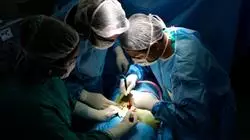
This program is based on the latest advances in educational technology, based on e-learning methodology"
Syllabus
The structure of the contents has been designed by a team of expert surgeons, which encompasses all the updates in spinal pathology.
This Professional master’s degree has a quality program adapted to the latest trends in the field of Spinal Surgery"
Module 1. Surgical Approaches to the Spine
1.1. Cervical Spine Approaches
1.1.1. Cervical Anatomy
1.1.2. Muscles and Anatomical Limits
1.1.3. Neurological Structures and Their Location
1.1.5. Anterior Approaches to the Cervical Spine
1.1.6. C1-C2 Transoral Approach
1.1.7. Lateral Approaches to the Cervical Spine
1.1.8. Which Approach to Perform: Right or Left?
1.1.9. Approaches to the Cervicothoracic Junction
1.1.10. Posterior Approaches to the Cervical Spine
1.1.11. Posterior Approach to the C1-C2 Joints
1.1.12. Posterior Cervical Foraminotomy
1.1.13. Complications of Cervical Spine Surgery
1.1.14. Bleeding
1.1.15. Durable Lesions
1.1.16. Alterations of Pharynx
1.1.17. Esophageal Injuries
1.1.18. Postoperative Management of Cervical Surgery Patients
1.2. Thoracic Spine Approaches
1.2.1. General Indications
1.2.2. Absolute and Relative Contraindications
1.2.3. Preoperative Planning
1.2.4. Anterior Approaches to Thoracic Spine
1.2.5. DIV-DXI Transthoracic Approach
1.2.6. Transpleural Anterior Approach DIII-DXI Louis
1.2.7. Thoracolumbar Junction Approaches
1.2.8. Transpleural-Retroperitoneal Approach
1.2.9. Extrapleural Approaches
1.2.10. Video-endoscopic Approach to the Thoracic Spine
1.2.11. Posterior and Posterolateral Approaches to the Thoracic Spine
Thoracic Disc Access
1.2.12. Costotransversectomy
1.2.13. Post-Operative Care
1.3. Lumbar Spine Approaches
1.3.1. Anterior Approach
1.3.2. L2-L5 Retroperitoneal Anterior Approaches
1.3.3. Extraperitoneal Anterior Approach with Median Incision for L2-L Levels
1.3.4. Anterior Pararectal Approach Retroperitoneal to L5-S1.
1.3.5. Laparoscopic Transperitoneal Approach to L5-S1.
1.3.6. Lateral Oblique Approach of the Lumbar Spine to L2-L5.
1.3.7. En Bloc Sacrectom
1.4. Lateral Approaches
1.4.1. Lateral Approach for Discectomies, Foraminotomies or XLIF Lateral Fusions
1.4.2. Microscopic or Minimally Invasive Lumbar Discectomy
1.5. Posterior Approach
1.5.1. Posterior Approaches to the Cervical Spine
1.5.2. Lumbar Paraspinous Spinal Cord Approaches
1.5.3. Foraminal Approach to the Lumbar Disc
1.6. Complications of Thoracolumbar and Lumbar Spine Approaches
Module 2. Cervical Spine Pathology
2.1. General Aspects. Cervical Pain Diagnosis
2.1.1. Anatomy and Biomechanics of the Cervical Spine, Applied to the New Surgical Techniques
2.1.2. Biochemical and Cellular Bases of Intervertebral Disc Degeneration
2.1.3. Advances in Diagnostic Imaging of Cervical Spine Diseases
2.1.4. Assessment of Neurological Compromise. Clinical and Neurophysiology
2.1.5. Posterior Access to the Craniovertebral Junction: Importance of Developing a Very Meticulous Technique
2.2. Therapeutic Overview Cervical Pain
2.2.1. Cervical Pain, Radiculopathy and Cervical Myelopathy. Pathophysiology and Natural History
2.2.2. Outcome Measurement Scales in Cervical Pathology
2.2.3. Semi-Invasive Treatment of Cervical Pain and Cervicobrachialgia
2.2.4. Role of Rehabilitation in the Treatment of Cervical Degenerative Processes. Different Protocols
2.3. Cervical Radicular Pain Surgery
2.3.1. Analyze the Causes and Incidence of Radicular Pain in the Cervical Spine
2.3.2. Justify the Different Indications for Surgery
2.3.3. Role of Posterior Approach and Microsurgery in Cervical Radiculopathy
2.3.4. Evidence on Conservative versus Surgical Treatment of Cervical Radiculopathy. Literature Review
2.3.5. Herniated Cervical Disc. Anterior or Posterior Approach
2.3.6. Fusion Techniques or Disc Prosthesis
2.3.7. Identify Factors Influencing the Regression of Cervical Hernia Symptoms
2.3.8. Assess the Surgical Options
2.3.9. Anticipate Possible Complications and Find a Return to Work Plan and Activity
2.3.10 Formulate a Plan for When Problems Arise at the Adjacent Level
2.4. Whiplash Syndrome. Whiplash
2.4.1. Pathophysiology, Anatomopathologic Alterations
2.4.2. Initial Assessment of the Patient. Prognostic Factors
2.4.3. Acute Phase Treatment. Treatment in the Sequelae Phase
2.4.4. Socioeconomic Impacts of Whiplash Syndrome
2.5. Cervical Degenerative Pathology
2.5.1. Multilevel Cervical Spondylosis
2.5.2. Anterior Approach
2.5.3. Posterior Approach
2.5.4. Adjacent Segment Degeneration. How To Act
2.5.5. Cervical Spine Deformities
2.6. Cervical Myelopathy
2.6.1. Manifestations, Causes and Natural History of Myelopathy
2.6.1.1. Compare the Clinical and Functional Presentation of Spondylotic Myelopathic Syndromes
2.6.1.2. Degrees of Disease Using Validated Scales
2.6.1.3. Anticipate Appearance of Clinical Signs and Consider Differential Diagnoses
2.6.1.4. Describe the Natural History of the Disease
2.6.1.5. Identify the Particular Clinical Presentation of Cervicocranial Myelopathy
2.6.2. Clinical and Surgical Decision Making in Myelopathy
2.6.2.1. Define a Treatment Plan for Patients with Myelopathy
2.6.2.2. Identify the Absolute and Relative Indications for Surgery in Spondylotic Myelopathy
2.6.2.3. Compare the Different Surgical Approaches in Myelopathy and Define a Rational Treatment Plan
2.6.2.4. Justification of the Anterior Route
2.6.2.5. Discuss the Role of Intraoperative Neuromonitoring in Cervical Myelopathy
2.6.2.6. Cervical Spondylotic Myelopathy Updating and Therapeutic Guidance
2.6.2.7. Management of Cervical Myelopathy Anterior Route. Multilevel Pathology
2.6.2.8. Use of Corpectomy or Boxes
2.6.2.9. Management of Cervical Myelopathy Posterior Route. Multilevel Pathology
2.6.2.10. Thoracic Scalene Gorge Syndrome
2.7. Cervical Spine Trauma
2.7.1. Imaging in Cervical Trauma
2.7.1.1. Selection of Appropriate Imaging to Detect Cervical Fracture
2.7.1.2. Evaluate Radiological Imaging Options
2.7.1.3. Select Appropriate CT or MRI Images
2.7.1.4. Differentiate Between Major and Minor Trauma of the Upper Cervical Spine
2.7.2. Occipito-Cervical Instability
2.7.2.1. Anatomy and Biomechanics of the Upper Cervical Spine
2.7.2.2. Types of Instabilities
2.7.2.3. Post-Traumatic
2.7.2.4. Congenital
2.7.3. Fractures of the Upper C0-C2 Region: Classification and Management
2.7.3.1. Define the Role of Ligaments in the Stability of the Cervical Spine
2.7.3.2. Classify in Relation to Subsequent Treatment
2.7.3.3. Occipital Condyle Fractures
2.7.3.4. Occipito-Cervical Dislocation, Occipito-Atloid Dislocation, or Attalanto-Axoid Instability. Atlas or C1 Fractures
2.7.3.5. Atlas or C2 Fractures
2.7.3.6. Traumatic Spondylolisthesis of C2
2.7.4. Subaxial Cervical Spine Traumas
2.7.4.1. Subaxial Cervical Spine Trauma. Classification and Management
2.7.4.2. Estimate Incidence and Classify using AO Classification
2.7.4.3. Use Other Classifications to Determine Treatment
2.7.4.4. Anticipate Errors in Diagnosis
2.7.4.5. Evaluate Non-Surgical Treatment Options and When They Are Appropriate
2.7.4.6. Recognize the Indications for Surgical Treatment
2.7.4.7. Recognize those Fractures that Require Urgent Treatment
2.7.4.8. Justify the Different Fracture Approaches, Anterior or Posterior Approach or Combined Anterior and Posterior Approaches
2.8. Inflammatory and Infectious Pathology in Cervical Spine
2.8.1. Current Treatment of Cervical Spine Infections
2.8.2. Rheumatoid Arthritis of the Cervical Spine
2.8.3. Cervical Instability in Down Syndrome
2.8.4. Pathology of the Craniocervical Region. Anterior Surgical Treatment
Module 3. Herniated Discs, Diagnosis and Treatment of Radicular Pain. Emerging Technologies for the Treatment of Low Back Pain
3.1. Epidemiology, Natural History and Imaging Findings in Radicular Pain
3.1.1. Use of the Usual Epidemiological Terms to Define and Prevalence Factors of Radicular Pain
3.1.2. Knowledge of the Natural History of Radicular Pain
3.1.3. Identify Contributing Fractures
3.1.4. Diagnosis of the Causes of Radicular Pain
3.1.5. Assess Why Disc Herniations Occur
3.1.6. Differentiate the Role of Diagnostic Imaging between Computerized Axial Tomography (CT) and Magnetic Resonance Imaging (MRI) for Radicular Pain
3.1.7. Interpretation of Images Using the Correct Nomenclature
3.2. Non-Surgical Action in the Treatment of Radicular Pain
3.2.1. Evaluate Non-Surgical Treatment Options for Radicular Pain
3.2.2. Explaining These Options to Patients
3.2.3. Identify Patients who are Susceptible to Non-Surgical Treatment
3.2.4. Differentiate Between Types of Analgesia. Analgesic Scale
3.2.5. Summarize the Roles of Rehabilitation and Physiotherapy
3.3. Surgery of Radicular Pain in the Lumbar Spine
3.3.1. Differences Between the Various Absolute and Relative Indications for Surgery
3.3.2. Identify Appropriate Timing for Surgery
3.3.3. Evaluate the Usual Surgical Techniques with Evidence Support
3.3.4. Compare Both Surgical and Non-Surgical Treatment Options
3.3.5. Formulate an Adequate Surgical Plan
3.3.6. Anticipate Possible Complications and Establish a Plan for Return to Work and Activity
3.4. Thoracic Myelopathy
3.4.1. Myelopathic Imaging: Techniques and Prognostic Indicators
3.4.2. Interpret MRI and CT Findings in Spondylotic Myelopathy
3.4.3. Recognize the Change of Signs in Different MRI Sequences and their Significance
3.4.4. Consider the Different Differential Diagnoses in Non-Tumor Pathology of the Spinal Cord
3.4.5. Know the Current Role of Myelography and Mielo-TAC in Myelopathy Imaging
3.4.5.1. Clinical and Surgical Decision Making in Thoracic Myelopathy
3.4.5.2. Management of the Risk-Benefit Balance in the Surgery of Patients with Thoracic Myelopathy
3.4.5.3. Compare the Different Approaches to Thoracic Myelopathy
3.5. Axial Low Back Pain
3.5.1. Natural History. Obstacles to Recovery and Aspects of Non-Surgical Treatment of Axial Pain
3.5.1.1. Anticipate Potential Obstacles to Recovery
3.5.1.2. Explain How Mood Can Be Used
3.5.1.3. How to Handle Catastrophic Ideas
3.5.1.4. Differentiate Between Acute and Chronic Low Back Pain
3.5.1.5. Assess the Different Options for the Non-Surgical Treatment of Low Back Pain
3.5.1.6. Summarize the Current State of the Evidence Regarding Surgical and Non-Surgical Management
3.5.2. How to Assess a Patient with Axial Pain?
3.5.2.1. Understand the Role of Natural History and Physical Examination in the Assessment of these Patients with Axial Pain
3.5.2.2. Decide on the Need for Imaging Studies
3.5.2.3. Select Patients with Axial Pain in Need of Advanced Diagnostic Techniques
3.5.2.4. Review the Role of Diagnostic Blocks and Discography in Patients with Axial Pain
3.5.2.5. Conservative Treatment of Low Back Pain
3.5.2.6. Opioid Practice and Perspective: Who is at Risk of Addiction?
3.5.2.7. Radiofrequency Ablation in Low Back Pain
3.5.2.8. Stem Cells and Intradiscal Lumbar Procedures
3.5.2.9. Implantable Therapies for Chronic Low Back Pain
3.5.3. Axial Lumbar Pain Surgery
3.5.3.1. Promote a Rational Alternative to Surgical Fusion
3.5.3.2. Evaluate Alternative Options
3.5.3.3. Select an Appropriate Approach
3.5.3.4. Check Current Evidence
Module 4. Degenerative Dorsolumbar Pathology Advances
4.1. Spinal Canal Stenosis and Degenerative Spondylolisthesis
4.1.1. Presentation, Clinical and Non-surgical Treatment of Lumbar Canal Stenosis
4.1.1.1. Knowledge of the Signs and Symptoms of Lumbar Spinal Stenosis (LSS)
4.1.1.2. Know the Clinical Characteristics and Natural History of Neurogenic Claudication
4.1.1.3. Classify Lumbar Spinal Stenosis
4.1.1.4. Assess Surgical and Non-Surgical Treatment Options
4.1.1.5. Know the Alternatives of Rehabilitation
4.1.2. Imaging of Lumbar Spinal Stenosis and Degenerative Spondylolisthesis
4.1.2.1. Describe the Different Imaging Techniques to Identify Lumbar Spinal Stenosis and Degenerative Spondylolisthesis
4.1.2.2. Classification and Degree of Lumbar Spinal Stenosis
4.1.2.3. Appreciate the Role of Complete and Functional Spine Radiographs in the Management of Patients with Lumbar Spinal Stenosis and Degenerative Spondylolisthesis
4.1.3. Surgical Treatment of Lumbar Spinal Stenosis
4.1.3.1. Formulate the Principles of Stenosis Surgery
4.1.3.2. Individualize the Surgical Technique for Each Patient
4.1.3.3. Recognize the Indications for Fusion in Patients with Lumbar Spinal Stenosis
4.1.4. Surgical Treatment of Degenerative Spondylolisthesis
4.1.4.1. Assess Surgical and Non-Surgical Treatment Options in Degenerative Spondylolisthesis
4.1.4.2. Summarize the Controversies in the Choice of Treatment in Degenerative Spondylolisthesis
4.2. Spondylolysis and Low-Grade Isthmic Spondylolisisthesis
4.2.1. Spondylolysis and Low-Grade Spondylolisisthesis
4.2.1.1. Epidemiological Aspects and Natural History
4.2.1.2. Describe the Signs and Symptoms of Spondylolysis and Low-Grade Spondylolisthesis
4.2.1.3. Formulate the Principles of its Therapeutic Management
4.2.1.4. Assess the Different Therapeutic Options
4.2.1.5. Anticipate Possible Complications of the Instrumentation and its Positioning
4.2.1.6. Analyze the Alternatives of Rehabilitation
4.3. Degenerative Deformity
4.3.1. Lumbar Degenerative Deformity
4.3.1.1. Describe the Pathogenesis and Natural History of Lumbar Degenerative Deformity
4.3.1.2. Explain the Concept of Spinal Balance and the Different Spinopelvic Parameters
4.3.1.3. Assess the Risk-Benefit Balance for Surgery and Potential Complications
4.3.1.4. Formulate a Surgical Plan for Degenerative Kyphoscoliosis
4.3.1.5. Pelvis Fixations
4.4. Advances in the Design of New Implants
4.4.1. Posterior or Posterolateral Instrumentations
4.4.2. Anterior Instrumentation
4.4.3. Interbody Implants
4.4.4. Disc Prosthesis
Module 5. Advances in Vertebral Deformities Treatment
5.1. Neuromuscular Scoliosis. Management and Advances
5.1.1. Classification, Generalities and Preoperative Planning of Neuromuscular Scoliosis
5.1.2. Assessment of Respiratory Function in Neuromuscular Scoliosis. Indications for the use of BiPAP Before or After Surgery
5.1.3. Anesthesia in Neuromuscular Patients
5.1.4. Intraoperative Monitoring
5.1.5. Use of Evoked Potentials in Patients with Non-Ambulatory Neuromuscular Scoliosis
5.1.6. Indication and Contraindication of the Anterior Approach in Neuromuscular Scoliosis
5.1.7. Surgical Treatment by Posterior Approach, Pedicle Screws and Instrumentation with Sublaminar Wires
5.1.8. Techniques and Indications for Lumbosacral Fixation
5.1.9. Indications for Growth Systems in Severe Neuromuscular Infantile Scoliosis
5.1.10. Evolution and Treatment of Neuromuscular Scoliosis in Adulthood
5.2. Congenital Scoliosis. Overview and Diagnosis
5.2.1. Classification of Congenital Scoliosis. Surgical Action Protocol
5.2.2. Alterations Associated with Vertebral Deformities. Pr-Operative Assessment
5.2.3. Neurosurgical Performance of Spinal Cord Disorders in Congenital Deformities
5.2.4. Surgical Strategy in Congenital Kyphosis. Classification and Types
5.2.5. Complex Congenital Scoliosis. Indications for Pedicular Subtraction Osteotomies
5.2.6. Hemivertebra Resection by Double Anterior Posterior Approach versus Posterior Approach
5.2.7. Treatment of Rib Malformations Associated with Congenital Malformations, VERTR Indications
5.2.8. Treatment and Evolution of Klippel-Feil Syndrome in Adulthood
5.3. Idiopathic Juvenile Scoliosis. Advances
5.3.1. Current State of Knowledge on the Etiology of Idiopathic Scoliosis
5.3.2. Natural History of Idiopathic Scoliosis after Maturation
5.3.3. Clinical Assessment of the Patient with Idiopathic Scoliosis
5.3.4. Cardiopulmonary Assessment
5.3.5. Review of Non-Fusion Techniques in Early Onset Scoliosis. What We Have Done and What's Next
5.3.6. Factors Predicting Progression During Growth
5.3.7. Conservative Treatment
5.3.7.1. Orthotic Treatment of Idiopathic Scoliosis
5.3.7.2. Observation as a Treatment
5.3.8. Surgical Management
5.3.8.1. Posterior Arthrodesis and Hybrid Instrumentation: Standard Technique
5.3.8.2. Posterior Arthrodesis with Thoracic Pedicle Screws
5.3.9. Treatment of Thoracolumbar Curves
5.3.9.1. Instrumented Anterior Arthrodesis
5.3.9.2. Instrumented Posterior Arthrodesis
5.3.10. Systematic Work in the Choice of Fusion Levels
5.3.11. Growth Bar Gradation. When and Which Options?
5.3.12. Are the Current Results of Instrumented Fusion in Adolescent Idiopathic Scoliosis Acceptable?
5.3.13. "Tethering" in Adolescent Idiopathic Scoliosis
5.3.14. Tricks and Errors to Limit Arthrodesis and Avoid Trunk Imbalance in Adolescent Idiopathic Scoliosis
5.3.15. Severe Untreated Deformities
5.3.16. Pediatric Reconstructive Surgery
5.3.17. Building Multidisciplinary Teams in the Care of Patients with Spinal Deformity
5.3.18. Clinical and Functional Results
5.3.18.1. Results of Idiopathic Scoliosis Surgery
5.3.18.2. Radiological Results of the Different Fusion Techniques
5.4. Adult Deformities
5.4.1. What is the Evidence for Conservative Treatment of Adult Scoliosis?
5.4.2. Toward a Classification Scheme in Adult Scoliosis that Predicts Quality Outcomes
5.4.3. How Does the Degenerative Process Affect Surgical Decisions/Approach?
5.4.4. Reconstruction of Sagittal Alignment. Lessons Learned over the last 10 Years
5.4.5. Surgical Management of Spinal Deformity in Frail Patients
5.4.6. Value of Databases and Predictive Models for Improving Results in Adult Scoliosis. Risk Calculators
5.4.7. Care of Patients with Adult Scoliosis
5.4.8. Failed Adult Scoliosis Surgery. Reconstruction
5.4.9. Cost-Effectiveness of Surgical Treatment of Adult Scoliosis Deformity
5.5. Reconstructive Surgery and Vertebral Osteotomies
5.5.1. Vertebral Osteotomies: Types and Historical Evolution
5.5.2. Reconstructive Surgery in Pediatric Spine: Causes and Prevention
5.5.3. Reconstructive Surgery in Adult Spine: Causes and Prevention
5.5.4. Reconstructive Surgical Strategy. Choice of Osteotomy
5.5.5. Cervical Spine and Cervical/Thoracic Hinge. Surgical Strategy
5.5.6. Reconstructive Surgery of Coronal Imbalance
5.5.7. Reconstructive Surgery of Sagittal Imbalance. Thoracic and Lumbar Osteotomies
5.5.7.1. Tri-Columnar Osteotomies. Pedicular Subtraction
5.5.7.2. Ponte Osteotomies. Smith-Petersen
5.5.7.3. Other Osteotomies
5.5.8. Lumbosacral Reconstructive Surgery. Spondylolisthesis Sacral/Pelvis Osteotomies
5.5.9. Optimize Safety in Reconstructive Spine Surgery
5.5.10. Outcome of Thoracolumbar Osteotomies in the Adult
Module 6. Spinal Tumors
6.1. General Information of Vertebral Tumors
6.1.1. Pathophysiology of Vertebral Tumors
6.1.2. Prevalence and Incidence
6.1.3. Form of Presentation and Common Symptoms of Spinal Tumors
6.1.4. Physical Examination and Laboratory Studies
6.1.5. Why are Spinal Tumors a Big Problem?
6.1.6. Common Radiotherapy Techniques for Spinal Tumors, their Indications and Special Technical Considerations
6.1.7. Effects of Chemotherapy on the Malignant Cells of These Tumors
6.2. Management of the Patient with Suspected Vertebral Tumor
6.2.1. Diagnostic Imaging and Percutaneous Biopsy
6.2.2. Principles and Approaches to Perform Biopsies
6.2.3. Histological Management of the Sample
6.3. Benign Primary Tumors
6.3.1. Main Benign Tumors of the Spine
6.3.2. Description and Indications for Percutaneous Surgery
6.3.3. Surgical Management
6.4. Primary Malignant Tumors of the Spine
6.4.1. Main Primary Malignant Tumors of the Spine
6.4.1.1. Multiple Myeloma and Plasmacytoma
6.4.1.2. Lymphoma
6.4.2. Oncologic and Surgical Staging
6.4.3. Low-grade and High-grade Malignant Tumors
6.4.4. Posterior Surgical Treatment. Technique of Resection in Bloc of Thoracolumbar and Cervical Tumors. Sacral Tumor Resections
6.4.5. Radiotherapy in Malignant Tumors. Indications and Results
6.4.6. Results and Complications of Surgery
6.5. Vertebral Metastases
6.5.1. Pathophysiology of Vertebral Metastases and Oncologic Management of the Patients
6.5.2. Main Metastatic Tumors in the Spine
6.5.2.1. Lung, Breast, Genitourinary, Gastrointestinal
6.5.2.2. Scales of Assessment and Prognosis
6.5.2.3. Oncologic Management. Radiotherapy Indications and Results
6.5.2.4. Pain Management in Patients with Vertebral Metastases
6.6. Surgical Management of Metastases
6.6.1. Application of the Treatment Protocol. Indications for Surgery of Vertebral Metastases
6.6.2. Percutaneous Treatment Vertebroplasty and Kyphoplasty
6.6.3. Palliative Versus Radical Treatment in Vertebral Metastases
6.6.4. Complications of Surgery and Medical Care. How to Anticipate and Manage Them
Module 7. Advances in Vertebral Fractures Treatment
7.1. Trauma of the Thoracolumbar Spine and Sacrum
7.1.1. Imaging in Thoracolumbar and Sacral Fractures
7.1.1.1. Use of the AO Classification
7.1.1.2. Selection of the Most Appropriate Images to Identify Major or Minor Trauma
7.1.1.3. Management and Use of Radiological Images
7.1.1.4. Define the Indications for Appropriate Use of CT or MRI
7.1.1.5. Recognize Special Circumstances that Compromise Spinal Cord Function
7.1.2. Thoracolumbar Spine Trauma; Classification and Management
7.1.2.1. Recognize the Signs and Symptoms of Thoracolumbar Fractures
7.1.2.2. Differentiate Between Denis, AO and TLICS Classifications
7.1.2.3. Explain the Role of Ligaments in Burst Fractures
7.1.2.4. Assess the Different Surgical Techniques: Anterior Approach including MIS Techniques or Posterior Approach including MIS Technique or Both Approaches
7.1.3. Sacral Fractures: Classification and Treatment
7.1.3.1. Description of Important Anatomical Aspects
7.1.3.2. Differentiate the Different Types of Sacral Fractures
7.1.3.3. Use of the AO Classification
7.1.3.4. Recognize the Signs and Symptoms of Sacral Fractures
7.1.3.5. Compare Surgical or Conservative Treatment
7.1.3.6. Assess the Correct Surgical Options
7.2. Cementation Techniques via MIS
7.2.1. Explanation of the Steps to Perform a Cementoplasty Technique, Including Correct Patient Positioning
7.2.2. Correct Positioning of the Fluoroscope
7.2.3. Placement of the Jamshidi Needles and their Exchange for the Working Cannula
7.2.4. Fixation with Cemented Screws via MIS. Indications
7.2.5. Explanation of the Steps to Performing a Pedicle Screw Fixation Technique and Performing a Cementoplasty, Including Correct Patient Positioning
7.2.6. Placement of Jamshidi Needles and Subsequent Tapping and Screw Placement
7.2.7. How the Cement is Injected into the Vertebrae and its Particularities
7.2.8. Placement of Percutaneous Bars
7.3. Fractures in Metabolic Spine Disorders and Pediatric Spine Fractures
7.3.1. Fractures in Ankylosing Spondylitis (AS): Characteristics and Treatment
7.3.1.1. Etiology of Ankylosing Spondylitis
7.3.1.2. Determine the Role of the Spine Surgeon in AS
7.3.1.3. Identify what Type of Imaging is Needed for its Diagnosis and Why
7.3.1.4. Formulating an Appropriate Treatment Plan for AS Fractures
7.3.1.5. Anticipate Difficulties in this Patient Population
7.3.2. Vertebral Osteoporotic Fractures. Diagnosis and Treatment
7.3.2.1. Define Osteoporosis
7.3.2.2. Description of the Medical Therapeutic Treatment of Osteoporosis
7.3.2.3. Know the Diagnosis of Osteoporotic Vertebral Fractures
7.3.2.4. Use of the AO Classification for Osteoporotic Vertebral Fractures
7.3.2.5. Assess the Different Surgical Alternatives
7.3.2.6. Recognize the Indications for Cementoplasty Procedures for Osteoporotic Vertebral Fractures
7.3.2.7. Recognize the Indications for Instrumentation of the Spine with or without Cementoplasty
7.3.3. Pediatric Spine Fractures. Characteristics and Treatment
7.3.3.1. Characteristics of Immature Cervical and Thoracolumbar Spine Fractures
7.3.3.2. Define SCIWORA/SCIWORET
7.3.3.3. Explain the Mechanism of Cervical Spine and Lumbar Apophysis Injuries
7.3.3.4. Determine the Appropriate Plan for Diagnosis and Treatment of Injuries
7.4. Post-traumatic Kyphosis
7.4.1. Prevention and Treatment of Post-traumatic Kyphosis
7.4.1.1. Discussion of the Reasons for Post-traumatic Kyphosis
7.4.1.2. Formulate Treatment Objectives
7.4.1.3. Explain How to Restore Sagittal Balance
7.4.1.4. Assess the Surgical Options
7.4.1.5. Justify the Approach by a Multidisciplinary Team
7.5. Diagnosis of Vertebromedullary Trauma
7.5.1. General Aspects
7.5.1.1. Vertebral Fractures with Neurological Compromise. Biomechanics. Stability Criteria. Diagnostic Tools
7.5.1.2. Differential Imaging Diagnosis of Vertebral Injury with Neurological Involvement
7.5.1.3. Clinical Assessment of Traumatic Spinal Cord Injury. Spinal Syndromes, ASIA Scale
7.5.1.4. Differentiation with other Spinal Cord Injuries. Determination of the Severity of the Spinal Cord Injury. Current Diagnostic Options in the Acute Phase
7.5.2. Spinal Shock and Incomplete Spinal Cord Injury Syndrome (SCI)
7.5.2.1. Pathophysiology of Traumatic Spinal Cord Injury. Differentiation from other Spinal Cord Injuries
7.5.2.2. Define the Different Types of Incomplete Spinal Cord Injury (SCI)
7.5.2.3. Classify SCI Using the ASIA Scale and Justify its Clinical and Surgical Relevance. Describe the Clinical Symptoms and Pathophysiology of Central Cord Syndrome
7.5.2.4. Initial Surgical Management of Traumatic Spinal Cord Injury: Early vs. Delayed Surgery
7.5.2.5. Define why Methylprednisolone Should Not Be Used in SCI (NACIS I-III)
7.5.2.6. Treatment of Neuropathic Pain and Spasticity
7.5.2.7. Treatment of Post-traumatic Syringomyelia and Late Deformity
7.5.2.8. Rehabilitation of the Spinal Cord Injured
7.5.2.9. Initial Adaptation to Spinal Cord Injury and Return and Social Participation
7.5.2.10. Current Clinical Application of Tissue Regeneration Therapies
7.5.3. Initial Management of Traumatic Spinal Cord Injury
7.5.3.1. Immobilization and Transport of the Critically Ill Patient with Traumatic Spinal Cord Injury
7.5.3.2. Timing and Initial Medical Management of Traumatic Spinal Cord Injury. Validity of the NASCIS Protocol. Importance of Specific Units
7.5.3.3. Variability of Surgical Treatment of Spinal Cord Injury in Spain
7.5.4. Surgical Management of Vertebro-Medullary Trauma
7.5.4.1. Surgical Treatment of Unstable C1-C2 Fractures
7.5.4.2. Treatment of Thoracolumbar Fractures with Neurological Compromise
7.5.4.3. Advantages of the Previous Route
7.5.4.4. Advantages of the Posterior Route
7.5.5. Surgical Management in Special Situations
7.5.5.1. Pediatric Spinal Cord Injury SCIWORA. Diagnosis and Treatment
7.5.5.2. Traumatic Neurological Injury in Patients with Cervical Myelopathy
7.5.5.3. Unstable Fractures in Patients with Ankylopoietic Spondylitis
7.5.5.4. Fractures with Neurological Injury in the Patient with Osteoporosis
7.5.5.5. Natural History of the Spinal Cord Injured. Complications. Prognostic Factors
7.5.5.6. Management of Heterotopic Ossification. Management of Pressure Ulcers
Module 8. Advances in Minimally Invasive Surgery
8.1. Cervical Spine
8.1.1. Minimally Invasive Surgical Techniques for the Treatment of Cervical Disc Herniation
8.1.2. Posterior Cervical Foraminotomy
8.1.3. Intervertebral Disc Replacement by Minimally Invasive Surgery
8.1.4. Posterior Cervical Fixation by Minimally Invasive Surgery
8.1.5. Fixation of Odontoid Fractures by Minimally Invasive Surgery
8.1.6. D-TRAX
8.2. Thoracic and Lumbar Spine
8.2.1. Minimally Invasive Surgical Techniques for the Treatment of Thoracic Disc Herniation
8.2.2. Endoscopic Techniques in the Management of Lumbar Disc Herniation
8.2.3. Lateral Extraforaminal Approach
8.2.4. Translaminar Approach
8.2.5. Transforaminal Approach
8.2.6. Nucleus Pulposus Replacement Technology
8.2.7. Translaminar Articular Facet Fusion Techniques with Screw and Other Devices
8.2.8. Microsurgical Decompression of Central and Lateral Canal Stenosis
8.2.9. Pedicle Screw Placement by Minimally Invasive Surgery
8.2.10. Posterior Approach Fusion Techniques. Minimally Invasive TLIF. Advantages and Disadvantages
8.2.11. Laparoscopic ALIF
8.2.12. Lateral Approaches for XLIF Intersomatic Arthrodesis. Technical Anatomy and Results
8.2.13. Sacroiliac Joint Fusion Percutaneous Access
8.3. Minimally Invasive Surgery in the Deformities
8.3.1. What are the Limits of Minimally Invasive Surgery in Deformity Correction? Indications
8.3.2. Realignment of the Anterior Spine
8.3.3. Posterior Correction Techniques
8.3.4. Posterior Percutaneous Fixation. Reduction Techniques
8.3.5. Temporary Fixation Technique
8.3.6. Indications for Minimally Invasive Techniques in Revision Surgery
8.3.7. Advantages and Disadvantages for Minimally Invasive Techniques in Revision Surgery
8.3.8. Complications in Previous Approaches and How to Avoid Them
8.3.9. Complications in Posterior Approaches and How to Avoid Them
8.4. Interspinous and Interlaminar Devices
8.4.1. Percutaneous Dynamic Stabilization Techniques with Interspinous Implants
8.4.2. Technical and Anatomical Considerations of Interspinous Implant Placement
8.4.3. Advances in Devices
8.5. Pain Treatment Techniques for Minimally Invasive Surgery
8.5.1. Radiofrequency Neurotomy of the Lumbar Articular Facets
8.5.2. Spinal Cord Electrostimulation for Chronic Pain
8.5.3. Epiduroscopy
8.6. Treatment of Fractures by Minimally Invasive Techniques
8.6.1. Role of Vertebroplasty and its Complications
8.6.2. Role of Kyphoplasty and its Complications
8.6.3. Other Percutaneous Treatment Techniques for Osteoporotic Vertebral Compression Fractures
Module 9. Spine Surgery in the Elderly
9.1. General Aspects of the Elderly Spine
9.1.1. Medical Aspects to be Taken into Consideration in the Elderly Patient Undergoing Spine Surgery
9.1.2. Anesthetic Aspects to be Taken into Cosideration in the Elderly Patient who is Undergoing Spine Surgery
9.2. Management of Vertebral Fractures in Elderly Patients
9.2.1. Conservative Treatment of Fractures in the Elderly
9.2.2. Odontoid Fractures in Elderly Patients: Functional and Quality of Life Outcomes of Patients With and Without Surgery
9.2.3. Fractures in Elderly Patients with Ankylopoietic Spondylitis
9.2.4. Quality of Life in Elderly Patients with Fractures
9.2.5. Surgical Treatment of Osteoporotic Compression Fractures in the Elderly
9.2.5.1. Role of Vertebroplasty
9.2.5.2. Kyphoplasty Role
9.2.5.3. Structural Osteoplasty Role
9.2.5.4. Vesselplasty
9.2.5.5. Use of Biologicals
9.3. Conservative Treatment of Spinal Pain in the Elderly
9.3.1. Rehabilitation in the Elderly Patient
9.3.2. Alternative Therapies. Yoga, Acupuncture. Aquagym, Pilates
9.3.3. Use of Spinal Injections in Spinal Pain
9.3.4. Use of Oral Medication. NSAIDs, Morphic. In Low Back Pain in the Elderly
9.4. Treatment of Tumors in Elderly Patients
9.4.1. Treatment of Bone Metastases in the Spine of the Elderly Patient
9.4.2. Minimally Invasive Approaches
9.5. Surgical Aspects of Scoliosis in Elderly Patients
9.5.1. The Sagittal Profile People Over 65 Years of Age: Characteristics and Radiographic Analysis
9.5.2. Clinical Evaluation of the Sagittal Plane: How to Integrate Sagittal Balance into Clinical Practice?
9.5.3. Scoliosis in Elderly Patients: Prevalence. Pathophysiology. Classification. Indications and Objective of the Surgery
9.5.4. Levels of Fusion in Scoliosis of Elderly Patients. Spinal Instrumentation
9.5.5. Dual Approach Versus All Posterior Approach in Scoliosis in the Elderly
9.5.6. Vertebral Osteotomies. How to Choose the Correct Surgical Approach
9.6. Specific Surgical Complications in the Elderly Patient
9.6.1. Coronal and Sagittal Imbalance. Flat Back Prevention. Treatment
9.6.2. Pseudarthrosis and Infection After Deformity Surgery in the Elderly Patient
9.6.3. Adjacent Disc/Segment Syndrome (Proximal and Distal)
9.6.4. Surgical Complications. How to Minimize Postoperative Complications. Who is at Risk and What is the Risk?
9.6.5. Proximal Junctional Kyphosis and Progressive Deformity. How to Minimize and Manage it
9.7. Other Degenerative Pathologies
9.7.1. Cervical Myelopathy in the Elderly
9.7.2. Degenerative Kyphosis: Influence of Osteoporotic Fractures
9.7.3. Lumbar Degenerative Stenosis and Spondylolisthesis
9.7.4. Thoracolumbar Spinal Cord Compression in the Elderly
9.7.5. Minimally Invasive Surgery in Patients Over 65 Years of Age
Module 10. Complications in Spine Surgery Miscellaneous
10.1. Neurological Complications in Spine Surgery
10.1.1. Dura Mater Tears
10.1.1.1. Conservative Management of Dural Tears
10.1.1.2. Primary Repair
10.1.1.3. Secondary Action
10.1.2. Nerve Root Injuries
10.1.2.1. Direct Injury to Nerves During Surgery
10.1.2.2. Peripheral Neuropathies due to Patient Positioning
10.1.3. Neurological Complications related to Bone Grafts
10.2. Vascular Complications
10.2.1. Vascular Injuries in Spine Surgery
10.2.2. Anterior Cervical Vascular Injuries
10.2.3. Thoracic Vascular Complications
10.2.3.1. Anterior Approach
10.2.3.2. Posterior Approach
10.2.4. Lumbar Vascular Complications
10.2.4.1. Anterior Approach
10.2.4.2. Posterior Approach
10.2.5. Other Vascular Complications
10.3. Spinal Infections
10.3.1. Main Pathogens in Spine Surgery
10.3.2. Causes of the Infections. Risk factors
10.3.3. Diagnostic and Imaging Tests
10.3.4. Spondylodiscitis
10.3.5. Post-Surgical Infections
10.3.6. Treatment Planning
10.3.6.1. Antibiotic Medical Treatment
10.3.6.2. Treatment of Surgical Wounds Vacuum Systems
10.4. Complications Derived from the Surgical Procedure
10.4.1. Failed Back Syndrome. Classification
10.4.1.1. Reasons for Failure of Surgical Instrumentation
10.4.1.2. Post-Operative Vertebral Instability
10.4.1.3. Post-Operative Deformities
10.4.1.4. Pseudarthrosis
10.4.2. Adjacent Level Diseases. Therapeutic Approach
10.4.3. Revision Surgery. Strategies
10.5. Evaluation and Treatment of Sacroiliac Pathology
10.6. Navigation and Robotics in Thoracolumbar Spine Surgery
10.7. Use of Bone Grafts in Spine Surgery
10.7.1. Autograft and Allograft
10.7.2. Demineralized Bone Matrix and Osteoconductive Ceramics
10.7.3. Biological Substitutes
10.7.4. Grafts in Revision Surgeries
10.7.5. Stem Cells and Cellular Bone Matrix
10.8. Evaluation and Monitoring Tools in Spine Surgery
10.8.1. Neurological Assessment Scales
10.8.2. SF-36, VAS, Oswestr
A unique, key and decisive experience to boost your professional development"
Professional Master's Degree in Spine Surgery
At the Faculty of Medicine of TECH Global University we have a Professional Master's Degree in Spine Surgery whose objective is to prepare health professionals to perform complex surgical interventions in this part of the body. It is also a postgraduate course scientifically endorsed by the Society for the Study of Spine Diseases (GEER) and by the Ibero-Latin American Spine Society (SILACO). In other words, it represents one of the most complete, updated and accredited academic programs in the online educational market.
The best postgraduate course in spine surgery
Medical professionals who take our Postgraduate Certificate will be able to establish biological, biomechanical, indication, procedure and results analysis criteria for the surgical treatment of the spine. Likewise, they will be able to diagnose, prevent and apply therapies against those pathologies that represent a serious and urgent disease because they compromise the life or functionality of patients. Finally, TECH students will be trained to understand the latest technological options in the management of the spine, through decisions, therapeutic planning, surgical techniques and perioperative care.