University certificate
The world's largest faculty of dentistry”
Why study at TECH?
A unique specialization program that will allow you to acquire advanced training in this field"

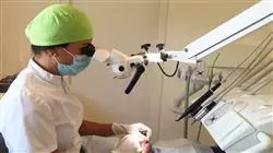
The Postgraduate diploma aims to cover the comprehensive training of the dentist in Endodontics with Optical Microscope, by providing the necessary skills to prepare them as highly qualified professionals in the field of Endodontics”
This online program aims to respond to the needs not only of its students, but also of society, anticipating the future demands of the environment. Change, the result of globalization and the imperatives of a new knowledge-based economy, requires ambitious modernization programs in the field of online training.
This training will be carried out in a balanced way, with a focus on endodontics, post-endodontic reconstruction and apical surgery with the intense involvement of anatomy, dental materials, radiology, the use of magnification, new technologies, and an interdisciplinary approach.
The knowledge acquired will allow the student to face working life from a more qualified position, giving them a clear advantage when it comes to finding a job, since they will be able to offer the application of the latest technological and scientific advances in the field of Endodontics.
The fundamental justification of the program is, therefore, to train a professional with adequate knowledge, skills, attitudes, values and competencies, who is able to serve society by satisfying its health demands, both in terms of prevention, diagnosis and treatment, in an ethical, efficient and safe manner. This professional must appreciate the need for lifelong professional development and continuing education, be able to efficiently utilize advances in knowledge and technology, and understand the central role of the patient in therapeutic decision making.
Expand your knowledge through the Postgraduate diploma in Endodontics with Optical Microscope, in a practical way and adapted to your needs”
This Postgraduate diploma in Endodontics with Optical Microscope contains the most complete and up-to-date scientific program on the market. The most important features of the program include:
- Clinical cases presented by experts in the different dental specialties. The graphic, schematic, and eminently practical contents with which they are created provide scientific and practical information on the disciplines that are essential for professional practice
- Latest information on Endodontics with an Optical Microscope
- Algorithm-based interactive learning system for decision-making in the presented clinical situations
- With special emphasis on evidence-based medicine and research methodologies in Endodontics with an Optical Microscope
- Content that is accessible from any fixed or portable device with an Internet connection
This Postgraduate diploma may be the best investment you can make when choosing a refresher program for two reasons: in addition to updating your knowledge in Endodontics with Optical Microscope, you will obtain a Postgraduate diploma from TECH Global University"
Forming part of the teaching staff is a group of professionals in the world of Dentistry, who bring to this course their work experience, as well as a group of renowned specialists, recognised by esteemed scientific communities.
The multimedia content developed with the latest educational technology will provide the professional with situated and contextual learning, i.e., a simulated environment that will provide immersive training program to train in real situations.
This program is designed around Problem Based Learning, whereby the physician must try to solve the different professional practice situations that arise during the course. For this reason, you will be assisted by an innovative, interactive video system created by renowned and experienced experts in the field of radiology with extensive teaching experience.
Increase your decision-making confidence by updating your knowledge with this Postgraduate diploma in Endodontics with Optical Microscope"

Don’t miss out on the opportunity to update your knowledge in Endodontics with Optical Microscope in order to improve your care of your patients"
Syllabus
The structure of the contents has been designed by a team of professionals knowledgeable about the implications of specialization in daily medical practice in who are aware of the relevance of the current relevance of training to be able to treat a patient with problems that require Endodontics with Optical Microscope and are committed to quality teaching through new educational technologies.
This Postgraduate diploma in Endodontics with Optical Microscope contains the most complete and up-to-date scientific program on the market”

Module 1. The Modern Concept of Endodontics
1.1. Reviewing the Concept of Dentinal Canal, Cementary Canal and Pulp Stump, Pulp Cap, or Differentiated Apical Periodontium
1.1.1. Dentinal Canal
1.1.2. Cementary Canal
1.1.3. Pulp Stump, Pulp Cap, or Differentiated Apical Periodontium
1.2. Reviewing the Concept of Root Cementum, Apical Foramen, Periodontal Membrane, and Alveolar Bone
1.2.1. Cementodentinal Junction
1.2.2. Root Apex
1.2.3. Root Cementum
1.2.4. Apical Foramen
1.2.5. Periodontal Membrane
Module 2. Pulpo-Periodontal Pathology and the Endoperiodontal Relations
2.1. Differential Diagnosis between Endodontic and Periodontal Lesions
2.1.1. General Considerations
2.1.2. The Pulpo-Periodontal Communication Pathways
2.1.3. Symptomatology and Diagnosis of Endoperiodontal Syndrome
2.1.4. Classification of Endoperiodontal Lesions
2.2. Endoperiodontal Lesions due to Root Abnormalities. Part I
2.2.1. General Considerations
2.2.2. Combined Endoperiodontal Lesions: Diagnosis
2.2.3. Combined Endoperiodontal Lesions: Treatment
2.3. Endoperiodontal Lesions due to Root Abnormalities. Part II
2.3.1. Pure Periodontal Lesions: Diagnosis
2.3.2. Pure Periodontal Lesions: Treatment
2.3.3. Conclusions
2.3.4. Other Treatment Options
2.4. Cracked Tooth Syndrome and Root Bursting. Part I
2.4.1. Crown Fracture without Pulp Involvement
2.4.2. Crown Fracture with Pulp Involvement
2.4.3. Crown Fracture with Pulp and Periodontal Involvement
2.4.4. Root Burst in an Endodontically Treated Tooth
2.5. Cracked Tooth Syndrome and Root Bursting. Part II
2.5.1. Fractura radicular por exceso de presión o fragilidad radicular
2.5.2. Root Fracture due to Excess Pressure or Root Fragility
2.5.3. Fracture due to Excessive Occlusal Contact or Overloading
2.6. Endoperiodontal Damage due to Accidents and Trauma
2.6.1. Crown-Root Fractures
2.6.2. Vertical and Horizontal Root Fractures
2.6.3. Contusion, Dental Luxation and Fracture of the Alveolar Process
2.6.4. Treatment of Alveolar-Dental Lesions
2.7. Endoperiodontal Lesions due to Resorption. Part I
2.7.1. Resorption due to Pressure
2.7.2. Resorption due to Pulp Inflammation or Internal Resorption
2.7.3. Non-Perforated Internal Resorption
2.7.4. Perforated Internal Resorption
2.7.5. Resorption due to Periodontal Inflammation
2.7.6. Inflammatory
2.7.7. Replacement, by Substitution or Ankylosis
2.7.8. Cervical Invasive
2.8. Endoperiodontal Lesions due to Resorption. Part II
2.8.1. Invasive Cervical Resorption in Endodontically Treated Teeth
2.8.2. Invasive Cervical Resorption without Pulp Involvement
2.8.3. Etiology and Prognosis of Cervical Resorption
2.8.4. Materials Used for the Treatment of Cervical Resorption
2.9. Periodontal Problems Related to Endodontic Surgery in Radicectomies, Hemisections, and Bicuspidations
2.9.1. Radisectomy or Root Amputation
2.9.2. Hemisection
2.9.3. Bicuspidization
Module 3. Retreatments
3.1. What is the cause of failure of an endodontically treated tooth?
3.1.1. Persistent or Secondary Endodontic Infections
3.1.2. Microbiology in the Root Filling Phase
3.2. Diagnosing Endodontic Failure
3.2.1. Clinical Evaluation of Root Canal Treatment
3.2.2. Radiographic Evaluation of Root Canal Treatment
3.2.3. Acceptable, Questionable, and Radiographically Unacceptable Root Canal Treatment
3.2.4. Diagnosing Apical Periodontitis with Cone Beam Computed Tomography (CBCT)
3.2.5. The Role of the Optical Microscope when We Need to Retreat a Tooth
3.2.6. Integration of Evaluative Factors in Determining the Outcome of Root Canal Treatment
3.3. Predisposing Factors for Post-Treatment Disease
3.3.1. Preoperative Factors that May Influence the Outcome of Root Canal Treatment
3.3.2. Intraoperative Factors that May Influence the Outcome of Root Canal Treatment
3.3.3. Postoperative Factors that May Influence the Outcome of Root Canal Treatment
3.4. Non-Surgical Clinical Retreatment
3.4.1. Preparing the Access Cavity
3.4.2. The Use of Ultrasound
3.4.3. Crown Removal
3.4.4. Removal of Bolts and/or Posts
3.4.5. Rotosonic VIbration
3.4.6. Ultrasound
3.4.7. Mechanical Option
3.4.8. Access to the Root Third
3.4.9. Gutta-Percha Solvents
3.4.10. Gutta-Percha Removal Techniques
3.4.11. Hedstroem Filing Technique
3.4.12. Techniques with Rotary Files
3.4.13. Removal via Ultrasound
3.4.14. Removal via Heat
3.4.15. Removal via Preheated Instruments
3.4.16. Removal with Files, Solvents, and Paper Cones
3.4.17. Paste Removal
3.4.18. Single Cone Gutta-Percha Removal with Solid Stem
3.4.19. Silver Tip Removal
3.4.20. Removal of Broken Instruments
Module 4. Endodontic Problems and Complications in Endodontics
4.1. Uncommon Root Anatomy in Different Teeth of the Dental Arch
4.1.1. Variations in the Root Anatomy of the Maxillary Incisors and Canines
4.1.2. Variations in the Root Anatomy of the Maxillary Premolars
4.1.3. Variations in the Root Anatomy of the Mandibular Incisors and Canines
4.1.4. Variations in the Root Anatomy of the Mandibular Premolars
4.2. Etiopathogenesis of Large Periapical Lesions and their Treatment in a Single Session
4.2.1. Pathologic Diagnosis of Granuloma
4.2.2. Anatomopathological Diagnosis of Cysts. Odontogenic Cysts
4.2.3. Bacteriological Considerations for Performing Endodontic Treatment of Large Periapical Lesions in a Single Session
4.2.4. Clinical Considerations for Performing Endodontic Treatment of Large Periapical Lesions in a Single Session
4.2.5. Clinical considerations on the Management of Fistulous Processes Associated with a Large Periapical Lesion
4.3. Treatment of Large Periapical Lesions in Multiple Sessions
4.3.1. Differential Diagnosis, Chamber Opening, Permeabilization, Cleaning, Disinfection, Apical Permeabilization, and Canal Drying
4.3.2. Intra-duct Medication
4.3.3. Temporary Crown Obutration (To close or not to close, that is the question)
4.3.4. Catheterization of the Fistulous Tract or Perforation of the Granuloma and Blind Scraping of the Apical Lesion of the Tooth
4.3.5. Guidelines for a Regulated Approach to a Large Periapical Lesion
4.4. Evolution in the Treatment of Large Periapical Lesions in Several Sessions
4.4.1. Positive Evolution and Treatment Control
4.4.2. Uncertain Evolution and Treatment Control
4.4.3. Negative Evolution and Treatment Control
4.4.4. Considerations on the Cause of Failure in the Conservative Treatment of Large Periapical Lesions
4.4.5. Clinical Considerations on Fistulous Processes in Relation to the Tooth of Origin
4.5. Location, Origin, and Management of Fistulous Processes
4.5.1. Fistulous Tracts Originating from the Anterosuperior Group
4.5.2. Fistulous Tracts Originating from the Maxillary Molars and Premolars
4.5.3. Fistulous Tracts Originating from the Anteroinferior Group
4.5.4. Fistulous Tracts Originating from the Mandibular Molars and Premolars
4.5.5. Cutaneous Fistulas of Dental Origin
4.6. The Problems of Maxillary First and Second Molars in Endodontic Treatment. The 4th Canal
4.6.1. Anatomical Considerations of the Maxillary First Molars of Children or Adolescents
4.6.2. Anatomical Considerations of Adult Maxillary First Molars
4.6.3. The Mesio-Buccal Root in the Maxillary First Molars. The 4th Canal or Mesio-Vesticulo-Palatine Canal and the 5th Canal
4.6.3.1. Ways to Detect the 4th Canal: See it Bleeding
4.6.3.2. Ways to Detect the 4th Canal: See its Entrance
4.6.3.3. Ways to Detect the 4th Canal: With a Manual File
4.6.3.4. Ways to Detect the 4th Canal: Using an Optical Microscope with Magnified Vision
4.6.3.5. Ways to Detect the 4th Canal: With a Mechanical File
4.6.4. The Disto-Buccal Root in the Maxillary First Molars
4.6.5. The Palatal Root in the Maxillary First Molars
4.7. The Problems of Mandibular First and Second Molars in Endodontic Treatment. 3 Ducts in the Mesial Root or the Intermediate Canal
4.7.1. Anatomical Considerations of the Mandibular First Molars of Children or Adolescents
4.7.2. Anatomical Considerations of Adult Mandibular First Molars
4.7.2.1. The Mesial Root in the Mandibular First Molars
4.7.2.2. The Distal Root in the Mandibular First Molars
4.7.3. Mandibular Molars with 5 Ducts
4.7.4. Anatomical Considerations of Adult Mandibular Second Molars
4.7.4.1. C-Shaped Canal
4.7.4.2. Molars with a Single Canal
4.7.5. Anatomical Considerations of the Mandibular Wisdom Teeth
Module 5. Surgery and Microsurgery in Endodontics
5.1. Surgical or Non-Surgical Retreatment. Decision-Making
5.1.1. Endodontic Surgery
5.1.2. Non-Surgical Retreatment
5.1.3. Surgical management
5.2. Basic Instruments
5.2.1. Scanning Tray
5.2.2. Anesthesia Tray
5.2.3. Rotary Instruments
5.2.4. Tipos de limas de endodoncia
5.3. Types of Endodontic Files
5.3.1. Incision through the Gingival Sulcus
5.3.2. Gingival Flap
5.3.3. Triangular Flap
5.3.4. Trapezoidal Flap
5.3.5. Modified Semilunar Incision
5.3.6. Semilunar Incision
5.4. Managing the Flap and Controlling Bleeding
5.4.1. Design of the Flap
5.4.2. Surgical Complication
5.4.3. General Considerations
5.4.4. Presurgical Considerations for Controlling Bleeding
5.4.5. Surgical Considerations for Controlling Bleeding
5.4.6. Local Anesthesia
5.4.7. Design and Elevation of the Flap
5.5. Techniques and Materials Used for Retropreparation and Retro-Obturation
5.5.1. Mineral Trioxide Aggregate (MTA)
5.5.2. Endodontic Application of MTA
5.5.3. Paraendodontic Surgery
5.5.4. Properties of MTA
5.5.5. Biodentine
5.6. Ultrasonic Tips and Optical Microscope as Essential Equipment
5.6.1. Types of Tips
5.6.2. Optical Microscope
5.6.3. Surgical Microscope (S.M.)
5.6.4. Appropriate Use of Instruments
5.6.5. Ultrasonic Devices and Designed Tips
5.7. The Maxillary Sinus and Other Anatomical Structures with which we Can Interact
5.7.1. Neighboring Anatomical Structures
5.7.2. Maxillary Sinus
5.7.3. Inferior Alveolar Nerve
5.7.4. Mental Foramen
5.8. Medication and Recommendations for Optimal Postoperative Care

A unique, key, and decisive training experience to boost your professional development”
Postgraduate Diploma in Endodontics with Optical Microscope
Boost your career in dentistry with our Postgraduate Diploma in Endodontics with Optical Microscopy program at TECH Global University! If you are a dentist with a passion for dental aesthetics and are looking to hone your skills in the area of endodontics, this specialization program is perfect for you. Our Postgraduate Diploma is taught in online mode, which gives you the flexibility to study from anywhere and adapt your schedule at your convenience. You will be able to access the contents of the program from your computer, tablet or cell phone, allowing you to advance in your studies at your own pace.
Enroll now and receive the best education
At TECH Global University, we understand the importance of constant updating in the field of esthetic dentistry and endodontics. Therefore, our program focuses on providing you with the latest techniques, tools and knowledge related to the use of the optical microscope in endodontic procedures. Our team of specialized dentistry teachers will be at your disposal to provide you with the necessary support throughout the program. You will be able to solve your doubts and receive personalized feedback to ensure a comprehensive and quality learning. One of the main advantages of our Postgraduate Diploma in Endodontics with Optical Microscopy is the opportunity to access practical demonstrations and real clinical cases. These experiences will allow you to strengthen your clinical skills and prepare you to face challenging cases in your professional practice. At the end of the program, you will obtain a university certification that will endorse your knowledge and skills in the area of endodontics with optical microscopy. This certification will give you a competitive advantage in the job market and will open doors to new professional opportunities in specialized dental clinics and high-demand care centers. Don't miss this opportunity to become an expert in optical microscope endodontics. Enroll in our Postgraduate Diploma program in Endodontics with Optical Microscopy at TECH Global University and take your career in cosmetic dentistry to the next level. Empower your professional future and provide your patients with the best dental treatments with the most advanced techniques!


