University certificate
The world's largest faculty of medicine”
Description
Through this Relearning-based Postgraduate diploma, you will develop advanced skills in the field of imaging and provide crucial findings to clarify forensic investigations"

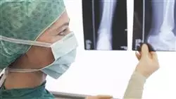
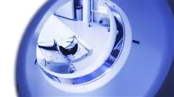
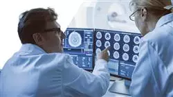
The Fourth Industrial Revolution has brought with it multiple technological advances, which have driven the development of high-resolution medical imaging equipment. Therefore, practitioners in the field of Forensic Radiology operate sophisticated machinery such as Computed Tomography to obtain detailed snapshots of bone and soft tissues. This enables medical professionals to contribute to the identification of unknown deceased individuals by comparing their previous medical records with anthropological and even odontological records. In this sense, specialists provide detailed radiological evidence that can be of great use both during forensic investigations and judicial proceedings.
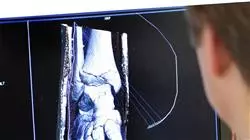
In this scenario, TECH has developed a complete Postgraduate diploma in Forensic Radiology in Pathologies by Compared Anatomy. Its purpose is to provide a high degree of specialization oriented to the characterization of bone and joint pathologies in radiological photographs. To make this possible, the academic itinerary will carry out an exhaustive analysis of the human skeleton that will allow graduates to identify anomalies such as the presence of relevant foreign objects in cases of traumatic deaths. At the same time, the syllabus will address the most common bone diseases in the forensic context, among which Osteoporosis, Rickets or Cancer stand out. Likewise, the didactic materials will offer the keys to detect signs of child abuse from the data obtained by means of tools such as MRI, X-rays or Axial Tomographies.
It should be noted that the approach of this program reinforces its innovative character. TECH offers a fully online educational environment, tailored to the needs of busy professionals who want to advance their careers. Through the Relearning methodology, based on the repetition of key concepts to fix knowledge and facilitate learning, flexibility is combined with a highly robust pedagogical approach. Additionally, specialists will have access to a library full of cutting-edge multimedia resources.
TECH's online methodology will allow you to choose the time and place to study, without hindering your professional work"
This Postgraduate diploma in Forensic Radiology in Pathologies by Compared Anatomy contains the most complete and up-to-date scientific program on the market. Its most notable features are:
- The development of practical cases presented by experts in Forensic Radiology
- The graphic, schematic and eminently practical contents with which it is conceived gather scientific and practical information on those disciplines that are indispensable for professional practice
- Practical exercises where the self-assessment process can be carried out to improve learning
- Its special emphasis on innovative methodologies
- Theoretical lessons, questions to the expert, debate forums on controversial topics, and individual reflection assignments
- Content that is accessible from any fixed or portable device with an Internet connection
You will incorporate into your daily practice the most innovative Ultrasound techniques to identify pathologies such as bone fractures, joint injuries or soft tissue inflammation"
The program’s teaching staff includes professionals from the field who contribute their work experience to this educational program, as well as renowned specialists from leading societies and prestigious universities.
The multimedia content, developed with the latest educational technology, will provide the professional with situated and contextual learning, i.e., a simulated environment that will provide immersive education programmed to learn in real situations.
This program is designed around Problem-Based Learning, whereby the professional must try to solve the different professional practice situations that arise during the course. For this purpose, students will be assisted by an innovative interactive video system created by renowned and experienced experts.
Do you want to specialize in Forensic Radiology of Pathologies in developing individuals? Achieve it with this syllabus in only 450 hours"

You will have access to a learning system based on repetition, with a progressive and natural teaching throughout the program"
Syllabus
Through this pathway, graduates will develop a detailed understanding of human skeletal anatomy. The syllabus will delve into the structural components of the Locomotor System, ranging from bones to joints. The syllabus will analyze the phases of biological maturation, so that specialists can establish age estimates. In tune with this, the program will emphasize the Forensic Radiology of the Child Skull, providing the usual traumas resulting from aggression. Therefore, experts will provide significant evidence that will contribute to the resolution of abuse investigations. The specialization will provide the keys to make the most of tools such as Axial Tomography or Magnetic Resonance Imaging.

You will have access to a study plan prepared by a distinguished teaching team, which will guarantee you a totally successful learning process"
Module 1. Forensic Radiology of the Non-Pathological and Non-Traumatic Human Skeleton
1.1. Forensic Radiology of the Locomotor System
1.1.1. Muscular System
1.1.2. Articular System
1.1.3. Skeletal System
1.2. Forensic Radiology of the Human Skeleton
1.2.1. Axial Skeleton
1.2.2. Appendicular Skeleton
1.2.3. Upper and Lower Extremities
1.3. Anatomical Plans and Axes of Movement in Forensic Investigation
1.3.1. Coronal Plan
1.3.2. Sagittal Plan
1.3.3. Transverse Plan
1.3.4. Bone Classification
1.4. Forensic Radiology of the Human Skull
1.4.1. Facial Bones
1.4.2. Neurocranium
1.4.3. Associated Pathologies
1.5. Forensic Radiology of the Spine
1.5.1. Cervical Vertebrae
1.5.2. Thoracic Vertebrae
1.5.3. Lumbar Vertebrae
1.5.4. Sacral Vertebrae
1.5.5. Associated Pathologies and Traumas
1.6. Forensic Radiology of the Coxal Bones
1.6.1. Ilium/Ischium/Sacral Complex
1.6.2. Public Symphysis
1.6.3. Associated Pathologies and Traumas
1.7. Forensic Upper Extremity Radiology
1.7.1. Long Bones
1.7.2. Bone Complexes of the Hands
1.7.3. Pathologies and Traumas
1.8. Forensic Radiology of the Lower Extremities
1.8.1. Long Bones
1.8.2. Bone Complexes of the Feet
1.8.3. Pathologies and Traumas
1.9. Forensic Pathologies and Traumas through Diagnostic Imaging
1.9.1. Congenital Diseases
1.9.2. Acquired Pathologies
1.9.3. Trauma and its Variants
1.10. Interpretation of Radiographic Images in the Forensic Field
1.10.1. Radiolucent Bodies
1.10.2. Radiopaque Bodies
1.10.3. Gray Scales
Module 2. Forensic Radiology of the Human Skeleton in Phases of Biological Maturation
2.1. Bone Physiopathology in the Forensic Context
2.1.1. Functions
2.1.2. Composition - Bone Tissue
2.1.3. Cellular Component
2.1.3.1. Bone-Forming Cells (Osteoblasts)
2.1.3.2. Bone Destroyers (Osteoclasts)
2.1.3.3. Mature Bone Cells (Osteocytes)
2.2. Osteogenesis in Individuals in the Forensic Context
2.2.1. Membranous Ossification Pathway
2.2.2. Chondral Ossification Pathway
2.2.3. Periosteum
2.3. Bone Vascularization in the Forensic Context
2.3.1. Main Pathway
2.3.2. Epiphyseal Pathway
2.3.3. Metaphyseal Pathway
2.3.4. Periosteal Arterial Pathway
2.4. Bone Growth in the Forensic Context
2.4.1. Width
2.4.2. Length
2.4.3. Associated Pathologies
2.5. Forensic Radiology of Pathologies in Developing Individuals
2.5.1. Congenital Diseases
2.5.2. Acquired Pathologies
2.5.3. Trauma and its Variants
2.6. Bone Diseases Through Diagnostic Imaging in the Forensic Context
2.6.1. Osteoporosis
2.6.2. Bone Cancer
2.6.3. Osteomyelitis
2.6.4. Osteogenesis Imperfecta
2.6.5. Rickets
2.7. Forensic Radiology of the Child Skull
2.7.1. Embryonic, Fetal and Neonatal Formation
2.7.2. Fontanelles and Fusion Phases
2.7.3. Facial and Dental Development
2.8. Forensic Radiobiological Osteology in the Adolescent
2.8.1. Sexual Dimorphism and Bone Growth
2.8.2. Bone Changes Resulting from Hormonal Action
2.8.3. Juvenile Growth Retardation and Metabolic Problems
2.9. Trauma and Categories of Childhood Fractures in Forensic Diagnostic Imaging
2.9.1. Frequent Traumas in Infantile Long Bones
2.9.2. Frequent Traumas in Infantile Flat Bones
2.9.3. Trauma Resulting from Aggression and Mistreatment
2.10. Radiology and Diagnostic Imaging Techniques in Forensic Pediatrics
2.10.1. Radiology for Neonates and Infants
2.10.2. Radiology for Children in Early Childhood
2.10.3. Radiology for Adolescents and Juveniles
Module 3. Radiodiagnosis of Pathologies Related to Forensic Investigation
3.1. Classification of Traumatic Fractures in the Forensic Context
3.1.1. Classification According to Skin Condition
3.1.2. Classification According to Location
3.1.3. Classification According to Fracture Trace
3.2. Stages of Bone Repair in the Forensic Context
3.2.1. Inflammatory Phase
3.2.2. Repair Phase
3.2.3. Remodelling Phase
3.3. Child Maltreatment and its Radiodiagnosis in a Forensic Context
3.3.1. Simple Radiography
3.3.2. Axial Tomography
3.3.3. Magnetic Resonance
3.4. Illegal Transport of Narcotics and Radiodiagnostics in a Forensic Context
3.4.1. Simple Radiography
3.4.2. Axial Tomography
3.4.3. Magnetic Resonance
3.5. Simple Radiographic Technique for Identification of Alterations within a Forensic Context
3.5.1. Cranial Pathologies
3.5.2. Thoracic Pathologies
3.5.3. Extremity Pathologies
3.6. Ultrasound Technique for Identification of Pathologies within a Forensic Context
3.6.1. Ultrasound
3.6.2. Obstetric
3.6.3. Wall
3.7. Computed Tomography and Identification of Pathologies in a Forensic Context
3.7.1. Cranial
3.7.2. Wall
3.7.3. Ultrasound
3.8. Magnetic Resonance Imaging and Pathology Identification in a Forensic Context
3.8.1. Cranial
3.8.2. Wall
3.8.3. Ultrasound
3.9. Diagnostic Angiography in a Forensic Context
3.9.1. Cranial
3.9.2. Ultrasound
3.9.3. Extremities
3.10. Virtopsia, Radiology in Forensic Medicine
3.10.1. Resonance
3.10.2. Tomography
3.10.3. Radiography

TECH's top priority is to help you acquire academic excellence and, therefore, boost your professional career. Join now!
Postgraduate Diploma in Forensic Radiology in Pathologies by Compared Anatomy
Forensic radiology plays a key role in the identification of bone pathologies in medico-legal investigations, and comparative anatomy offers a unique perspective by comparing human bone structures with those of other species. Are you passionate about this innovative field? This Postgraduate Diploma created by TECH Global University provides you with the perfect opportunity to immerse yourself in this exciting field of study. Through our online program, you will explore a wide range of pathologies, from fractures and tumors to degenerative diseases, using advanced imaging tools. You will also learn to interpret and analyze radiological images to identify bone pathologies and anomalies using anatomical comparison techniques between different species. As a result, you will become an expert in the identification of bone pathologies by means of comparative anatomy.
Get qualified with a Postgraduate Diploma in Forensic Radiology in Pathologies by Comparative Anatomy
The teaching team is composed of experts in forensic radiology, comparative anatomy and forensic medicine, who will guide you throughout your education and provide you with a unique perspective based on their practical experience in the field. In addition to the technical aspect, our program also focuses on developing communication and teamwork skills, as interdisciplinary collaboration is essential in the field of forensic radiology. You will learn to communicate your findings clearly and concisely in both written reports and oral presentations. Upon successful completion of the program, you will be prepared for key roles in the field of forensic radiology and medico-legal investigation, including forensic expert witness, forensic radiologist and more. Our program will provide you with a valuable, internationally recognized credential that will open doors in the exciting world of forensic science. Enroll now and take the first step toward an exciting future in forensic radiology!


