University certificate
The world's largest faculty of medicine”
Why study at TECH?
You will be able to update your knowledge on Obstetric and Gynecological Ultrasound through the best Hybrid professional master’s degree”
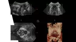
In an era marked by technology, the field of Gynecology has benefited, especially in the diagnostic field. In this sense, ultrasound devices have improved image quality and resolution, as well as their dimensions, which facilitate their use in any clinical space. These benefits have a direct impact on the detection of pathologies and better patient follow-up.
For this reason, specialists are constantly updating their technical skills in this field. Therefore, this 12-month Hybrid professional master’s degree in Obstetric and Gynecological Ultrasound was born, designed and developed by an excellent teaching team with extensive experience in this medical field.
This is a program that will lead the graduates to obtain an effective update on the use of ultrasound for the assessment of certain gynecological diseases, for the performance of echocardiographic and neurosonographic studies. All this, through innovative multimedia didactic material and clinical case studies, accessible 24 hours a day, from any digital device with an Internet connection.
The culmination of this program is the practical phase, which will allow the professionals to carry out a practical stay of 3 weeks in a first level health space in this field. In this unique experience, students will be able to integrate all the concepts covered in the theoretical phase directly and with real patients.
The physicians are faced with an unparalleled academic option that adapts to their daily personal activities and at the same time provides a direct response to their needs for updating their skills in the field of Obstetric and Gynecological Ultrasound.
Make the most of this opportunity to surround yourself with expert professionals and learn from their work methodology"
This Hybrid professional master’s degree in Obstetric and Gynecological Ultrasound contains the most complete and up-to-date scientific program on the market. Its most notable features are:
- Development of more than 100 clinical cases presented by gynecologists and obstetricians, experts in ultrasound techniques in pregnant patients or patients with gynecological pathologies
- Its graphic, schematic and eminently practical contents with which they are conceived, collect scientific and assistance information on those medical disciplines indispensable for professional practice
- Patient assessment and application of the latest international recommendations when detecting fetal anomalies or pathologies that seriously affect women's health
- Comprehensive systematized action plans for the main pathologies in the field of gynecology
- Presentation of practical workshops on diagnostic and therapeutic techniques in the gynecological patients
- An algorithm-based interactive learning system for decision-making in the clinical situations presented throughout the course
- Practical clinical guides on approaching different pathologies
- With a special emphasis on evidence-based medicine and research methodologies in Obstetric and Gynecological Ultrasound
- All of this will be complemented by theoretical lessons, questions to the expert, debate forums on controversial topics, and individual reflection assignments
- Content that is accessible from any fixed or portable device with an Internet connection
- In addition, you will be able to carry out a clinical internship in one of the best hospitals in the world
Take an intensive 3-week internship in a prestigious center and acquire all the knowledge to grow personally and professionally”
In this proposal for a Hybrid professional master’s degree, of a professionalizing nature and blended learning modality, the program is aimed at updating nursing professionals who perform their functions in intensive care units, and who require a high level of qualification. The content is based on the latest scientific evidence and is organized in a didactic way to integrate theoretical knowledge into nursing practice. The theoretical-practical elements allow professionals to update their knowledge and help them to make the right decisions in patient care.
Thanks to its multimedia content elaborated with the latest educational technology, they will allow the Gynecology professional to obtain a situated and contextual learning, that is to say, a simulated environment that will provide an immersive learning programmed to train in real situations. This program is designed around Problem-Based Learning, whereby the professional must try to solve the different professional practice situations that arise throughout the program. For this purpose, the students will be assisted by an innovative interactive video system created by renowned experts.
Access, whenever and wherever you want to the most innovative didactic material through any digital device with an Internet connection"
Get an effective update on ultrasound techniques in obstetrics and gynecology from the best specialists in this field"
Teaching Planning
The syllabus of this program will allow the graduates to be aware of the most rigorous scientific literature on the detection of gynecological pathologies through the use of ultrasound devices, as well as the techniques used with this technology for the follow-up of pregnant patients. An academic itinerary that will end with a practical stay of 120 hours with an excellent team of professionals with a deep experience in this field.
A lot of scientific readings with which you will be able to further extend the information provided in this program”
Module 1. Ultrasound. Normal study in gynecology
1.1. Normal anatomy in gynecology
1.1.1. Normal anatomy in gynecology
1.1.2. Normal gynecologic ultrasound anatomy: structures and anatomical landmarks to consider
1.1.3. Ultrasound technique: systematics of the exploration
1.1.4. Language and description of normality and pathology in the ultrasound technique
1.2. Physical principles of ultrasound. Technical Aspects
1.2.1. Basic Principles of Ultrasound Physics
1.2.2. Creation of the image from ultrasounds
1.2.3. Characteristics of the ultrasound image
1.2.4. Optimization of the gynecological ultrasound
1.2.5. Recognition and correction of artifacts
1.3. Types of ultrasound probes in gynecologic ultrasonography
1.3.1. Types of transducers
1.3.2. Advantages and disadvantages of the different probes and approaches
1.4. Technical principles of Doppler. Technical Aspects
1.4.1. Physical principles of Doppler
1.4.2. Main indications of Doppler in gynecologic ultrasound examination
1.4.3. Optimization of the Doppler technique
1.5. Technical principles of 3D/4D ultrasound. Technical aspects and usefulness
1.5.1. Basic principles of 3-4D ultrasound
1.5.2. Application of the 3-4D technique in gynecology
1.5.3. Systematics of the technique by structures: volume acquisition
1.5.4. Navigation, reconstruction and rendering of the ultrasound volume
1.5.5. Optimization of the reconstruction: default modes
1.5.6. Principles of multiplanar reconstruction or TUI
1.6. Use of ultrasound in gynecologic and obstetric emergencies
1.6.1. Applicability of ultrasound in gynecologic and obstetric emergencies
1.6.2. Systematic use of ultrasound technique in gynecological emergencies
1.6.3. Ultrasound in the differential diagnosis of acute abdomen
1.6.4. Ultrasound in the differential diagnosis of metrorrhagias
1.6.5. Ultrasound report in urgent gynecologic pathology
1.6.6. Limitations of ultrasonography: complementary techniques to be requested
1.7. Management of offline volumes
1.7.1. Presentation of different software
1.7.2. Volume storage
1.7.3. Recovery of volumes in the offline software
1.7.4. Two-dimensional plane navigation and optimization
1.7.5. 2D plane navigation: in time and space
1.7.6. Three-dimensional reconstruction
1.7.7. Optimization of the three-dimensional image
1.8. Complementary Techniques: Sonohysterography / hysterosonosalpingography
1.8.1. Basic principles of exploration
1.8.2. Systematic of the technique
1.8.3. Hysterosonography: technique, interpretation of the image and preparation of the report
1.8.4. Hysterosonosalpingography: technique, interpretation of the image and preparation of the report
1.9. Lines of research in gynecologic ultrasonography
1.9.1. Status of artificial intelligence applied to gynecologic ultrasound
1.9.2. The current and future role of ultrasound in the evaluation of the gynecologic oncologic patient
1.9.3. Elastography in gynecology
1.9.4. Ultrasound in the diagnosis and management of severe genital atrophy and genitourinary syndrome
Module 2. Pathology of the endometrium, myometrium and cervix
2.1. Ultrasound in benign endometrial pathology
2.1.1. Endometrial ultrasound normality: qualitative and quantitative assessment
2.1.2. Ultrasonography, endometrium and variation with the menstrual cycle
2.1.3. Three-dimensional technique in endometrial assessment
2.1.4. Description and terminology according to IETA group
2.1.5. Ultrasound in the assessment of endometrial hyperplasia
2.1.6. Ultrasound in the assessment of endometrial polyps
2.2. Ultrasonography of malignant endometrial pathology
2.2.1. Introduction: Endometrial Cancer
2.2.2. Ultrasound characteristics of endometrial cancer
2.2.3. Systematic local assessment of endometrial cancer
2.2.4. Systematic assessment of extraendometrial disease
2.2.5. Ultrasound in the assessment of recurrence of endometrial cancer
2.3. Gynecological ultrasound after abortion: Retention of conception debris / Sd. Asherman's syndrome
2.3.1. Endometrial ultrasound normality after complete miscarriage
2.3.2. Ultrasound in the diagnosis and follow up of the remnants of conception
2.3.3. Ultrasound in the assessment and follow-up of uterine synechiae
2.4. Ultrasound in the diagnostic study of fibroids
2.4.1. Definition and general aspects of myomas
2.4.2. Types of fibroids: classifications and implications
2.4.3. Description and ultrasound classification
2.4.4. Types of myoma degeneration
2.4.5. Ultrasound Characteristics: Doppler technique and three-dimensional reconstruction
2.4.6. Ultrasound monitoring of the patient with uterine myomatosis
2.4.7. Differential diagnosis, limitations of the technique and complementary explorations
2.5. Ultrasound in the therapeutic approach to myomas
2.5.1. Ultrasound in the treatment of myomas with radiofrequency
2.5.2. Ultrasound in the treatment of fibroids with high-frequency ultrasound (HIFU)
2.6. Ultrasonography in the assessment of malignant myometrial pathology
2.6.1. General information on malignant myometrial tumors
2.6.2. Ultrasound differential diagnosis of uterine sarcomas
2.6.3. Limitation of ultrasonography in the diagnosis of uterine sarcomas complementary tests
2.7. Adenomyosis
2.7.1. Basic concepts about adenomyosis
2.7.2. Ultrasound characteristics of the normal myometrium
2.7.3. Ultrasonographic characteristics of adenomyosis using the MUSA system
2.7.4. Report of the ultrasound description of the findings in the clinical report
2.7.5. Correlation of the pathological anatomy with the ultrasound assessment of the myometrial-endometrial junction
2.7.6. Limitations of ultrasound and complementary tests in the diagnosis and follow-up of adenomyosis
2.8. Ultrasound study in the evaluation of the cervix
2.8.1. Ultrasound anatomy of the normal cervix
2.8.2. Ultrasound characteristics and description of cervical tumors
2.8.3. Role of ultrasonography in the initial staging of cervical cancer
2.8.4. Role of ultrasonography in extracervical disease in cervical cancer
2.8.5. Ultrasonography in the follow-up of the patient with cervical cancer: evaluation of treatment and assessment of recurrences
2.9. Ultrasound study in the assessment of the vagina and vulva
2.9.1. Current evidence for ultrasound assessment of the vagina and vulva
2.9.2. Ultrasound applications
2.9.3. Systematic technique and findings
2.10. Ultrasound study in pediatric age
2.10.1. Introduction to the most frequent pediatric pathology
2.10.2. Normal ultrasound in the pediatric and adolescent patient
2.10.3. Recommended routes of approach: advantages and disadvantages
2.10.4. Ultrasound of precocious puberty
2.10.5. Ultrasonographic findings in intersexuality
2.10.6. Hematocolpos secondary to imperforate hymen
Module 3. Ovarian Pathology, Endometriosis and Pain
3.1. Ultrasonography in the assessment of benign ovarian pathology
3.1.1. Normal ovarian ultrasound anatomy
3.1.2. Generalities and classifications of benign ovarian pathologies
3.1.3. Systematic in the evaluation and ultrasound description of annexial pathology: ultrasound criteria of benignity
3.1.4. Types of tumors and ultrasound characteristics
3.1.5. Ovarian torsion: sonographic findings
3.2. Ultrasonography in the assessment of malignant ovarian pathology
3.2.1. Introduction and generalities malignant ovarian lesions
3.2.2. Classification and ultrasound systematics according to IOTA
3.2.3. Types of tumors and ultrasound characteristics
3.2.4. Ultrasonography in regional and distant staging of ovarian neoplasms
3.2.5. Limitations of ultrasound and complementary tests
3.2.6. Ultrasound in the follow-up and recurrence of patients with a history of ovarian neoplasia
3.2.7. Borderline tumors and ultrasound
3.3. Ultrasound study of tubal pathology
3.3.1. Ultrasound of normal tubes
3.3.2. Ultrasound findings in patients with hydrosalpinx
3.3.3. Ultrasonographic findings in patients with pelvic inflammatory disease
3.3.4. Malignant tubular pathology
3.4. Ultrasonography in the assessment of pelvic congestion syndrome
3.4.1. Definition, diagnosis and therapeutic approach
3.4.2. Ultrasound findings in patients with pelvic congestion syndrome
3.4.3. Complementary imaging tests
3.5. Ultrasonography in the diagnosis of ovarian endometriosis
3.5.1. Definition, repercussions and diagnosis
3.5.2. Systematic of the ultrasound technique
3.5.3. Ultrasound findings in patients with Ovarian Endometriosis
3.5.4. Differential Diagnosis and Complementary Tests
3.6. Ultrasonography in the diagnosis of extraovarian endometriosis
3.6.1. Definition, repercussions and diagnosis
3.6.2. Systematic of the ultrasound technique
3.6.3. Ultrasound assessment of the pelvis by structures and compartments
3.6.4. Assessment of extrapelvic implants: umbilical implants, trocar or on cesarean scars
3.6.5. Complementary imaging tests
3.7. Ultrasound in the patient with chronic pain in gynecology
3.7.1. Introduction and generalities
3.7.2. Ultrasound findings in gynecologic patients with chronic pain
3.7.3. Ultrasound in the local treatment of gynecologic patients with chronic pain
3.8. Ultrasound in breast pathology
3.8.1. Breast ultrasound anatomy
3.8.2. Systematics of the technique and probes
3.8.3. Ultrasonography in the assessment of benign breast pathology
3.8.4. Ultrasonography in the assessment of malignant breast pathology
3.9. Interventional ultrasound
3.9.1. Definition
3.9.2. Applications of interventional ultrasound in gynecology
3.9.3. Paracentesis technique
3.9.4. Technique of ultrasound-guided drainage of tuboovarian abscesses
3.9.5. Technique of alcoholization of endometriomas
3.9.6. Breast abscess drainage technique
Module 4. Reproduction and pelvic floor
4.1. Ultrasound in the diagnosis of infertility
4.1.1. Antral follicle count
4.1.2. Screening for tubal pathology
4.1.3. Endometrial pathology screening
4.2. Uterine Malformations
4.2.1. Classification of uterine malformations
4.2.2. Differential Diagnosis
4.2.3. T-shaped uterus
4.3. 3D ultrasound in reproduction
4.3.1. Introduction
4.3.2. 3D ultrasound of the ovary
4.3.3. 3D ultrasound of the uterus
4.4. Ultrasound in the treatment of reproduction
4.4.1. Ultrasonography in controlled ovarian stimulation for IVF
4.4.2. Ultrasonography for embryo transfer
4.4.3. Ultrasonography in programmed coitus
4.4.4. Ultrasonography in artificial insemination
4.5. Ultrasound anatomy of the pelvic floor
4.5.1. Normal ultrasound anatomy of the pelvic floor
4.5.2. Probes and approach routes: pros and cons of each one of them
4.5.3. Ultrasound Technique: How to obtain the two-dimensional reference plane
4.5.4. Dynamic Ultrasound: Rest, retention and Valsalva in the different structures of the pelvic floor
4.5.5. Improvement of the technique: to the conquest of an excellent image avoiding artifacts
4.5.6. Three-dimensional acquisition technique of the urogenital hiatus for the assessment of the levator pellucidis muscle
4.5.7. Volume storage and offline management
4.6. The role of ultrasound in the assessment of genital prolapse
4.6.1. Normal position of the pelvic organs: normal two-dimensional ultrasound plan
4.6.2. Assessment of the anterior compartment: technique and considerations
4.6.3. Assessment of the mid-technical compartment and considerations
4.6.4. Assessment of the posterior compartment: technique and considerations
4.6.5. Ultrasound findings and therapeutic implications
4.7. The role of ultrasound in the assessment of urinary incontinence
4.7.1. Ultrasound recognition of the anatomical structures involved in continence
4.7.2. Assessment of postvoid residual and its implications
4.7.3. Assessment of urethral slippage and implications
4.7.4. Ultrasound in the diagnosis of stress urinary incontinence
4.7.5. Ultrasound in the diagnosis of urgency urinary incontinence
4.8. The role of ultrasound in the assessment of obstetric trauma
4.8.1. Transperineal ultrasound technique in the assessment of the anal sphincter
4.8.2. Intravaginal ultrasound technique in the assessment of the anal sphincter
4.9. Usefulness of ultrasound in the post-surgical control of pelvic floor surge
4.9.1. Ultrasound characteristics of meshes in pelvic floor surgeries
4.9.2. Ultrasonography in the post-surgical follow-up of incontinence meshes
4.9.3. Ultrasound in the post-surgical follow-up of prolapse meshes
Module 5. First Trimester Ultrasound
5.1. Protocol for the study of first trimester ultrasound, normalcy
5.1.1. Gestational age and dating
5.1.2. Anatomic examination
5.1.3. Measurement of markers of aneuploidy
5.1.4. Placenta, uterus and adnexa
5.2. Uncertain Location Pregnancy
5.2.1. Differential Diagnosis
5.2.2. Blood Biochemistry
5.2.3. Action Protocol
5.3. Early gestation (trophoblastic disease, amnios, vesicles, etc.)
5.3.1. Gestational sac
5.3.2. Yolk vesicle
5.3.3. Amniotic and chorionic cavity
5.3.4. Embryo
5.3.5. Early embryonic development
5.3.6. Early pathology
5.3.7. Findings of poor gestational prognosis
5.4. Ultrasound markers of first trimester chromosomopathy
5.4.1. Introduction
5.4.2. Nuchal translucency
5.4.3. Nasal bone
5.4.4. Venous ductus
5.4.5. Tricuspid Regurgitation
5.5. Other first trimester ultrasound markers (angulopathy, trans intracranial, uterine, etc.)
5.5.1. Intracranial translucency
5.5.2. Frontomaxillary angle
5.5.3. Retrononasal triangle
5.5.4. Uterine arteries
5.6. Diagnosable morphologic pathology in the first trimester
5.6.1. Cranial and central nervous system pathology
5.6.2. Face
5.6.3. Skeletal System
5.6.4. Thorax and neck
5.6.5. Heart
5.6.6. Abdomen
5.6.7. Urinary System
5.7. First trimester aneuploidy screening
5.7.1. History of aneuploidy screening
5.7.2. Blood Biochemistry
5.7.3. Ultrasound markers
5.7.4. Study Protocol
5.8. Fetal DNA in maternal blood (also in twins)
5.8.1. History of fetal DNA
5.8.2. Methods of Analysis
5.8.3. Practical Aspects
5.8.4. Fetal fraction and absence of result
5.8.5. Fetal DNA in twins
5.8.6. Microdeletions
5.8.7. Interpretation of results and protocol
5.9. First trimester preeclampsia screening
5.9.1. History of preeclampsia screening
5.9.2. Types of screening
5.9.3. Components of screening
5.9.4. Available calculators
5.9.5. Cut-off points and prevention
5.9.6. Follow-up in high risk of preeclampsia
5.10. Invasive Techniques
5.10.1. Amniocentesis
5.10.2. Chorion Biopsy
5.10.3. Multiple Gestation
5.11. Basic genetics in obstetrics
5.11.1. Genetic Concepts
5.11.2. Mendelian Genetics
5.11.3. Non-Mendelian genetics
5.11.4. Prenatal genetic testing
Module 6. Second Trimester Ultrasound
6.1. Protocol for the study of second trimester ultrasound, normality
6.1.1. Gestational age and second trimester dating
6.1.2. Skull and central nervous system
6.1.3. Limbs and spine
6.1.4. Thorax and heart
6.1.5. Abdomen
6.1.6. Genitourinary system
6.2. Assessment of the placenta and umbilical cord
6.2.1. Abnormalities of placental shape, location and insertion
6.2.2. Placental tumors
6.2.3. Vascular anomalies and hematomas
6.2.4. Abnormalities of the cord
6.3. Spectrum of placenta accreta
6.3.1. Classification
6.3.2. Ultrasound Diagnosis
6.3.3. Magnetic Resonance
6.3.4. Management
6.4. Cervical assessment. Risk of Premature Delivery
6.4.1. Measurement Techniques
6.4.2. Risk of Premature Delivery
6.4.3. Recommendations of scientific societies
6.5. Ultrasound markers of second trimester chromosomopathy
6.5.1. History of second trimester markers
6.5.2. Likelihood ratio
6.5.3. Ultrasound markers
6.5.4. Management
6.6. Malformations of the abdomen and abdominal wall
6.6.1. Umbilical Hernia
6.6.2. Omphalocele
6.6.3. Gastroschisis
6.6.4. Bladder exstrophy
6.6.5. Other abdominal wall anomalies
6.6.6. Abdominal cysts
6.6.7. Gastrointestinal Pathology
6.7. Malformations of the face, neck and thorax
6.7.1. Facial malformations
6.7.2. Malformations of the neck
6.7.3. Thoracic Malformations
6.8. Spinal malformations
6.8.1. Hemivertebra
6.8.2. Neural Tube Defects
6.8.3. Sacrococcygeal Teratomas
6.8.4. Flow regression sequence
6.9. Limb malformations
6.9.1. Skeletal Dysplasias
6.9.2. Clubfoot
6.9.3. Reductional alterations
6.9.4. Arthrogryposis
6.10. Genitourinary malformations
6.10.1. Renal agenesis
6.10.2. Obstructive Pathology
6.10.3. Renal ectopias
6.10.4. Multicystic and polycystic kidney
6.10.5. Other renal anomalies
6.10.6. Adrenal abnormalities
6.10.7. Bladder anomalies
6.10.8. Genital abnormalities
6.11. Fetal Hydrops
6.11.1. Definition
6.11.2. Ultrasound abnormalities
6.11.3. Etiology
6.11.4. Management
6.11.5. Prognosis
6.11.6. Associated Complications
6.11.7. Recurrence
Module 7. Third Trimester Ultrasound
7.1. Protocol for the study of third trimester ultrasound, normality
7.1.1. Gestational age and third trimester dating
7.1.2. Objectives of third trimester ultrasound
7.1.3. Ultrasound systematics
7.2. Diagnostic malformation pathology in the third trimester
7.2.1. Introduction
7.2.2. Most frequent malformations
7.3. Estimation of fetal growth
7.3.1. Definitions
7.3.2. Estimation of fetal weight. Bio-Meters
7.3.3. Normality curves and percentiles
7.4. Doppler study in third trimester ultrasound
7.4.1. Umbilical Artery
7.4.2. Middle Brain Artery
7.4.3. Venous ductus
7.4.4. Uterine arteries
7.4.5. Others
7.5. Growth disturbances (PEG and CIR)
7.5.1. Introduction
7.5.2. Small for gestational age fetus
7.5.3. Intrauterine growth retardation
7.6. Hemodynamics and fetal impairment in intrauterine growth retardation
7.6.1. Fetal hemodynamics
7.6.2. Biophysical Profile
7.6.3. Fetal Monitoring
7.7. Fetal Macrosomia
7.7.1. Introduction
7.7.2. Risk Factors
7.7.3. Diagnosis
7.7.4. Complications
7.7.5. Management
7.8. Intrapartum ultrasound
7.8.1. Technique
7.8.2. Station evaluation
7.8.3. Head attitude evaluation
7.8.4. Indications
7.9. Abnormalities of amniotic fluid
7.9.1. Introduction
7.9.2. Oligohydramnios
7.9.3. Polyhydramnios
7.9.4. Management
Module 8. Multiple Gestation
8.1. Introduction and embryology
8.1.1. Introduction
8.1.2. Embryology
8.1.3. Classification
8.2. Ultrasound Diagnosis Screening for aneuploidy in multiple pregnancies
8.2.1. Introduction
8.2.2. Ultrasound Diagnosis
8.2.3. Dating
8.2.4. First trimester aneuploidy screening
8.3. Two-chorionic twin gestation
8.3.1. Introduction
8.3.2. Follow-up of normoevolutionary two-chorionic gestation
8.3.3. Termination of normoevolutionary bicurial gestation
8.4. Normal monochorionic twin gestation
8.4.1. Introduction
8.4.2. Follow-up of nomoevolutionary monochorionic pregnancy
8.4.3. Termination of normoevolutionary monochorionic gestation
8.5. Complicated monochorionic gestation (TAPS, TRAP, TFF)
8.5.1. TAPS
8.5.2. TRAP
8.5.3. TFF
8.5.4. Discordant structural malformation
8.6. Growth retardation in twin gestation (monochorionic and bicorionic)
8.6.1. Introduction
8.6.2. Delayed growth in two-chorionic gestation
8.6.3. Growth retardation in monochorionic gestation
8.7. Prevention and screening for preeclampsia
8.7.1. Introduction
8.7.2. First trimester preeclampsia screening
8.7.3. Prevention of preeclampsia in twin gestations
8.8. Screening for preterm delivery in twin gestation
8.8.1. Introduction
8.8.2. Cervical assessment, evidence
8.8.3. Prevention of Prematurity
8.9. Fetal reduction in twin gestation
8.9.1. Fetal reduction in monochorionic gestation
8.9.2. Risks of fetal reduction
8.10. Fetal abortion in twin gestation
8.10.1. Introduction
8.10.2. Fetal abortion in two-chorionic gestation
8.10.3. Fetal death in monochorionic gestation
Module 9. Fetal Echocardiography
9.1. Normal Fetal Echocardiography
9.1.1. Indications for Fetal Echocardiography
9.1.2. Fetal Echocardiography Technique
9.1.3. Measurement of Cardiac Structures. Z-score
9.2. Cardiac Functional Study Normality
9.2.1. Pathophysiology of Cardiac Dysfunction
9.2.2. Functional Echocardiography Technique
9.2.3. Advanced Techniques
9.3. Septal Defects
9.3.1. Atrial Septal Defects
9.3.2. Defects of the Interventricular Septum
9.3.3. Atrioventricular Septal Defects
9.3.4. Double Inlet Single Ventricle
9.4. Right Heart Defects
9.4.1. Tricuspid Pathology
9.4.2. Pulmonary Stenosis
9.4.3. Complete Septal Pulmonary Atresia
9.5. Left Heart Defects
9.5.1. Mitral Pathology
9.5.2. Aortic Stenosis
9.5.3. Aortic Coarctation
9.5.4. Interrupted Aortic Arch
9.6. Conotruncal Anomalies
9.6.1. Tetralogy of Fallot
9.6.2. Main Artery Transposition
9.6.3. Double Outlet Right Ventricle
9.6.4. Truncus Arteriosus
9.7. Venous Return Abnormalities
9.7.1. Superior Vena Cava Abnormalities
9.7.2. Inferior Vena Cava Filter Abnormalities
9.7.3. Persistence of the Right Umbilical Vein
9.7.4. Venous Ductus Venosus Agenesis
9.8. Abnormalities of Cardiac and Situs Position
9.8.1. Situs Abnormalities
9.8.2. Heterotaxy Syndromes
9.9. Cardiac Rhythm Abnormalities
9.9.1. Irregular Rhythms
9.9.2. Bradycardias
9.9.3. Tachycardias
Module 10. Fetal neurosonography
10.1. Fetal neurosonography. Normality
10.1.1. Indications for fetal neurosonography
10.1.2. Technique of fetal neurosonography
10.1.3. Measurement of Brain Structures
10.2. Alterations of the Head Circumference and the Skull
10.2.1. Microcephaly
10.2.2. Macrocephaly
10.2.3. Encephalocele
10.2.4. Other Alterations
10.3. Ventriculomegaly
10.3.1. Ultrasound Diagnosis
10.3.2. Etiology
10.3.3. Associated Anomalies and Study
10.3.4. Prognosis
10.3.5. Recurrence
10.4. Midline Anomalies
10.4.1. Corpus Callosum Anomalies
10.4.2. Absence of cavum septi pellucidi
10.4.3. Holoprosencephaly
10.5. Posterior Fossa Anomalies
10.5.1. Dandy Walker Malformation
10.5.2. Megacisterna Magna
10.5.3. Blake's Cyst
10.5.4. Vermis Hypoplasia
10.5.5. Other Anomalies
10.6. Cystic Pathology of the Central Nervous System
10.6.1. Choroid Plexus Cyst
10.6.2. Connatal Cyst
10.6.3. Arachnoid Cyst
10.6.4. Other Alterations
10.7. Ischemic/Hemorrhagic Pathology of the Central Nervous System
10.7.1. Porencephaly
10.7.2. Schizencephaly
10.7.3. Other Ischemic and Hemorrhagic Injuries
10.8. Tumors of the Central Nervous System and Vascular Anomalies
10.8.1. Teratoma
10.8.2. Tuberous Sclerosis
10.8.3. Aneurysm of Galen's Vein
10.8.4. Thrombosis of Dural Venous Sinuses
10.9. Sulcation Anomalies
10.9.1. Introduction
10.9.2. Lissencephaly
10.9.3. Hemimegalencephaly
10.10. Magnetic Resonance Imaging in the Study of the Central Nervous System
10.10.1. Introduction
10.10.2. Indications
10.10.3. Adequate Gestational Age for Fetal MRI
10.10.4. Utility of fetal MRI in the study of the nervous system
You have a library of teaching resources 24 hours a day, 7 days a week”
Hybrid Professional Master's Degree in Obstetric and Gynecologic Ultrasound
Do you want to master the art of obstetric and gynecological ultrasound? Then TECH Global University's Hybrid Professional Master's Degree is designed for you. Get ready to delve into a rigorous and exciting academic program that will provide you with the skills and knowledge you need to become an expert in this vitally important field. You will have access to top-notch virtual classroom education, combined with in-person internships at renowned medical organizations. At TECH, we pride ourselves in providing you with a comprehensive and balanced learning experience that will allow you to acquire practical skills under the supervision of highly qualified professionals. Our outstanding teaching team is comprised of leaders in the field of obstetric and gynecologic ultrasound, who will guide you throughout the program. They will provide you with personalized support, sharing their knowledge and clinical experiences to enrich your learning. During the program, you will explore in depth the theoretical and practical fundamentals of obstetric and gynecologic ultrasound. From the study of anatomy and physiology to image interpretation and pathology diagnosis, you'll become an expert in this cutting-edge technique
Expand your knowledge in Obstetric and Gynecologic Ultrasound
Our institution is at the forefront of medical education, offering state-of-the-art technology, cutting-edge resources and a stimulating learning environment. Plus, you'll become part of a diverse and global academic community, interacting with students and professionals from around the world. Upon completion of the program, you will receive a prestigious certificate attesting to your skills and knowledge in obstetric and gynecologic ultrasound. This degree will open doors to exciting opportunities in hospitals, clinics, research centers and more, where you can apply your skills and make a real difference in the health of women and their babies. Don't miss this unique opportunity to become an expert in obstetric and gynecologic ultrasound - enroll in TECH Global University's Hybrid Professional Master's Degree in Obstetric and Gynecologic Ultrasound and take a leap toward success in your medical career.