University certificate
Scientific endorser

The world's largest faculty of medicine”
Introduction to the Program
With this Postgraduate diploma, based on Relearning, you will address the significant radiological images to clarify causes of deaths during criminal investigations"

More and more healthcare entities are calling for the incorporation of specialists with a high degree of specialization inRadiology of Trauma with firearms and explosives. These physicians are in charge of analyzing and interpreting radiological images to evaluate internal injuries caused by the impact of projectiles from objects such as guns, rifles, rifles and explosives. In this way, they determine the trajectory of the bullets inside the victim's body and the sequence of events that led to the victim's death. In addition, these professionals translate their findings into detailed reports that can be presented as scientific evidence in different legal proceedings.
For this reason, TECH implements a Forensic Radiology in Bone Trauma in Forensic Radiology in Bone Trauma that will delve into the various injury patterns generated by firearms, as well as the characterization of wounds. The didactic materials will delve into the most innovative radiological techniques for the study of blunt weapon injuries. Therefore, experts will acquire advanced skills to master modern tools such as Magnetic Resonance Imaging, Computerized Axial Tomography or X-rays. At the same time, the syllabus will deal with the Virtual Autopsy procedure, so that graduates can examine the tissues and internal organs of the body without the need to make incisions or physical dissections on the corpses.
TECH offers a 100% online educational environment, tailored to the needs of health professionals seeking to advance their careers. In addition, it uses the revolutionary Relearningmethodology, consisting of the repetition of key concepts to fix knowledge and facilitate learning. Therefore, the combination of flexibility and a robust pedagogical approach makes the university program highly accessible. The only thing experts will need is a device with Internet access to enter the virtual platform and enjoy an educational experience that will take their professional practice to a higher level.
The 100% online methodology of this university program will allow you to obtain an optimized learning without leaving your own home"
This Postgraduate diploma in Forensic Radiology in Bone Trauma contains the most complete and up-to-date scientific program on the market. The most important features include:
- The development of practical cases presented by experts in Forensic Radiology
- The graphic, schematic and eminently practical contents with which it is conceived gather scientific and practical information on those disciplines that are indispensable for professional practice
- Practical exercises where the self-assessment process can be carried out to improve learning
- Its special emphasis on innovative methodologies
- Theoretical lessons, questions to the expert, debate forums on controversial topics, and individual reflection assignments
- Content that is accessible from any fixed or portable device with an Internet connection
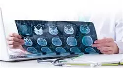
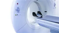
You will handle Computed Axial Tomography and obtain detailed images in cross sections of the body to detect even internal hemorrhages"
The program’s teaching staff includes professionals from the field who contribute their work experience to this educational program, as well as renowned specialists from leading societies and prestigious universities.
The multimedia content, developed with the latest educational technology, will provide the professional with situated and contextual learning, i.e., a simulated environment that will provide immersive education programmed to learn in real situations.
This program is designed around Problem-Based Learning, whereby the professional must try to solve the different professional practice situations that arise during the course. For this purpose, the student will be assisted by an innovative interactive video system created by renowned and experienced experts
You will have at your disposal the most advanced radiological techniques for the study of injuries caused by sharp weapons. You will make the most accurate diagnoses"

This program will make you a more complete professional by equipping you with the most effective resources to meet today's challenges in radiological image interpretation"
Why study at TECH?
TECH is the world’s largest online university. With an impressive catalog of more than 14,000 university programs available in 11 languages, it is positioned as a leader in employability, with a 99% job placement rate. In addition, it relies on an enormous faculty of more than 6,000 professors of the highest international renown.

Study at the world's largest online university and guarantee your professional success. The future starts at TECH”
The world’s best online university according to FORBES
The prestigious Forbes magazine, specialized in business and finance, has highlighted TECH as “the world's best online university” This is what they have recently stated in an article in their digital edition in which they echo the success story of this institution, “thanks to the academic offer it provides, the selection of its teaching staff, and an innovative learning method aimed at educating the professionals of the future”
A revolutionary study method, a cutting-edge faculty and a practical focus: the key to TECH's success.
The most complete study plans on the university scene
TECH offers the most complete study plans on the university scene, with syllabuses that cover fundamental concepts and, at the same time, the main scientific advances in their specific scientific areas. In addition, these programs are continuously being updated to guarantee students the academic vanguard and the most in-demand professional skills. In this way, the university's qualifications provide its graduates with a significant advantage to propel their careers to success.
TECH offers the most comprehensive and intensive study plans on the current university scene.
A world-class teaching staff
TECH's teaching staff is made up of more than 6,000 professors with the highest international recognition. Professors, researchers and top executives of multinational companies, including Isaiah Covington, performance coach of the Boston Celtics; Magda Romanska, principal investigator at Harvard MetaLAB; Ignacio Wistumba, chairman of the department of translational molecular pathology at MD Anderson Cancer Center; and D.W. Pine, creative director of TIME magazine, among others.
Internationally renowned experts, specialized in different branches of Health, Technology, Communication and Business, form part of the TECH faculty.
A unique learning method
TECH is the first university to use Relearning in all its programs. It is the best online learning methodology, accredited with international teaching quality certifications, provided by prestigious educational agencies. In addition, this disruptive educational model is complemented with the “Case Method”, thereby setting up a unique online teaching strategy. Innovative teaching resources are also implemented, including detailed videos, infographics and interactive summaries.
TECH combines Relearning and the Case Method in all its university programs to guarantee excellent theoretical and practical learning, studying whenever and wherever you want.
The world's largest online university
TECH is the world’s largest online university. We are the largest educational institution, with the best and widest online educational catalog, one hundred percent online and covering the vast majority of areas of knowledge. We offer a large selection of our own degrees and accredited online undergraduate and postgraduate degrees. In total, more than 14,000 university degrees, in eleven different languages, make us the largest educational largest in the world.
TECH has the world's most extensive catalog of academic and official programs, available in more than 11 languages.
Google Premier Partner
The American technology giant has awarded TECH the Google Google Premier Partner badge. This award, which is only available to 3% of the world's companies, highlights the efficient, flexible and tailored experience that this university provides to students. The recognition as a Google Premier Partner not only accredits the maximum rigor, performance and investment in TECH's digital infrastructures, but also places this university as one of the world's leading technology companies.
Google has positioned TECH in the top 3% of the world's most important technology companies by awarding it its Google Premier Partner badge.
The official online university of the NBA
TECH is the official online university of the NBA. Thanks to our agreement with the biggest league in basketball, we offer our students exclusive university programs, as well as a wide variety of educational resources focused on the business of the league and other areas of the sports industry. Each program is made up of a uniquely designed syllabus and features exceptional guest hosts: professionals with a distinguished sports background who will offer their expertise on the most relevant topics.
TECH has been selected by the NBA, the world's top basketball league, as its official online university.
The top-rated university by its students
Students have positioned TECH as the world's top-rated university on the main review websites, with a highest rating of 4.9 out of 5, obtained from more than 1,000 reviews. These results consolidate TECH as the benchmark university institution at an international level, reflecting the excellence and positive impact of its educational model.” reflecting the excellence and positive impact of its educational model.”
TECH is the world’s top-rated university by its students.
Leaders in employability
TECH has managed to become the leading university in employability. 99% of its students obtain jobs in the academic field they have studied, within one year of completing any of the university's programs. A similar number achieve immediate career enhancement. All this thanks to a study methodology that bases its effectiveness on the acquisition of practical skills, which are absolutely necessary for professional development.
99% of TECH graduates find a job within a year of completing their studies.
Postgraduate Diploma in Forensic Radiology in Bone Trauma
Forensic radiology plays a crucial role in the investigation of bone trauma cases, helping to identify injuries, determine cause of death and reconstruct events prior to death. Are you passionate about forensic science and looking to specialize in the field of radiology for bone trauma investigation? This Postgraduate Diploma developed by TECH Global University is ideal for you. The program offers you the perfect opportunity to acquire advanced knowledge in this highly specialized field. Through our online program, you will immerse yourself in the study of advanced imaging techniques, such as X-rays, computed tomography (CT), magnetic resonance imaging (MRI) and more. You will also learn how to interpret and analyze radiological images to identify fractures, trauma injuries and other bone abnormalities that can be crucial in criminal and medico-legal investigations.
Get qualified with a Postgraduate Diploma in Forensic Radiology in Bone Trauma
The program's teaching team is made up of experts in forensic radiology and forensic medicine, who will guide you throughout your education and provide you with a unique perspective based on their practical experience in the field. In addition to the technical aspect, our program also focuses on developing communication and teamwork skills, as interdisciplinary collaboration is essential in the field of forensic radiology. As such, you will learn to communicate your findings clearly and concisely, both in written reports and presentations. Upon successful completion of the program, you will be prepared for key roles in the field of forensic radiology, including forensic expert witness, forensic radiologist, criminal investigator and more. The program you acquire will provide you with a valuable, internationally recognized credential that will open doors in the exciting and challenging world of forensic science. Take the opportunity to advance your career and become an expert in forensic radiology. Enroll now and take the first step toward an exciting future in bone trauma investigation!


